J Vasc Bras. 2012;11(4):277-285. Abstract
Background: Ulcer is a severe complication from chronic venous insuiciency; thus, its pathophysiology needs to be deeply understood. Venous Doppler ultrasonography is the most appropriate complementary imaging study, enabling the study of the supericial and deep venous system, the diameter and low of the veins. Recent studies have suggested that popliteal vein relux is an important factor for the development of ulceration.
Objective: To evaluate the venous relux of the femoropopliteal segment in patients with venous ulcers.
Study design: Prevalence study.
Methods: Were enrolled 104 patients with 118 lower extremities with venous ulcers. Patients underwent Doppler ultrasonography of the afected limb showing the venous relux of the femoropopliteal segment and popliteal vein diameter. Primary variable was venous relux in the femoropopliteal segment; and secondary variable was diameter of the popliteal vein.
Results: Venous relux in the femoropopliteal segment was observed in 56 of the 118 limbs with venous ulcer (47.45%) in 104 patients. he mean diameter of the popliteal vein was 1.14 cm, whereas 0.6 cm was the normal mean diameter of the population.
Conclusion: Venous relux in the femoropopliteal segment is a major factor in assessing the prognosis of these patients. Increased diameter of the popliteal vein relects the magnitude of venous insuiciency.
Keywords: venous ulcer; Doppler ultrasonography; popliteal vein.
Resumo
Contexto: Como a úlcera é uma grave complicação da insuiciência venosa crônica, é necessário o conhecimento amplo de sua isiopatologia. A ultrassonograia Doppler venosa é o exame complementar mais adequado, que possibilita o estudo do sistema venoso supericial e profundo, sua anatomia e isiologia. Trabalhos recentes valorizam o reluxo em Veia Poplítea como importante fator para o desenvolvimento deste quadro clínico.
Objetivo: Avaliar o reluxo em segmento venoso femoropoplíteo em pacientes com úlcera varicosa.
Tipo de Estudo: Estudo de prevalência.
Métodos: Cento e quatro pacientes apresentando 118 membros inferiores com úlcera varicosa. Procedimentos: exame de ultrassonograia Doppler venosa do membro acometido, observado o reluxo no segmento venoso femoropoplíteo e diâmetro da Veia Poplítea. Variáveis: Primária: reluxo no segmento venoso femoropoplíteo. Secundária: diâmetro da Veia Poplítea.
Resultados: A presença de reluxo no segmento venoso femoropoplíteo foi observada em 56 (47,45%) dos 118 membros com úlcera varicosa, examinados em 104 pacientes. O diâmetro médio da Veia Poplítea foi de 1,14 cm, sendo o diâmetro médio normal da população 0,6 cm.
Conclusão: O reluxo venoso no segmento venoso femoropoplíteo é um importante fator na avaliação do prognóstico destes pacientes, o aumento de diâmetro da Veia Poplítea relete a magnitude da insuiciência venosa.
Palavras-chave: úlcera varicosa; ultrassonograia Doppler; Veia Poplítea.
Doppler ultrasonography of the femoropopliteal segment in
patients with venous ulcer
Avaliação do segmento venoso femoropoplíteo pela ultrassonografia Doppler em pacientes
com úlcera varicosa
Jesus Antonio de Carvalho Abreu1, Guilherme Benjamin Brandão Pitta2, Fausto Miranda Júnior3
Study carried out at Ambulatório de Cirurgia Vascular e no Laboratório de Ecografia Vascular do Hospital Getúlio Vargas, Avenida Frei Serafim, sem número, Teresina PI, hospital terciário da rede pública do Estado do Piauí.
hesis submitted to the Federal University of São Paulo, Paulista School of Medicine, to obtain the title of Master of Science.
1 Master in cardiovascular surgery, Federal University of São Paulo (UNIFESP) - São Paulo (SP), Brazil. Specialist in vascular surgery by the Brazilian Society of Angiology and Vascular Surgery. 2 PhD in cardiovascular surgery (UNIFESP) - São Paulo (SP), Brazil. Associate Professor at the State University of Medical Sciences of Alagoas (UNCISAL).
3 Full Professor of Vascular Surgery, Department of Surgery, Paulista School of Medicine (EPM), Federal University of São Paulo (UNIFESP) - São Paulo (SP), Brazil.
Financial support: None.
his study evaluated the prevalence of relux in a femoropopliteal venous segment and the diameter of the PV in patients with venous ulcers.
Methods
Patients with venous ulcer conirmed by physical examination were recruited to undergo DUS scanning. he study included 104 patients and 118 limbs with venous ulcers, distributed as follows: 14 patients had ulcers in both legs; all ulcers were classiied as CEAP C6. Patients were both men and women with no occlusions in the femoropopliteal segment (FPS), and all signed an informed consent term ater receiving the necessary information from the author. Data were collected using a standardized form.
A formula to deine sample size for inite populations22
was used, and the result was 103 individuals for a 2% proportion of venous ulcer in the population (p = 0.02), 98% of individuals without venous ulcer in the population (q = 0.98), 1.96 constant corresponding to the 95% conidence index and p = 0.05 (95%Z = 1.96) and d = 0.027 constant corresponding to the population density in the city of Teresina, Brazil, which has 800,000 inhabitants (N).
Sample size estimation (size determination formula)22
(Equation 1):
× × ×
= =
× − + × × 2
2 ( 1) 2 103
z p q N
n
d N Z p q 1
DUS scanning was performed according to service routine: the patient was standing and facing the examiner; the limb to be examined was slightly bent and externally rotated; and weight was supported on the contralateral limb. he exam was performed using a color digital scanner (Logic® 500, GE), Windows® operating system, and
4- to 10-MHz linear transducers. he venous system was evaluated, with special attention to the FPS, as well as the anteroposterior and latero-lateral aspects of the PV, whose measurements should be added up and divided by two to calculate mean diameter in each patient.
Venous relux at the saphenofemoral junction (SFJ) was evaluated while the patient performed the Valsalva maneuver, and, in the other segments, while the examiner applied manual compression of the muscles distal to the transducer to produce and detect low during the DUS study of the lower limbs in real time. Flow usually stops ater these maneuvers; any time of up to 1 second may be classiied as physiological for the deep veins, and for supericial and perforating veins, of up to 0.5 seconds. Perforating veins with a diameter greater than 0.35 cm are also classiied as having insuiciency23.
Introduction
he venous system of lower limbs has a deep and a supericial system, and the two are connected by the perforating veins, which direct the low from the supericial to the deep veins towards the heart. he deep system is responsible for draining 85% of the blood low of lower limbs, whereas the supericial system drains, with the help of valves that block relux, about 15% of the low in the ascending direction when in a standing position1. here
are 90 and 200 valves in the venous system of each lower limb; they are usually bicuspids and direct the low to the heart2. he physiological consequences of valve lesions are
relux and persistent venous distension due to retrograde pressure, particularly when standing3.
A venous leg ulcer, deined as an open wound between the knee and the ankle, usually at the level of the malleoli and which does not healed in four weeks4, is the most serious
lesion in chronic venous insuiciency (CVI) of the lower extremities5 and afects 1% to 2% of the world population6,7.
CVI is a set of signs and symptoms whose major physiopathological factor is chronic venous hypertension in the lower limb, commonly caused by valve incompetence that may afect the supericial, deep and perforating veins, together or separately7-10.
he relative importance of relux in several sites of the supericial and deep systems in the physiopathology of venous ulcers remains unclear11. In 10% to 50% of the
patients with venous ulcers, incompetence is limited only to the supericial veins11, whereas incompetent communicating
veins are found in 70% to 100% of the cases12. Numerous
studies showed that deep vein relux plays an important role in cases of ulceration13-15.
he popliteal vein (PV) is a component of the deep venous system, and its incompetence has been associated with diiculties in venous ulcer healing; studies have suggested that a PV with competent valves acts as a barrier against infrapatellar deep venous relux12,13,16-18.
Color Doppler ultrasound (DUS) is a noninvasive and painless method without collateral efects that may be performed as many times as necessary. It may be used to conirm a diagnosis by evaluating both vein diameter and anatomy, and it has a speciic advantage over other methods in the accurate determination of venous disease distribution and extent19. Diameter and venous relux
measurements evaluated by DUS are reliable. he shape of vein cross section remains practically the same when in an anatomic position20. DUS examination is part of both the
followed by leg heaviness in 90 (86.53%), pruritus in 89 (85.7%), burning sensation in 83 (79.80%), cramps in 83 (79.80%), fatigue in 76 (73.07%), and paresthesia in 67 (64.42%).
Ulcer diameter, measured in centimeters in two directions as the longer and the shorter distances between the internal margins, ranged from 1 to 12 centimeters, and mean diameter was 3 centimeters. hese indings were used to calculate the area of each ulcer, which was then classiied as small, when the area was smaller than 10 cm2 (83 cases,
70.33%), medium, 10 to 100 cm2 (32, 27.11%), and large,
greater than 100 cm2 (3, 2.5%).
In the analysis of previous deep venous thrombosis (DVT), 67 (64.42%) patients denied it, 20 (19.23%) reported having had at least one episode, and 17 (16.63%) did not know.
Total or segmental GSV insuiciency was seen in 98 (83.05%) limbs.
Perforating vein insuiciency was found in 79 (66.94%) limbs. Mean number of insuicient perforating veins was 2.5 for each patient.
Discussion
Chronic venous insuiciency (CVI) with leg ulceration, a disease found all over the world, has physical, social, economic and emotional consequences that may oten lead to anxiety and depression, depending on its duration and the socioeconomic and cultural characteristics of the population under study24. Ulcerations are deined as CEAP
C6 - open venous ulcers25.
Morbidity among patients with venous ulcers, as well as absenteeism and poor quality of life24,26,27, is signiicant,
particularly in the northeastern region of Brazil, where the hot climate and the exhausting working load for most of the population contribute to the worsening of this disease.
Doppler ultrasound (DUS), when combined with an evaluation of clinical characteristics, may be useful in making an accurate diagnosis of both functional and anatomic venous changes28-34, and the CEAP25 classiication
provides an accurate diagnosis for diferent groups under study10.
he comparisons with other studies revealed that there is a great variation in FPS relux, although most authors found a mean value similar to the one reported here28-34.
Hemodynamic characteristics and the main relux points in the genesis of venous ulcers suggest that supericial venous system insuiciency is the most frequent cause of trophic lesions secondary to venous disease35,36.
hose patients that have wounds that do not heal with he primary variable was FPS relux, the secondary,
PV diameter, and complementary data were sex, age, time of ulcer activity, occupation, heredity, use of contraceptive hormones and form of administration, clinical conditions, ulcer diameter, deep vein thrombosis (DVT), great saphenous vein (GSV) insuiciency, perforating vein insuiciency.
Results
Fourteen patients were excluded from the study: two did not sign the informed consent term; eleven, who had lower extremity ulcers and varicose veins, also had comorbidities that might explain the lesion, such as collagenosis (ive), ischemia (four), ankle brachial pressure lower than 0.9, and tegumentary leishmaniasis (two) diagnosed by means of biopsy and histopathology; and one patient with a psychiatric disorder, who was not cooperative. he study, therefore, included 104 patients, 14 with bilateral ulcers, at a total of 118 limbs.
Relux in the FPS was found in 56 limbs (47.45%). Mean PV diameter in this study was 1.14 cm: the smallest diameter was 0.62 cm, and the largest, 2.01 cm. Mean diameter in the population according to the literature ranges from 0.5 to 0.7 cm. In this study, the normal diameter was deined as 0.6 cm20.
Sixty nine patients (75.96%) were women, and the women to men ratio were 3:1.
Patient age ranged from 31 to 80 years, and mean age was 53 years: in the group of patients 30 to 39 years old, there were 15 patients (15.38%), in the 40 to 49, twenty-three (22.11%), in the 50 to 59, thirty-one (30.76%), in the 60 to 69, twenty-eight (27.88%); in the 70 to 79, ive (4.80%) and in the 80 to 89 group, two (1.92%).
he most prevalent occupation was farming, for 54 patients (51.92%), followed by 22 patients (21.15%) whose occupation was housekeeping, that is, they were housewives; 8 were salespeople (7.69%); there were also 4 seamstresses (3.84%), 3 civil servants (2.88%), 2 cooks (1.92%), 2 mechanics (1.92%), 2 teachers (1.92%), 2 hairdressers (1.92%), 2 masons (1.92%), 1 stevedore (0.96%), 1 baker (0.96%), and 1 health visitor (0.96%).
In the item about heredity, patients were asked whether their irst degree relatives had varicose veins corresponding to CEAP clinical class 2 (C2) to 6 (C6), and 80 (76.92%) of the patients answered airmatively.
he use of contraceptives was denied by 73 (92.4%) of the patients, and 6 (7.6%) reported using them, all orally.
venous ulcers and suggested that supericial venous system insuiciency is the most frequent cause of trophic lesions secondary to venous disease, in agreement with other studies.35,36 hose patients whose lesions do not heal with
clinical treatment, medication, compression or surgery of the supericial veins are candidates to valve reconstruction of the deep vein systems, in which case the PV has an important role in prognosis.35,37,41 Sukuvatykh et al. found
relux insuiciency in infrapatellar deep veins of the microcirculation, a condition that favors the appearance of trophic ulcers36. Rosales et al. performed PV reconstruction
in 19 patients that did not respond to clinical treatment or conventional varicose vein surgery and found that healing occurred in up to three months in 13.68% of the patients42.
Other groups of authors and surgeons, such Labas and Ohradka or Raju and Fredericks, have, for a long time, recommended FPS valve reconstruction as an essential procedure for the healing of refractory lesions14,43.
Mean PV diameter in our study was 1.14 cm, and mean diameter in the population, according to the literature, ranges from 0.5 to 0.7 cm; there is little variation of the cross-sectional diameter of veins when measured in the same position using DUS20.
Salles-Cunha et al. studied the changes in FPS diameter to plan the placement of an endovascular vein valve and found values of 8.4 mm to 9.7 mm for PV with venous hypertension due to valve insuicient. hey classiied their results as fundamentally important for the planning of this type of treatment44.
PV diameter also seems to afect the area of venous ulcer, as shown in Table 5, which shows that a mean increase in PV diameter is followed by a corresponding increase in the venous ulcer area, which suggests that the greater the PV diameter, the greater the ulcer size.
he analysis of sex revealed that our indings were similar to those described in the literature6,7,13,39. A recent
study found a proportion that is similar to the one we found clinical treatment, medication, compression or surgery of
the supericial veins are candidates for valve reconstruction of the deep venous system, and the PV plays an important role in prognosis35,37-39. Relux insuiciency in infrapatellar
deep veins of the microcirculation favors the appearance of trophic ulcers35. Carpentier et al. performed PV valve
reconstruction in 19 patients that had not responded to clinical treatment or conventional varicose vein surgery, and in 13 (68%) cases healing was complete in three months40. According to other authors and surgeons, FPS
valve reconstruction is essential for the healing of refractory lesions14,34.
his study demonstrated the association between venous relux and venous distension, as shown in Table 1, as the increase in PV diameter was followed by a greater relux proportion, both in supericial and deep veins. In the calf, PV has a relevant role, because this is the point of convergence of the venous system at this level. herefore, the increase in its diameter is a sign of the magnitude of venous insuiciency20 (Table 1).
A recent clinical trial used PV diameter as one of its main evaluation parameters and compared compression treatment and medication in patients with chronic venous insuiciency. he authors found a decrease in PV diameter when treatment was successful, which also afected patient symptoms38.
In this study, FPS relux was found in 56 (47.45%) of the limbs, but not in 62 (52.55%). Other authors found deep venous insuiciency in 9.3% to 85.5% of their cases33,
a very large variation between the results of several studies, although most had a mean value similar to the one found in our study23,30-33. Relux was more frequent in the supericial
veins, as shown by GSV in Table 6, than in the deep veins, whereas the frequency of relux in the FPS and GSV increased as PV diameter increased, as seen in Table 1.
Sukovatykh et al. studied the hemodynamic characteristics and the main relux points in the genesis of
Table 1. Distribution of PV diameter according to FPS and GSV relux in 118 limbs.
Popliteal vein diameter
Femoropopliteal
segment relux P Great saphenous vein relux p
With Without With Without
0.60 to 0.90 7 10 0.233 12 6 0.079
0.91 to 1.20 29 37 0.163 57 8 0.001
1.21 to 1.50 16 12 0.227 24 4 0.001
1.51 to 1.80 3 3 - 5 1
1.81 to 2.10 1 0 - 0 1
-Total 56 62 - 98 20
in 80% of the cases, edema in 65.5%, heaviness in 53.3%, cramps in 53%, lipodermatosclerosis in 39%, supericial thrombophlebitis in 33.5%, venous ulcer in 32%, eczema in 22% and cellulitis in 12.5%46. heir percentage of signs
and symptoms was below the ones that we found, but their patients were classiied as CEAP 2 to 6, whereas our study included only the clinical phase of CEAP 6 and, therefore, more advanced disease (Table 4).
A study using a questionnaire compared 288 patients with small or great FV insuiciency and 550 individuals in the general population to investigate, speciically, muscle cramps. he incidence of calf cramps was signiicantly greater in the group with venous insuiciency than in the general population (91% and 75%)52. heir rate was
also greater than the one we found in this study, even if compared with the general population, and the most plausible explanation was the better muscle conditions, maybe as a result of the fact that most were farmers and walked frequently during their daily activities.
Venous ulcers are the most advanced stage of venous disease and are a relex of chronic venous hypertension associated with inlammation. heir characteristics, particularly their surface area, are parameters to estimate severity and healing time. Most of the last venous ulcers found in this study had an area below 10 cm2 and were
classiied as small. hey accounted for 70% of all cases, followed by the 27% of the cases that were mid-sized ulcers, measuring 10 to 100 cm2, and 2.5% of large ulcers, with an
and suggested that heredity and gestations are responsible for this proportion (Table 2)39.
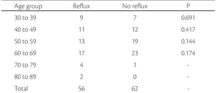
Varicose veins progress spontaneously, and younger patients have a lower percentage of varicose veins than individuals older than 60 years. he prevalence of venous ulcer follows that trend and also increases with age in both sexes: its greatest prevalence is found around age 80 years40,45,46. his study did not ind patients younger than 30
years with venous ulcers, and age distribution peaked at 60 to 69 years, but mean age was 53 years. Most studies in the literature found a higher mean age, which may be explained by the longevity of their populations, as most studies have been conducted in Europe or the USA. In another study, treatment of 202 patients with the Penang venous disease in Malaysia, who formed a group of great ethnic diversity comprising, for example, Chinese, Malays and Indians in a region where socioeconomic conditions are similar to those found in the northeastern region of Brazil, mean age was 52 years46 (Table 3).
In all the occupations found in our study, people spend long periods of time standing or under high temperatures, which contributes to the progression of venous disease, as reported in similar studies47-49. In Malaysia, the most
frequent occupations among patients with varicose veins were: homemakers (43%), blue collar workers (19%) and salespeople (12%). he highest rate of venous ulcers was found among indigenous people, and 32.5% of all patients were treated, and the cause suggested by the authors in their discussion was their low body mass index and low socioeconomic status46.
Genetic inheritance as an etiological factor of primary varicose veins is practically a consensus, but the form of gene transmission remains hypothetical. he explanation most oten found is polygenic inheritance50, which is
afected by individual and environmental factors and the price that humans pay for adopting a biped position and walking on rigid loors51. Inheritance is estimated at 50%,
more frequent when the mother is afected and more serious when inherited from the father51. A prospective analysis
of 67 patients and their parents, in a study that included spouses and their parents as controls at a total of 402 individuals, found development of varicose veins in 90% of the cases whose parents were both afected, 25% among men and 62% among women when one of the parents was afected, and in 20% of the cases when parents did not have the disease, which conirmed the hypothesis of autosomal dominant transmission with variable penetrance51.
A study conducted in Malaysia investigated symptoms of patients that sought treatment for varicose veins, and the authors found, in decreasing order of severity, pain
Table 2. Distribution of 118 limbs according to sex in a group of
pa-tients with venous ulcer examined to investigate FPS relux.
Sex Relux No relux p
Women 12 16 0.227
Men 45 45
-Total 57 61
FPS – Femoropopliteal segment.
Table 3. Distribution of 118 limbs according to age in a group of
pa-tients with venous ulcer examined to investigate FPS relux.
Age group Relux No relux P
30 to 39 9 7 0.691
40 to 49 11 12 0.417
50 to 59 13 19 0.144
60 to 69 17 23 0.174
70 to 79 4 1
-80 to 89 2 0
-Total 56 62
he frequency of relux in the supericial veins is greater when combined with deep vein relux. Labropoulus et al. found that ulcer incidence increases when there is also extended relux to the supericial veins, and that 47% of the patients with symptomatic post-thrombotic syndrome had supericial and deep venous insuiciency23.
Most studies recommend surgery of the supericial system as the adequate treatment in case compression and medications fail57,58; it is only in the most refractory cases
that the hypothesis of procedures in the deep system are examined.
he efective contraction of the calf muscles may generate pressures greater than 300 mmHg, which promotes blood low. To ensure the direction of low, veins have valves that prevent relux during this cycle of muscle contraction and relaxation59. he PV is a component of the deep venous
system, and its incompetence has been strongly associated with diiculties in the healing of venous ulcers. Studies suggested that a PV with competent valves acts as a barrier against infrapatellar deep venous relux12,15.
Mean PV diameter is relevant because the venous systems converges at this point in this level; an increase in diameter indicates the magnitude of venous insuiciency and is an important parameter of response to either clinical, surgical, conventional or endovascular treatment.
Data about sex, age, ulcer time of activity, occupation, heredity, use of contraceptive hormones and form of administration, clinical conditions, ulcer diameter, presence of relux, history of DVT, GSV insuiciency and insuiciency of perforating veins have a particular importance in characterizing the sample under study according to demographic, socioeconomic and cultural traits, which makes it possible to establish critical analyses of the environment where patients live and make comparisons with populations with diferent characteristics.
Conclusions
he frequency of venous relux in the femoropopliteal segment in patients with venous ulcer in this study was 47.45%, and patients with relux had greater morbidity, a factor that should be included in the evaluation of their prognosis.
Mean diameter of the popliteal vein in the group under study was 1.14 cm, about twice the mean diameter in the general population, which is indicative of the magnitude of venous insuiciency. his increase results from greater retrograde pressure when standing and from dilatation of the veins that converge at this level, which is associated, in area larger than 100 cm2. hese indings are similar to those
reported by other authors and can be used to estimate patient prognosis, response to treatment and time necessary for healing41,53,54. In the study conducted by Milic et al., venous
ulcer area smaller than 20 cm2 was one of the factors of a
good prognosis of healing ater compression treatment53.
Margolis et al., however, used the parameters of an ulcer larger than 5 cm2 or at least 6 months of time it was
open to estimate prognosis of time it would take to heal ater compression treatment54. Chaby et al. also evaluated
venous ulcer surface area and found that the larger the lesion, the worst the prognosis; however, they did not divide their sample into groups55. Ioannou et al. evaluated 519
patients and 798 limbs with CVI, sixty with venous ulcers, classiied as CEAP 5 and 6. Twenty-six patients in the latter group, that is, 43.3% of the sample, had post-thrombotic syndrome; their venous ulcers were more diicult to heal and the efect of relux elimination in the supericial system ater surgery was not clear56.
Table 4. Distribution of number of associated symptoms in a group of
104 patients with venous ulcer.
Symptoms Patients
1 3
2 3
3 5
4 20
5 10
6 23
7 40
Total 104
Table 5. Distribution of venous ulcer area according to femoropopliteal
segment relux and mean popliteal vein diameter in 118 lower limbs. Venous ulcer area Relux
(FPS)
No relux
(FPS) p
Mean diameter (PV)
<10 cm² 40 43 0.371 1.09
10 to 100 16 16 - 1.19
>100 cm² 0 3 - 1.31
FPS – Femoropopliteal segment. PV – popliteal vein.
Table 6. Distribution of 118 lower limbs according to femoropopliteal
segment relux and great saphenous vein relux in a group of 104 pa-tients with venous ulcer.
Incompetent venous segment Limbs with relux
Femoropopliteal 20
Great saphenous vein 60
scanning. Br J Surg. 1994;81:39-41. PMid:8313114. http://dx.doi. org/10.1002/bjs.1800810112
16. O’Donnell Junior TF, Mackey WC, Shepard AD, Callow AD. Clinical, hemodynamic, and anatomic follow-up of direct venous reconstruction. Arch Surg. 1987;122:474-82. http://dx.doi. org/10.1001/archsurg.1987.01400160100016
17. Bauer G. he etiology of leg ulcers and their treatment by resection of the popliteal vein. J Int Chir. 1948;8:937-67.
18. Shull KC, Nicolaides AN, Fernandes e Fernandes J, et al. Signiicance of popliteal relux in relation to ambulatory venous pressure and ulceration. Arch Surg. 1979;114:1304-6. PMid:496632. http://dx.doi. org/10.1001/archsurg.1979.01370350106012
19. Nicolaides NA. From symptoms to leg edema: eicacy of Dalon 500mg. Angiology. 2003; 54(S1)33-44. http://dx.doi. org/10.1177/000331970305400105
20. Lurie F, Ogawa T, Kistner RL, Eklof B. Changes in venous lumen size and shape do not afect of volume low measurements in healthy volunteers and patients with primary chronic venous insuiciency. J Vasc Surg. 2002;35:522-526. PMid:11877702. http:// dx.doi.org/10.1067/mva.2002.121565
21. Van Bemmelen PS, Beach K, Bedford G, et al. Quantitative segmental evaluation of venous valvular relux with ultrasound scanning. J Vasc Surg. 1989;10(4):425-431. PMid:2677416.
22. Fleming MD, Berkebile JS, Hofer RM. Computer Aided Analysis of LANDSAT-l MSS Data: A Comparison of hree Approaches Including a Modiied Clustering Approach. Proceedings of the 1975 Symposium on Machine Processing of Remotely Sensed Data, Purdue University, West Lafayette, Indiana.
23. Labropoulus N, Leon M, Nicolaides AN, et al. Venous relux in patients with ulceration with and other symptoms. J Vasc Surg. 1994;20:20-6. http://dx.doi.org/10.1016/0741-5214(94)90171-6
24. Souza Nogueira G, Rodrigues Zanin C, Miyazaki MC, Pereira de Godoy JM. Venous leg ulcers and emotional consequences. Int J Low Extrem Wounds. 2009;8(4):194-6. PMid:19934181. http:// dx.doi.org/10.1177/1534734609350548
25. Eklof B, Rutherford RB, Bergan JJ, et al. Revision of the CEAP classiication for chronic venous disorders: consensus statement. J Vasc Surg. 2004;40(6):1248-52. PMid:15622385. http://dx.doi. org/10.1016/j.jvs.2004.09.027
26. Bergqvist D, Lindholm C, Nelzen O. Chronic leg ulcers: the impact of venous disease. J Vasc Surg. 1999;29(4):752-5. http://dx.doi. org/10.1016/S0741-5214(99)70330-7
27. Browse NL, Burnand KG, Irvine AT, Wilson NM. Úlcera venosa: diagnóstico. In: Browse NL, Burnand KG, Irvine AT, Wilson NM, editors. Doenças Venosas. Rio de Janeiro: Di-Livros; 2001. p. 485-520.
28. Fan CM. Epidemiology and pathophysiology of varicose veins. Tec Vasc Inter Radiol. 2003;3:108-10. http://dx.doi.org/10.1053/S1089-2516(03)00060-X
29. Labropoulos N, Giannoukas AD, Nicolaides AN, Ramaswami G, Leon M, Burke P. New insights the pathophysiologic condition of venous ulceration with color-low duplex imaging: implications for treatment? J Vasc Surg. 1995;22(1):45-50. http://dx.doi.org/10.1016/ S0741-5214(95)70087-0
this study, with the diameter of the corresponding venous ulcer (Table 5).
References
1. Van Bemmelen PS, Beach K, Bedford G, et al. he mechanisms of venous valve closure. Its relationship to the velocity of reverse low. Arch Surg. 1990;125:617-619. PMid:2184798. http://dx.doi. org/10.1001/archsurg.1990.01410170063013
2. Strandness Junior DE, hiele B. Selected Topics in Venous Disorders. Mount Kisco: Futura; 1981. chapt. 2, 491p.
3. Zwiebel WJ. Insuiciência Venosa crônica, veias varicosas e mapeamento da veia safena. In: Zwiebel WJ. Introdução à ultra-sonograia vascular. 3. ed. Rio de Janeiro: Editora Revinter; 1996. p. 319-327.
4. Wilson E. Just briely prevention and treatment of leg ulcers. Health Trends. 1989; 2:97.
5. National Guideline Clearinghouse. Management of chronic venous leg ulcers. A national clinical guideline. http://www.guideline.gov. Acessado: 3/17/2012.
6. Allam MJ, Ruckley CV, Harper DR, Dale JJ. Chronic ulceration of the leg: extent of the problem and provision of care. Br Med J (Clin Res Ed). 1985; 290(6485):1855-6. http://dx.doi.org/10.1136/ bmj.290.6485.1855
7. Mafei FHA, Magaldi C, Pinho SZ, et al. Varicose veins and chronic venous insuiciency en Brazil: prevalence among 1755 inhabitants of country town. J Epidemiol. 1986;15:210.
8. Fowkes FG, Evans CJ, Lee AJ. Prevalence and risk factors of chronic venous insuiciency. Angiology. 2001;52(Suppl 1):S5-15. PMid:11510598. http://dx.doi.org/10.1177/000331970105200102
9. Ad Hoc Committee American venous Forum: Classiication and grading of chronic venous desease in the lower limbs. A Consensus Statement. J Cardiovasc Surg. 1997;38:437-41.
10. Vanhoutte PM, Corcaud S, Montrion C. Venous disease: From pathophysiology to quality of life. Angiology. 1997;48:559-567. PMid:9242153. http://dx.doi.org/10.1177/000331979704800702
11. Sethia KK, Darke SG. Long saphenous incompetence as a cause of venous ulceration. Br J Surg. 1984;71:754-5. http://dx.doi. org/10.1002/bjs.1800711006
12. Wilson NM, Rutt DL, Browse NL. Repair and replacement of deep vein valves in the treatment of venous insuiciency. Br J Surg. 1991;78:388-94. PMid:2032094. http://dx.doi.org/10.1002/ bjs.1800780404
13. McEnroe CS, O’Donnell Junior TF, Mackey WC. Correlation of clinical indings with venous hemodynamics in 386 patients with chronic venous insuiciency. Am J Surg. 1988;156:148-52. http:// dx.doi.org/10.1016/S0002-9610(88)80377-5
14. Raju S, Fredericks R. Valve reconstruction procedures for nonobstructive venous insuiciency: rationale, techniques, and results in 107 procedures with two- to eight-year follow-up. J Vasc Surg. 1988;7:310-10.
43. Labas P, Ohradka B. Anti-relux surgery of the popliteal vein. Bratisl Lek Listy. 1998;99(2):116-8.
44. Salles-Cunha SX, Shuman S, Beebe HG. Planning endovascular vein valve implantation: signiicance of vein size variability. J Vasc Surg. 2003;37(5):984-90. PMid:12756343. http://dx.doi.org/10.1067/ mva.2003.245
45. Takahashi PY, Chandra A, Cha SS, Crane SJ. A predictive model for venous ulceration in older adults: results of a retrospective cohort study. Ostomy Wound Manage. 2010;56(4):60-6. PMid:20424293 PMCid:2975563.
46. Murli NL, Navin ID. Classical varicose vein surgery in a diverse ethnic community. Med J Malaysia. 2008;63(3):193-8. PMid:19248688.
47. Tuchsen BF, Krause N, Hannerz H, Kristensen TS. Standing at work and varicose veins. Scand J Work Environ Health. 2000;26:414-20. PMid:11103840. http://dx.doi.org/10.5271/sjweh.562
48. Ziegler S, Eckhardt G, Stoger R, Machula J, Rudiger HW. High prevalence of chronic venous disease in hospital employees. Wien Klin Wochenschr. 2003;115(15-16):575-9. PMid:14531170. http:// dx.doi.org/10.1007/BF03040451
49. Kamber V, Widmer LK, Munst G. Prevalence. In: Widmer LK, editor. Peripheral Venous Disorders. Prevalence and Sociomedical Importance. Bern: Hans Huber Publishers; 1978. chapt. 4, p. 43-50.
50. Matousek V, Prerovsky I. A contribution to the problem of the inheritance of primary varicose veins. Hum Hered. 1974;24:225-35. http://dx.doi.org/10.1159/000152655
51. Cornu-Trenard A, Boivin P, Baud NM, et al. Importance of the familial factor in varicose disease: Clinical study of 134 families. J-Derm Surg Oncol. 1994;20:318-326.
52. Hirai M. Prevalence and characteristics of muscle cramps in patients with varicose veins. Vasa. 2000;29(4):269-73. PMid:11141650. http://dx.doi.org/10.1024/0301-1526.29.4.269
53. Milic DJ, Zivic SS, Bogdanovic DC, Karanovic ND, Golubovic ZV. Risk factors related to the failure of venous leg ulcers to heal with compression treatment. J Vasc Surg. 2009;49(5):1242-7. PMid:19233601. http://dx.doi.org/10.1016/j.jvs.2008.11.069
54. Margolis DJ, Berlin JA, Strom BL. Which venous leg ulcers will heal with limb compression bandages? Am J Med. 2000;109(1):15-9. http://dx.doi.org/10.1016/S0002-9343(00)00379-X
55. Chaby G, Viseux V, Ramelet AA, Ganry O, Billet A, Lok C. Refractory venous leg ulcers: a study of risk factors. Dermatol Surg. 2006;32(4):512-9. PMid:16681658. http://dx.doi.org/10.1111/j.1524-4725.2006.32104.x
56. Ioannou CV, Giannoukas AD, Kostas T, et al. Patterns of venous relux in limbs with venous ulcers. Implications for treatment. Int Angiol. 2003;22(2):182-7. PMid:12865885.
57. O’Donnell TF, Iafrati MD. he small saphenous vein and other ‘neglected’ veins of the popliteal fossa: a review. Phlebology. 2007;22(4):148-55. http://dx.doi.org/10.1258/026835507781477172
58. Pittaluga P, Chastanet S, Locret T, Barbe R. he efect of isolated phlebectomy on relux and diameter of the great saphenous vein: a prospective study. Eur J Vasc Endovasc Surg. 2010;40(1):122-8. PMid:20434375. http://dx.doi.org/10.1016/j.ejvs.2010.03.031
59. Recek C. he venous relux. Angiology. 2004;55(5):541-8. PMid:15378117. http://dx.doi.org/10.1177/000331970405500510
30. Abbade LPF. Úlcera venosa do membro inferior: avaliação clínica e pelo mapeamento dúplex venoso [tese]. Botucatu: Universidade Estadual Paulista, Faculdade de Medicina de Botucatu; 2006.
31. Wong JK, Duncan JL, Nichols DM. Whole-leg duplex mapping for varicose veins: observations on patterns of relux in recurrent and primary legs, with clinical correlation. Eur J Vasc Endovasc Surg. 2003;25(3)267-75. PMid:12623340. http://dx.doi.org/10.1053/ ejvs.2002.1830
32. Garcia-Gimeno M, Rodriguez-Camarero S, Tagarro-Villalba S, et al. Duplex mapping of 2036 primary varicose veins. J Vasc Surg. 2009;49(3):681-9. PMid:19268773. http://dx.doi.org/10.1016/j. jvs.2008.09.062
33. Labropoulos N, Gasparis AP, Pefanis D, Leon LR, Tassiopoulos AK. Secondary chronic venous disease progresses faster than primary. J Vasc Surg. 2009;49(3):704-10. PMid:19268774. http://dx.doi. org/10.1016/j.jvs.2008.10.014
34. Myers KA, Ziegenbein RW, Zeng GH, Mattews PG. Duplex ultrasonography scanning for chronic venous disease: patterns of venous relux. J Vasc Surg. 1995;21(4):605-12. http://dx.doi. org/10.1016/S0741-5214(95)70192-3
35. Sukovatykh BS, Belikov LN, Akatov AL, Itinson AI, Sukovatykh MB. Role of blood reluxes in the genesis of venous trophic disorders in patients with chronic venous insuiciency. Angiol Sosud Khir. 2007;13(2):73-8. PMid:18004263.
36. Sukovatykh BS, Akatov AL, Itinson AI, Sukovatykh MB. Hemodynamic characteristics and priority of blood reluxes in genesis of trophic ulcers in patients with varicose disease of lower extremities. Vestn Khir Im I I Grek. 2006;165(4):38-41. PMid:17120420.
37. Brittenden J, Bradbury AW, Allan PL, Prescott RJ, Harper DR, Ruckley CV. Popliteal vein relux reduces the healing of chronic venous ulcer. Br J Surg. 1998;85(1):60-2. PMid:9462385. http:// dx.doi.org/10.1046/j.1365-2168.1998.00552.x
38. Porto CL, Milhomens AL, Pires CE, et al. Changes on venous diameter and leg perimeter with diferent clinical treatments for moderate chronic venous disease: evaluation using Duplex scanning and perimeter measurements. Int Angiol. 2009;28(3):222-31. PMid:19506542.
39. Krasinski Z, Sajdak S, Staniszewski R, et al. Pregnancy as a risk factor in development of varicose veins in women. Ginekol Pol. 2006;77(6):441-9. PMid:16964695.
40. Carpentier PH, Maricq HR, Biro C, et al. Prevalence, risk factors, and clinical patterns of chronic venous disorders of lower limbs: a population-based study in France. J Vasc Surg. 2004;40:650-9. PMid:15472591. http://dx.doi.org/10.1016/j.jvs.2004.07.025
41. Abbade LP, Lastoria S, De Almeida Rollo H, Stolf HO. A sociodemographic, clinical study of patients with venous ulcer. Int J Dermatol. 2005;44(12):989-92. PMid:16409260. http://dx.doi. org/10.1111/j.1365-4632.2004.02276.x
Correspondence Fausto Miranda Júnior
EPM-UNIFESP Rua Estela, 515 bloco G cj 81, Paraiso CEP 04011-002 – São Paulo(SP), Brazil E-mail: fmiranda@apm.org.br