ABSTRACT
Glucocorticoids have a major role in determining adipose tissue metabolism and distribution. 11β-hydroxysteroid dehydrogenase type 1 (11βHSD1) is a NADPH-dependent enzyme highly expressed in the liver and adipose tissue. In most intact cells and tissues it functions as a reductase (to convert inactive cortisone to active cortisol). It has been hypothesized that tissue-specific deregulation of cortisol metabolism may be involved in the complex pathophysiology of the metabolic syndrome (MS) and obesity. Transgenic mice overexpressing 11βHSD1 in adipose tissue develop obesity with all features of the MS, whereas 11βHSD1-knockout mice are protected from both. The bulk of evidences points to an overexpression and increased activity of 11βHSD1 also in human adipose tissue. However, 11βHSD1 seems to adjust local cortisol concentrations inde-pendently of its plasma levels. In Cushing’s syndrome, 11βHSD1 is downregu-lated and may not be responsible for the abdominal fat depots; it also undergoes downregulation in response to weight loss in human obesity. The nonselective 11βHSD1 inhibitor carbenoxolone improves insulin sensitivity in humans, and selective inhibitors enhance insulin action in diabetic mice liver, thereby lower-ing blood glucose. Thus, 11βHSD1 is now emerging as a modulator of energy partitioning and a promising pharmacological target to treat the MS and dia-betes. (Arq Bras Endocrinol Metab 2007;51/8:1397-1403)
Keywords:11β-hydroxysteroid dehydrogenase type 1; Adipose tissue; Corti-sol; Cortisone; Metabolic syndrome; Obesity; Cushing’s syndrome
RESUMO
Expressão da 11β-Hidroxisteróide Desidrogenase Tipo 1 no Tecido Adiposo na Síndrome de Cushing e na Obesidade.
Os glicocorticóides (GC) têm papel importante na determinação do metabolis-mo e da distribuição do tecido adiposo. A 11β-hidroxisteróide desidrogenase tipo 1 (11βHSD1) é uma enzima dependente de NADPH, altamente expressa nos tecidos hepático e adiposo. Em muitas células e tecidos intactos, ela fun-ciona como redutase (convertendo cortisona em cortisol). Postula-se que uma desregulação tecido-específica do cortisol estaria envolvida na complexa fisiopatologia da síndrome metabólica (SM) e obesidade. Ratos que super-expressam 11βHSD1 no tecido adiposo desenvolvem obesidade e todas as ca-racterísticas da SM, enquanto ratos knockoutpara 11βHSD1 são protegidos.
Evidências apontam para uma super-expressão e aumento da atividade 11βHSD1 também no tecido adiposo humano. Entretanto, a 11βHSD1 parece ajustar a concentração local de cortisol independente da sua concentração séri-ca. Na síndrome de Cushing, a expressão da 11βHSD1 é regulada para baixo, não devendo ser a causa dos depósitos de gordura visceral; em obesos, há também regulação para baixo em resposta à perda de peso. A carbenoxolona, um inibidor não seletivo da 11βHSD1, melhora a sensibilidade insulínica em humanos e inibidores seletivos aumentam a sensibilidade insulínica hepática e melhoram o controle glicêmico em ratos diabéticos. Assim, a 11βHSD1 está emergindo como um modulador da compartimentalização de energia e um alvo farmacológico promissor para o tratamento da SM e do diabetes. (Arq Bras Endocrinol Metab 2007;51/8:1397-1403)
Descritores:11β-hidroxisteróide desidrogenase tipo 1; Tecido adiposo; Corti-sol; Cortisona; Síndrome metabólica; Obesidade; Síndrome de Cushing
atualização
DANIELAESPÍNDOLA-ANTUNES
CLAUDIOE. KATER
Division of Endocrinology and Metabolism, Department of Medicine, Federal University of São Paulo (UNIFESP/EPM), São Paulo, SP.
THE INTRIGUING HISTORY OF
11β-HYDROXYSTEROID DEHYDROGENASE
T
HIRTY YEARS AGO(1977), Stanley Ulick described a syndrome characterized by hypertension, hypokalemia and reduced plasma renin activity (1), typical manifestations of mineralocorticoid excess. Interestingly, blood and urine levels of all known min-eralocorticoids were normal. Accordingly, this condi-tion was termed “syndrome of apparent mineralocor-ticoid excess” (AME). The defect did not appear to be in the renin–angiotensin–aldosterone system, but instead in the peripheral metabolism of cortisol (F), as shown later (2).In the late 1980s, 11β-hydroxysteroid dehydro-genase (11βHSD) was described as an enzyme respon-sible for inactivation of physiologically active F to inert cortisone (E). This process occurs mainly at the kidney level, conferring protection to the unselective miner-alocorticoid receptor (MCR). Congenital deficiency of 11βHSD is responsible for the AME syndrome that could be reproduced by exaggerated licorice ingestion (glycirrhyzinic acid, the active principle of licorice, inhibits 11βHSD). In both conditions, F acts as a potent mineralocorticoid in the kidney tubule, activat-ing the MCR similarly to aldosterone, but at a higher concentration.
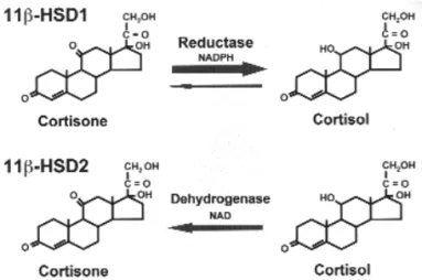
At that time, however, 11βHSD had an unex-plained two-way function: to convert F into E and, conversely, E into F. Early on, two isoforms of 11βHSD were cloned and characterized: 11βHSD type 1 (11βHSD1) and 11βHSD type 2 (11βHSD2) (3,4). The former is a low-affinity NADP(H)-depen-dent dehydrogenase/oxoreductase (cortisol to corti-sone and vice-versa, respectively), which was originally identified in the liver, but that is also highly expressed in adipose, gonadal, and central nervous system tis-sues. By contrast, 11βHSD2 is a high-affinity NAD-dependent dehydrogenase, whose sole action is to pro-tect the non-selective MCR in the kidney, colon, and salivary glands from cortisol occupancy. 11βHSD1 was first regarded by Lakshmi and Monder (3) as a weaker version of 11βHSD2, because it showed dehy-drogenase activity in tissues homogenates and micro-somes, however evidence has now accumulated to show that in most intact cells and organs 11βHSD1 catalyses the reverse reaction (figure 1) (5).
It has been hypothesized that tissue-specific dysregulation of cortisol metabolism may be involved in the complex pathophysiology of the metabolic syn-drome (MS) and simple obesity. 11βHSD1 is ex-pressed in both adipocyte and preadipocyte (6); based
on the evidence that glucocorticoids induce adipocyte differentiation in vitro (7), adipose autocrine genera-tion of F from E may be implicated in the pathogene-sis of central obesity and its associated metabolic com-plications. In support to this hypothesis, transgenic mice overexpressing 11βHSD1 in adipose tissue devel-op obesity with all the features of the MS (8); con-versely, 11βHSD1-knockout mice are protected from obesity and MS (9). Aside some controversies, the bulk of evidences show that 11βHSD1 mRNA and activity are upregulated in human obesity.
CUSHING’S SYNDROME AND METABOLIC SYNDROME: HIGH CIRCULATING LEVELS VERSUSHIGH TISSUE CONCENTRATIONS
OF GLUCOCORTICOIDS
Cushing’s syndrome: Effects of chronic exposure to high concentrations of circulating cortisol
In 1932, Harvey Cushing reported on eight patients with adrenal hyperplasia associated with pituitary basophilic adenomas (10), defining a new clinical entity that now bears his name: “Cushing’s disease”. His meticulous description gives information about the deleterious effects of cortisol excess. Cushing’s syndrome (CS) caus-es hypertension in more than 90% of the cascaus-es, central obesity in more than 80%, osteoporosis in 50% (11), in addition to other typical signs and symptoms (12).
jects with endogenous or exogenous Cushing’s syn-drome develop a central obesity pattern that is rever-sible upon treatment or glucocorticoid withdrawal. Cortisol augments directly or indirectly the total mass of adipose tissue and redistributes it from peripheral to central depots (13). Glucocorticoids also regulate multiple processes in the adipose tissue. They influ-ence fat cell size, so that enlarged abdominal fat cells are seen in CS (14); promote differentiation of human preadipocytes into mature adipocytes, increasing fat cell number (7); and activate lipolyses, releasing free fatty acids into circulation. Chronically, however, as seen in CS, lipoprotein lipase activity is elevated 2–3 fold with a low lipolytic capacity (14).
Moreover, part of the glucocorticoid actions is regulated at a pre-receptor level by 11β-HSD1. This enzyme is co-localized with the glucocorticoid recep-tor in several cells, including adipose tissue and liver, where it is ideally positioned to amplify glucocorticoid action. Bujalska et al. (6) cultured omental adipose stromal cells with cortisol and showed an increased activity of 11β-HSD1. This observation led to the speculation that 11β-HSD1 is upregulated in the vis-ceral adipose tissue of subjects with Cushing’s syn-drome. However, recent data evidence the opposite, as will be discussed below. 11β-HSD1 seems not to be the cause of the metabolic syndrome, but instead a modulator of energy partitioning (15).
Obesity and metabolic syndrome:
Increased adipose tissue concentrations of cortisol
Since the original description of “Syndrome X” by Gerald Reaven in 1988 (16), obesity has been associ-ated, to some extent, to abnormalities in the hypo-thalamus–pituitary–adrenal (HPA) axis. Similarities between Cushing’s syndrome and the clinical features of the metabolic syndrome (visceral obesity, hyper-glycemia, hypertension, and dyslipidemia) led to the hypothesis that obesity may be associated with gluco-corticoid excess in the latter.
Correlations between abdominal fat and hyper-activity of the HPA axis have been found, although there is considerable controversy in the literature. The proposed alterations include resistance to the negative feedback (impaired suppression) exerted by low oral dexamethasone or intravenous doses of glucocorti-coids (17-19); elevated diurnal ACTH levels and altered pulsatile secretory ACTH dynamics (20), hyperesponsiveness to different peptides (CRH, AVP) (21), and increased cortisol production rate, as mea-sured by stable isotope ratio (22).
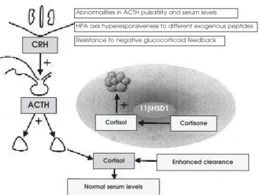
The increased HPA axis drive and production of cortisol observed in obese subjects is counterbal-anced by enhcounterbal-anced urinary excretion rate of free corti-sol and its metabolites and also by its enhanced periph-eral clearance (21-23), resulting in normal (or even low) blood levels (figure 2). This has been recently explained by observations that there is a tissue-specific deregulation of cortisol metabolism in human obesity in which 11βHSD1 activity is upregulated in adipose tissue and underegulated in the liver, resulting in lower plasma cortisol levels with a compensatory rise in ACTH and cortisol production (see below).
11β-HSD1 IN OBESITY AND METABOLIC
SYNDROME
In vitrostudies and animal models
Omental adipose stromal cells treated with cortisol and insulin show increased 11βHSD1 reductase activ-ity (generation of active cortisol from inactive corti-sone), suggesting that obesity may reflect a “Cushing’s disease of the omentum” (6). These cells in early stage of differentiation show a switch from dehydrogenase (inactivation of cortisol into cortisone) to reductase activity, ensuring autocrine generation of cortisol, which will induce adipocyte differentiation (7). Thus, 11βHSD1 seems to play a key role in adipocyte differ-entiation and in regulating adipose tissue depots.
Series of studies in animal models of obesity have shown increased 11βHSD1 mRNA expression Figure 2.Abnormalities in the central regulatory control and in the peripheral metabolism of cortisol in obesity. Increased hypothalamus–pituitary–adrenal axis (HPA) drive appears to result from elevated peripheral cortisol clearance, the bal-ance of which yields normal plasma levels. Peripherally, mRNA expression and enzymatic activity of 11βHSD1 is increased in the adipose tissue (inset).
Abnormalities in ACTH pulsatility and serum levels
Resistance to negative glucocorticoid feedback
CRH
ACTH
Cortisol
Cortisol
Cortisone
Enhanced clearence
Normal serum levels
HPA axis hyperesponsiveness to different exogenous peptides
and activity in adipose tissue. Transgenic mice overex-pressing 11βHSD1 in adipose tissue develop obesity and other features of the metabolic syndrome, pre-senting with elevated intra-adipose glucocorticoid concentrations and higher glucocorticoid receptor alpha (GRα) expression (24). In contrast, 11β HSD1-knockout mice are protected from obesity and its metabolic complications (9). Overexpression of 11βHSD1 in the liver does not induce obesity or hyperglycemia, although there are changes in serum lipids, insulin sensitivity and blood pressure (25). Besides, 11βHSD1 appears to be involved in the homeostatic adaptation to macronutrient intake; it undergoes downregulation in adipose tissue of high-fat feeding mice (26).
In rodents, pharmacologic inhibition of 11βHSD1 is effective in enhancing hepatic insulin sen-sitivity and lowering blood glucose in diabetic mice (27) and inducing weight loss in obese mice (28).
Human studies
The bulk of evidences points both to an overexpres-sion and an increased activity of 11βHSD1 in subcu-taneous (SAT) and visceral adipose tissue (VAT) of obese subjects, although biopsies of the omentum were conducted in but a few studies. Several groups have shown higher 11βHSD1 mRNA expression in obese compared to non-obese subjects (29-32), although not all studies agree (33). Direct in vivo mea-surements using microdialysis in SAT also suggest an increase in the conversion rate of cortisone to cortisol (34). Moreover, 11βHSD1 mRNA expression posi-tively correlates with obesity (body mass index and abdominal circumference), body composition, insulin resistance (30-32), resistins and other cytokines, as TNFα, IL-6, and leptin (35).
The whole body 11βHSD1 activity reflects main-ly hepatic expression. Initial studies that relied on mea-surements of cortisol-to-cortisone metabolites in urine (23,36) should be taken with caution as indicative of 11βHSD1 activity, because several other cortisol and cortisone metabolizing enzymes are deregulated in obe-sity (36). Of greater importance is the finding of reduced hepatic 11βHSD1 activity measured by the conversion of orally administered cortisone to cortisol (23,37). Thus, 11βHSD1 upregulation in obesity seems not to be a gen-eralized process. In both the whole body and the splanchnic circulation there are no differences between obese and lean subjects regarding cortisol regeneration rates (as measured by [2H4]-cortisol tracer), presumably because an upregulation in adipose tissue is counterbal-anced by a downregulation in the liver (15).
Polymorphisms in the 11βHSD1 gene were identified in an attempt to clarify the basis for the increased activity of adipose tissue 11βHSD1 in obesi-ty. In two populations, polymorphisms were associat-ed with an increasassociat-ed risk of diabetes and hypertension, but not obesity (38,39). A polymorphism was also found that predicts lower 11βHSD1 expression and protection against diabetes (40).
11β-HSD1 IN CUSHING’S SYNDROME
In view of this background, it has been speculated that visceral fat depot in Cushing’s syndrome (CS) was due to increased 11βHSD1 activity. However, there is only one published study to date on CS that shows the opposite. Mariniello et al. (41) found no differences in 11βHSD1 mRNA expression between CS patients and normal weight controls, although 11βHSD1 mRNA was 13-fold higher in obese subjects. Recent observa-tions performed by our group (42) are in agreement with Mariniello group’s data (41). Our data in patients with CS also establish the absence of correlation between 11βHSD1 mRNA expression in VAT and salivary free cortisol (42). In CS, 11βHSD1 expression may be downregulated by chronic exposure to cortisol levels with a compensatory upregulation of GRα(42). 11βHSD1 is emerging as a key component in homeostatic adaptation, rather than the cause of fatty-acid accumulation in adipose tissue. Recent studies suggest that the enzyme is influenced by the nutri-tional status (15); accordingly, its lack of increase in CS may suggest a protective mechanism against the metabolic complications. Indeed, when the opposite occurs, e.g., weight loss in simple obesity, 11βHSD1 undergoes upregulation (43), although this is not a universal finding (32). Thus, there are some evidences that 11βHSD1 adjusts local cortisol concentrations independently of its circulating levels.
REGULATION OF 11β-HSD1 EXPRESSION
rosigli-tazone, a PPARγ agonist, did not change 11βHSD1 mRNA expression and activity in human SAT, at least acutely.
In addition, recent research has begun to address the question of 11βHSD1 regulation in healthy subjects, and suggest that the enzyme is influ-enced by the nutritional status (15). A single mixed meal induces a rise in whole body rates of regeneration of cortisol by 11βHSD1 (46), an effect that seems mediated by hyperinsulinemia (47).
11β-HSD1 AS A THERAPEUTIC TARGET
FOR THE METABOLIC SYNDROME AND DIABETES
Inhibition of 11βHSD1 shows a considerable promise as a therapeutic target to the metabolic syndrome and type 2 diabetes. It offers a key advantage over other strategies for manipulating glucocorticoid action, in that circulating cortisol levels and the response to stress are not impaired. Ideally, it would reduce gluco-corticoid action selectively within the metabolically active tissues such as adipose and liver, without blunt-ing the negative feedback regulation at the HPA axis. 11βHSD1 is important for glucocorticoid action also in the brain, in addition to HPA axis regulation (48). To avoid hypothalamic interference by specific
11βHSD1 inhibitors, such drugs must not cross the blood-brain barrier (49).
Pharmacologic inhibition of 11βHSD1 with the anti-ulcer drug carbenoxolone improves insulin sensitivity in healthy human subjects (50) and in pa-tients with type 2 diabetes (51). However, carbeno-xolone is neither selective nor very potent and does not appear to inhibit 11βHSD1 in adipose tissue. Aryl-sulphonamidothiazoles were the first 11βHSD1 selec-tive inhibitors to be synthesized; in diabetic mice, they enhance insulin action in the liver, thereby lowering blood glucose concentrations (27).
To date approximately eighteen pharmaceutical companies and other organizations have filled patents for 11βHSD1 inhibitors (15). Results of clinical stud-ies with novel potent inhibitors are therefore eagerly awaited.
CONCLUSION
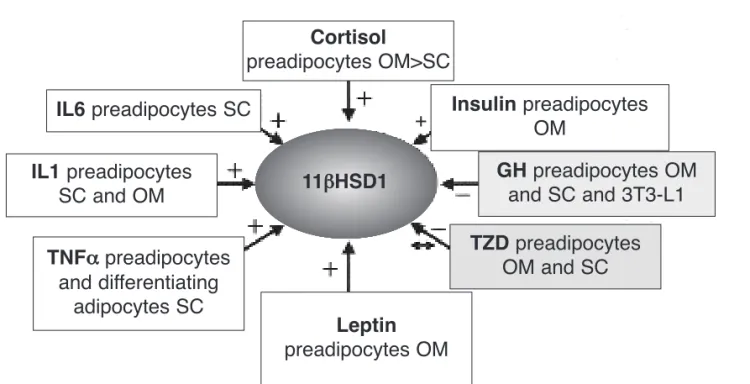
Several controversies on 11βHSD1 expression and activ-ity in human adipose tissue remain unsolved. However, there are accumulating evidences that intra-adipocyte generation of cortisol contributes for the development of features of the MS and type 2 diabetes. Several groups have shown upregulation of 11βHSD1 in obesity and its correlation with abdominal circumference, BMI, and Figure 3.Some of the factors involved in the complex adipose tissue regulation of 11βHSD1 enzyme. [+], [-], [↔]: increase, inhibit, or do not change expression, respectively.
OM: omental adipose tissue, SC: subcutaneous adipose tissue, IL-6: interleukyne 6, IL-1: interleukyne 1, TNFα: tumor necrosis factor-α, TZD: thiazolidinediones, GH: growth hormone.
Cortisol
preadipocytes OM>SC
IL6
preadipocytes SC
IL1
preadipocytes
SC and OM
TZD
preadipocytes
OM and SC
GH
preadipocytes OM
and SC and 3T3-L1
Insulin
preadipocytes
OM
TNFα
preadipocytes
and differentiating
adipocytes SC
Leptin
preadipocytes OM
+
insulin resistance. In Cushing’s syndrome, 11βHSD1 is downregulated, suggesting that it may adjust local corti-sol concentrations independently of its plasma levels. 11βHSD1 seems also to play a role in the complex pathophysiology of the MS and in energy partitioning. Future clinical studies are envisioned to prove the effica-cy of selective 11βHSD1 inhibition.
REFERENCES
1. New MI, Levine LS, Biglieri EG, Pareira J, Ulick S. Evidence for an unidentified steroid in a child with apparent mineralo-corticoid hypertension. J Clin Endocrinol Metab 1977;44:924-33.
2. Ulick S, Levine LS, Gunczler P, Zanconato G, Ramires LC, Raul W, et al. A syndrome of apparent mineralocorticoid excess associated with defects in the peripheral metabolism of cortisol. J Clin Endocrinol Metab 1979;49:757-64. 3. Lakshmi V, Monder C. Purification and characterization of the
11β-dehydrogenase component of the rat liver 11β -hydroxys-teroid dehydrogenase complex. Endocrinology 1988; 123:2390-8.
4. Low SC, Assaad SN, Rajan V, Chapman KE, Edwards CR, Seckl JR. Regulation of 11β-hydroxysteroid dehydrogenase by sex steroids in vivo: further evidence for the existence of
a second dehydrogenase in rat kidney. J Endocrinol 1993; 139:27-35.
5. Seckl JR, Walker BR. 11β-hydroxysteroid dehydrogenase type 1 as a modulator of glucocorticoid action: from metabolism to memory. Trends Endocrinol Metab 2004;15:418-24. 6. Bujalska IJ, Kumar S, Stewart PM. Does central obesity reflect
“Cushing’s disease of the omentum”? Lancet 1997;349: 1210-3.
7. Bujalska IJ, Kumar S, Hewison M, Stewart PM. A switch in the dehydrogenase to reductase activity of 11β-hydroxysteroid dehydrogenase type 1 upon differentiation of human omen-tal adipose stromal cells. J Clin Endocrinol Metab 2002;87:1205-10.
8. Masuzaki H, Yamamoto H, Kenyon CJ, Elmquist JK, Morton NM, Paterson JM, et al. Transgenic amplification of glucocor-ticoid action in adipose tissue causes high blood pressure in mice. J Clin Invest 2003;112:83-90.
9. Morton NM, Paterson JM, Masuzaki H, Holmes MC, Staels B, Fievet C, et al. Novel adipose tissue-mediated resistance to diet-induced visceral obesity in 11β-hydroxysteroid dehydro-genase type 1 deficient mice. Diabetes 2004;53:931-8. 10. Cushing H. The basophil adenomas of the pituitary body and
their clinical manifestations (pituitary basophilism). Bull
Johns Hopkins Hosp 1932;50:137-95.
11. Stewart PM. Tissue-specific Cushing’s syndrome, 11β -hydrox-ysteroid dehydrogenase and the redefinition of corticosteroid hormone action. Eur J Endocrinol 2003;149:163-8.
12. Stewart PM. The adrenal cortex. In: Larsen PR, Kronenberg HM, Melmed S, Polonsky KS, Foster DW, Wilson JD.
Williams textbook of endocrinology. 10th ed.
Philadel-phia: Saunders, 2003. pp. 491-539.
13. Lönn L, Kvist H, Ernest I, Sjöström L. Changes in body com-position and adipose tissue distribution after treatment of women with Cushing’s syndrome. Metabolism 1994;43: 1517-22.
14. Rebuffe-Scrive M, Krotkiewski M, Elfverson J, Bjorntorp P. Muscle and adipose tissue morphology and metabolism in Cushing’s syndrome. J Clin Endocrinol Metab 1988;67: 1122-8.
15. Walker BR. Extra-adrenal regeneration of glucorticoids by 11β-hydroxysteroid dehydrogenase type 1: physiological reg-ulator and pharmacological target for energy petitioning.
Proc Nutr Soc 2007;66:1-8.
16. Reaven GM. Role of insulin resistance in human disease.
Dia-betes 1988;37:1595-607.
17. Ljung T, Andersson B, Bengtson BA, Bjorntorp P, Marin P. Inhibition of cortisol secretion by dexamethasone in relation to body fat distribution: a dose response study. Obes Res 1996;4:277-82.
18. Jessop DF, Dallman MF, Flaming D, Lightman SL. Resistance to glucocorticoid feedback in obesity. J Clin Endocrinol
Metab 2001;86:4109-14.
19. Pasquali R, Ambrosi B, Armanini D, Cavagnine F, Uberti ED, Del Rio G, et al. Cortisol and ACTH response to oral dexam-ethasone in obesity and effects of sex, body fat distribution, and dexamethasone concentrations: a dose-response study.
J Clin Endocrinol Metab 2002;87:166-75.
20. Ljung T, Holm G, Friberg P, Bjorn A, Bengtsson B-A, Svens-son J, et al. The activity of the hypothalamic-pituitary-adren-al-axis and the sympathetic nervous system in relation to waist-hip circumference in men. Obes Res 2000;8:487-95. 21. Pasquali R, Cantobelli S, Casimirri F, Capelli M, Bortoluzzi L,
Flamia R, et al. The hypothalamic pituitary adrenal axis in obese women with different patterns of body fat distribution.
J Clin Endocrinol Metab 1993;77:341-6.
22. Purnell JQ, Brandon DD, Isabelle LM, Loriaux DL, Samuels MH. Association of 24h-cortisol production rates, cortisol binding globulin, and plasma free cortisol levels with body composition, leptin levels, and aging in adult men women. J
Clin Endocrinol Metab 2004;89:281-7.
23. Rask E, Olsson T, Soderberg S, Andrew R, Livingstone DE, Johnson O, et al. Tissue-specific dysregulation of cortisol metabolism in human obesity. J Clin Endocrinol Metab 2001;86:1418-21.
24. Masuzaki H, Paterson JM, Shinyama H, Morton NM, Mullins JJ, Seckl JR, et al. A transgenic model of visceral obesity and the metabolic syndrome. Science 2001;294:2166-70. 25. Paterson JM, Morton NM, Fievet C, Kenyon CJ, Holmes MC,
Staels B, et al. Metabolic syndrome without obesity: hepatic overexpression of 11β-hydroxysteroid dehydrogenase type 1 in transgenic mice. Proc Nat Acad Sci USA 2004; 101:7088-93. 26. Morton NM, Ramage L, Seckl JR. Down-regulation of adipose tissue 11β-hydroxysteroid dehydrogenase type 1 by high fat feeding in mice: a potential adaptive mechanism counteract-ing metabolic disease. Endocrinology 2004;145:2707-12. 27. Alberts P, Engblom L, Eddling N, Forsgren M, Klingstron G,
Larsson C, et al. Selective inhibition of 11β-hydroxysteroid dehydrogenase type 1 decreased blood glucose concentrations in hyperglycaemic mice. Diabetologia 2002;45:1528-32. 28. Hermanowski-Vosatka A, Blakovec JM, Cheng K, Chen HY,
Hernandez, Koo GC, et al. 11β-HSD1 inhibition ameliorates metabolic syndrome and prevents progression of atheroscle-rosis in mice. J Exp Med 2005;202:517-27.
29. Paulmyer-Lacroix O, Boullu S, Oliver C, Alessi MC, Grino M. Expression of the mRNA coding for 11β-hydroxysteroid dehydrogenase type 1 in adipose tissue from obese patients: an in situhybridization study. J Clin Endocrinol Metab
2001;87:2701-5.
30. Wake DJ, Rask E, Livingstone, Sodeberg S, Olsson T, Walker BR. Local and systemic impact of transcriptional up-regula-tion of 11β-hydroxysteroid dehydrogenase type 1 in adipose tissue in human obesity. J Clin Endocrinol Metab 2003;88:3983-8.
31. Lindsay RS, Wake JW, Saraswathy N, Bunt J, Livingstone DEW, Permana PA, et al. Subcutaneous adipose 11β -hydrox-ysteroid dehydrogenase type 1 activity and messenger ribonucleic acid levels are associated with adiposity and insu-linemia in Pima Indians and Caucasians. J Clin Endocrinol
Metab 2003;88:2738-44.
32. Engeli S, Bohnke J, Feldpausch M, Gorzelniak K, Heintze U, Janke J, et al. Regulation of 11β-HSD genes in human adi-pose tissue: influence of central obesity and weight loss.
Obes Res 2004;12:9-17.
33. Tomlinson JW, Sinha B, Bujalska I, Hewison M, Stewart PM. Expression of 11β-hydroxysteroid dehydrogenase type 1 in adipose tissue is not increased in human obesity. J Clin
34. Sandeep TC, Andrew R, Homer NZM, Andrews RC, Smith K, Walker BR. Increased in vivo generation of cortisol in adipose tissue in human obesity and the effects of the 11β -hydroxys-teroid dehydrogenase type 1 inhibitor carbenoxolone.
Dia-betes 2005;54:872-9.
35. Tomlinson JW, Moore J, Cooper MS, Bujalska I, Shahmanesh M, Burt C, et al. Regulation of the expression of 11β -hydrox-ysteroid dehydrogenase type 1 in adipose tissue-specific induction by cytokines. Endocrinology 2001;142:1982-9. 36. Andrew R, Philips DIW, Walker BR. Obesity and gender
influ-ence cortisol secretion and metabolism in man. J Clin
Endocrinol Metab 1998;83:1806-9.
37. Stewart PM, Boulton A, Kumar S, Clark PMS, Shackleton CHL. Cortisol metabolism in human obesity: impaired cortisone-cortisol conversion in subjects with central adiposity. J Clin
Endocrinol Metab 1999;84:1022-7.
38. Franks PW, Knowler WC, Nair S, Koska J, Lee Y-H, Lindsay RS, et al. Interaction between an 11β-HSD1 gene variant and birth era modifies the risk of hypertension in Pima Indians.
Hypertension 2004;44:681-8.
39. Nair S, Lee YH, Lindsay RS, Walker BR, Tataranni PA, Bogar-dus C, et al. 11β-hydroxysteroid dehydrogenase type 1: Genetic polymorphisms are associated with type 2 diabetes in Pima Indians independently of obesity and expression in adipocyte and muscle. Diabetologia 2004;47:1088-95. 40. Draper N, Echwald SM, Lavery GG, Walker EA, Fraser R,
Davies E, et al. Association studies between microsatellite markers within the gene encoding for human 11β -hydroxys-teroid dehydrogenase type 1 and body mass index, waist to hip ratio, and glucocorticoid metabolism. J Clin Endocrinol
Metab 2002;87:4984-90.
41. Mariniello B, Ronconi V, Rilli S, Bernante P, Boscoro M, Man-tero F, et al. Adipose tissue 11β-hydroxysteroid dehydroge-nase type 1 expression in obesity and Cushing’s syndrome.
Eur J Endocrinol 2006;155:435-41.
42. Espíndola-Antunes D, Goto EM, Guimarães AO, Pesquero JB, Silva-Júnior JA, Kater CE. Quantitative mRNA expression of 11β-hydroxysteroid dehydrogenase type 1 and glucocorticoid receptor alpha in visceral adipose tissue of patients with Cushing’s syndrome and obese and non-obese controls.
Proc Endocr Soc Meeting 2007; [abstract].
43. Tomlinson JW, Moore JS, Clarck PMS, Holder G, Shake-speare L, Stewart PM. Weight loss increases 11β -hydroxys-teroid dehydrogenase type 1 expression in human adipose tissue. J Clin Endocrinol Metab 2004;289:2711-6.
44. Tomlinson JW, Walker EA, Bujalska IJ, Draper N, Lavery GG, Cooper MS, et al. 11β-hydroxysteroid dehydrogenase type 1: a tissue specific regulator of glucocorticoid response.
Endocr Rev 2004;25:831-66.
45. Wake DJ, Stimson RH, Tam GD, Homer NZM, Andrew R, Carpe F, et al. Effects of peroxisome proliferator-activated receptor αand γagonists in 11β-hydroxysteroid dehydroge-nase type 1 in subcutaneous adipose tissue in men. J Clin
Endocrinol Metab 2007;92:1848-56.
46. Basu R, Singh R, Basu A, Johson CM, Rizza RA. Effect of nutri-ent ingestion on total-body and splanic cortisol production in humans. Diabetes 2006;55:667-74.
47. Wake DJ, Homer NZD, Andrew R, Walker BR. Acute in vivo
regulation of 11β-hydroxysteroid dehydrogenase type 1 by insulin and intralipid infusions in humans. J Clin
Endocrinol Metab 2006;91:4682-8.
48. Harris HJ, Kotelevtsev Y, Mullings JJ, Seckl JR, Holmes MC. Intracellular regeneration of glucocorticoids by 11β -hydrox-ysteroid dehydrogenase (11β-HSD)-1 plays a key role in reg-ulation of the hypothalamic-pituitary-adrenal axis: analysis of 11β-HSD-1-deficient mice. Endocrinology 2001;142:114-20. 49. Stulnig TM, Waldhausl W. 11β-hydroxysteroid dehydroge-nase type 1 in obesity and type 2 diabetes. Diabetologia 2004;47:1-11.
50. Walker BR, Connacher AA, Lindsay RM, Webb DJ, Edwards CRW. Carbenoxolone increases hepatic insulin sensitivity in man: a novel role for 11-oxosteroid reductase in enhancing glucocorticoid receptor activation. J Clin Endocrinol
Metab 1995;80:3155-9.
51. Andrews RC, Rooyackers O, Walker BR. Effects of the 11β -hydroxysteroid dehydrogenase inhibitor carbenoxolone on insulin sensitivity in men with type 2 diabetes. J Clin
Endocrinol Metab 2003;88:285-91.
Address for correspondence:
Claudio E. Kater
Laboratório de Esteróides
Disciplina de Endocrinologia — UNIFESP Rua Pedro de Toledo 781, 13ºandar 04062-023 São Paulo, SP