TREATMENT OF HEPATIC ENCEPHALOPATHY:
TARGETING THE GUT-LIVER-BRAIN AXIS
Summary of Presentations from the Norgine Sponsored
Satellite Symposium, UEG Week, Berlin, Germany,
12
th–16
thOctober 2013
Chairperson
Rajiv Jalan
1Speakers
Agustin Albillos,
2Flemming Bendtsen
31. UCL Institute for Liver and Digestive Health, Royal Free Hospital, London, UK 2. Dept. of Gastroenterology and Hepatology, Hospital Universitario Ramón y Cajal,
Universidad de Alcalá, CIBERehd, Madrid, Spain
3. Dept. of Gastroenterology, Hvidovre Hospital, University of Copenhagen, Denmark
Disclosure: The speakers received honorarium from Norgine for participating in this symposium.
Acknowledgements: Writing assistance has been provided by Trilogy Writing and Consulting Ltd.
Support: The publication of this article was funded by Norgine. The views and opinions expressed are those of the authors and not necessarily of Norgine.
Citation: EMJ Gastroenterol. 2013;1:20-29.
Introduction
Professor Rajiv Jalan
The pathogenesis of hepatic encephalopathy (HE) involves the interaction between two pathophysiological mechanisms: ammonia detoxiication and inlammation. The neuropathology of HE is characterised by astrocyte dysfunction and swelling due to ammonia detoxiication. The enzyme glutamine synthetase, which is located mainly in astrocytes, protects neurons by absorbing excess ammonia and glutamate, converting it to glutamine. The concentration of ammonia is increased in patients with hepatic failure when compared with healthy individuals, clearly indicating the signiicance of ammonia in the pathogenesis of HE, and as an important target for treatment. Bacterial translocation is a signiicant factor in driving the progression of cirrhosis, hepatic ibrosis, compensated and decompensated cirrhosis and the recurrence of HE (Figure 1). Bacterial translocation appears to be a key event in the transition from well-compensated to decompensated cirrhosis (or acute on chronic liver failure). This transition manifests in severe
levels of HE, and probably contributes directly to the ‘second hit’ which is inlammation.
Gut Bacterial Translocation in Cirrhosis
Professor Agustin Albillos
Intestinal microlora and gut bacterial translocation (GBT) have been implicated in the pathogenesis of spontaneous bacterial infections and in the progression of cirrhosis. Bowel decontamination with quinolones or luoroquinolones has been shown to improve survival in patients with decompensated cirrhosis. Non-absorbable antibiotics such as rifaximin reduce the rate of spontaneous bacterial infection, the rate of portal hypertension related complications, and improve survival.1,2
A positive mesenteric lymph node culture suggests increased passage of enteric bacteria due to increased intestinal permeability, intestinal bacterial overgrowth, or both. More importantly, it also indicates an inability of the immune system to destroy the translocated bacteria. According to the previous deinition, GBT was present in about 30-40% of patients with cirrhosis and ascites and in cirrhotic rats with ascites. This demonstrates that bacterial translocation is present in advanced decompensated cirrhosis when severe liver insuiciency has already developed.
Most of the translocated bacteria belong to the common intestinal microbiota, which signiies that there is a disruption of the intestinal barrier in cirrhosis. The intestinal barrier is composed of three interrelated layers, the external composed by mucous and bacterial microlora, the epithelial cells and the sub epithelial where interactions take place between the bacteria and the immune system.5 The integrity of the epithelial cells is the most important layer of defence against microbiota. Abnormalities in any of these levels of defence have been advocated to explain the high rate of GBT of cirrhosis.
Intestinal bacterial overgrowth, increased intestinal permeability and impaired immune system response indicate changes in each of the three layers of the gut barrier in cirrhosis. In cirrhosis, there are abnormalities in the function and structure of the intestinal mucosa, involving tight junction proteins, which lead to increased permeability of macromolecules.3 There are also qualitative (dysbiosis) and quantitative changes in gut microbiota that indicate intestinal overgrowth, which is associated with most episodes of bacterial translocation.3 This is mainly attributed to the presence of intestinal hypomotility in cirrhosis, although impaired immunity can also contribute. However, other elements are necessary for GBT to develop; these are predominantly linked to the hepatic insuiciency that is found in cirrhosis. The exact mechanism is unclear, but it is possibly related to impaired immune function considering the role of the liver in innate immune function. There might also be contribution of the neuroendocrine abnormalities present in liver cirrhosis, speciically sympathetic nervous system hyperactivity and changes in bile low and composition. Therefore, GBT in liver cirrhosis is
Figure 1. Inlammation in hepatic encephalopathy. Gut dysbiosis
Altered intestinal permeability Reduced intestinal defences
Reduced immunological surveillance Liver injury
Hepatic ibrosis
Priming of multiple organs Immune dysfunction Compensated cirrhosis
Increased bacterial translocation
Second hit
Infection, bleeding, diuretics, alcohol, Na Decompensated cirrhosis
Hepatic encephalopathy
ACLF
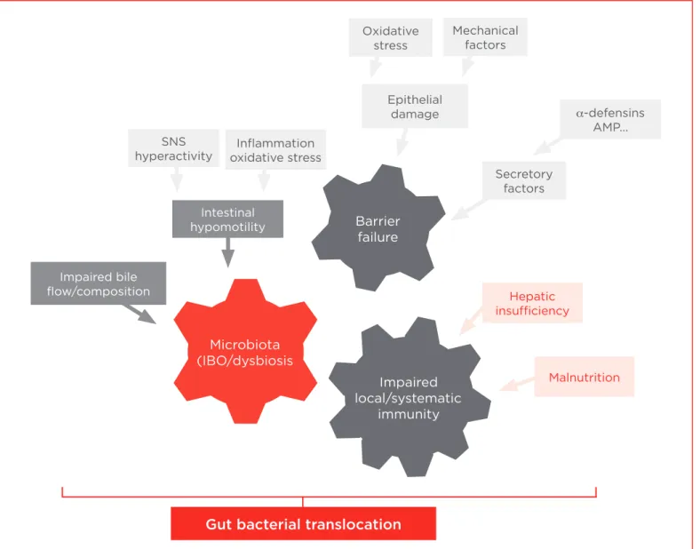
the result of damage at diferent levels of the intestinal barrier, i.e. changes in microbiota, changes in the integrity of the epithelium and impaired immunity (Figure 2).
In the absence of overt infection, GBT contributes to cirrhosis progression by inducing an activation of the immune system at the systemic level.2 Systemic inlammation results from the production and release of pathogen-associated molecular patterns that activate speciic receptors on the surface of immune cells. This results in the production of proinlammatory lymphokines and monokines, and circulating immune system cells in cirrhosis produce proinlammatory cytokines. This mechanism modulates the clinical expression of cirrhosis, for example the modulation of the activity of astrocytes leading to the neurological changes of cirrhosis or the regulation of vascular tone. Persistent activation of immune
cells worsens the immunodeiciency of cirrhosis because it leads to the exhaustion and death of the cells (Figure 3). This situation can be reversed by reducing the enteric bacterial load with non-absorbable or poorly absorbable antibiotics.6 This was shown in a study where bowel decontamination with antibiotics improved the dendritic cell function of the intestinal lamina propria of cirrhotic rats with bacterial translocation.
Multiple intestinal damage in cirrhosis involves diferent potential therapeutic targets. These include bile acids, farnesoid X receptor (FXR) agonists for impaired bile low or composition, beta-blockers for intestinal hypomotility, antioxidants for inlammation oxidative stress and antibiotics, and probiotics for microbiota. The most efective treatment is controlling the microbiota with non-absorbable antibiotics to prevent spontaneous bacterial infection. The use of other speciic
Figure 2. Mechanisms of gut bacterial translocation in advanced cirrhosis. Gut bacterial translocation
Microbiota (IBO/dysbiosis
Barrier failure
Impaired local/systematic
immunity
Epithelial damage
Secretory factors
α-defensins AMP... Oxidative
stress
SNS hyperactivity
Mechanical factors
Inlammation oxidative stress
Impaired bile low/composition
Intestinal hypomotility
Malnutrition Hepatic
targeted agents (FXR agonists, antioxidants and probiotics) has only been studied in experimental settings; therefore, eicacy data in patients are not available. However, the use of beta-blockers in cirrhotic patients has been shown to be beneicial by reducing portal pressure and GBT.7 This was demonstrated in cirrhotic rats; propranolol was shown to accelerate intestinal transit and lower intestinal bacterial overgrowth and GBT to mesenteric lymph nodes.
In summary, GBT in cirrhosis is the pathophysiological hallmark of spontaneous bacterial infections. GBT contributes to further decompensation in already decompensated cirrhosis by driving a persistent activation of the inlammatory immune system and exacerbating immunodeiciency. As the pathogenesis of GBT in cirrhosis involves damage at diferent levels of the intestinal barrier, there are multiple potential targets for the control of GBT. Although most targets have only been tested in an experimental setting, it has been shown that bowel decontamination improves outcomes in patients with decompensated cirrhosis. Furthermore, beta-blockers improve survival by reducing the variceal bleeding risk and bacterial translocation.
Clinical Consequences of
Bacterial Translocation – Does Gut
Decontamination Improve Outcome?
Professor Flemming Bendtsen
Bacterial translocation is associated with cirrhosis and portal hypertension and contributes to splanchnic vasodilation and systemic vasodilation. Therefore, clinicians should focus on bacterial translocation in patients with ascites and decompensated liver disease. As a consequence of portal hypertension and possibly GBT, patients may develop an increased hepatic venous pressure gradient (HVPG) resulting in the formation of oesophageal varices. Furthermore, systemic inlammatory response leads to disease progression, with an associated increased risk of infection. Structural and functional changes in the gut mucosa, bacterial overgrowth in the small intestine, impairment of defence mechanisms, and decreased gut motility can all lead to bacterial translocation.
Bacterial decontamination is believed to modify the risk of complications of portal hypertension. There are no large clinical trials that evaluate clinical endpoints such as death, development of variceal bleeding, hepatorenal syndrome, and
Figure 3. The consequences of hepatic insuiciency that contribute to GBT in cirrhosis.
Immune function of the liver • ‘Blood’ ilter
• Secondary lymphoid organ • ‘Secretor’ of molecules of the innate immune response Hepatic
insuiciency Impaired synthetic ability
malnutrition
Impaired immune function
Neuroendocrine abnormalities: SNS hyperactivity Impaired bile
other complications of cirrhosis. However, there are trials that evaluate surrogate markers such as the efect on haemodynamics and inlammation, vasoactive hormones, and inlammatory markers. Albillos et al.2 evaluated lipopolysaccharide binding protein (LBP) levels in cirrhotic patients with marked immune and haemodynamic derangement. Patients were randomised to either norloxacin (which targets most of the Gram-negative bacteria in the intestine) or to placebo. A further sub-division of patients was made into those that had signs of inlammation with an increased LBP at baseline, and those with normal LBP. The patients with increased LBP had more severe signs of derangement in their haemodynamic evaluations; these included decreased blood pressure (BP) and higher pulse rate leading to a hyperdynamic circulation with increased cardiac output. Hepatic venous catheterisation was performed to measure HVPG. In patients with a high LBP randomised to norloxacin, 4 weeks’ treatment had a beneicial efect on BP and systemic vascular resistance when compared with baseline measurements. However, no efect was seen on HVGP in this study. Norloxacin demonstrated a clear efect on LBP (a marker of bacterial translocation) whereas no efect was seen with placebo (Figure 4).
The results of this study concur with those of a randomised crossover trial8 in which patients with cirrhosis were randomised to placebo or norloxacin for 28 days, and then crossed over to the treatment not previously received. A clear efect was seen on systemic vascular resistance, but no efect was seen on HVPG in patients receiving norloxacin. Furthermore, the study evaluated blood low and found that BP increased and systemic vascular resistance increased in patients receiving norloxacin. These studies demonstrate that treatment with norloxacin generates a signiicant beneicial change in systemic haemodynamic parameters, but not in splanchnic haemodynamic parameters.
Although the non-absorbable antibiotic norloxacin does not appear to have an efect on splanchnic haemodynamics, recent studies indicate that rifaximin might reduce HVPG.9 Patients were given rifaximin 1200 mg daily for 29 days, HVPG was measured at baseline and at day 29. The results showed a decrease in HVPG. However, this study was uncontrolled and it is unclear whether this limitation skewed the results, or whether there is a true treatment efect. Unlike norloxacin, rifaximin has a broader microbial spectrum and has an efect on Gram-positive bacteria.
Figure 4. The efect of 4 weeks’ treatment with norloxacin on LBP and systemic vascular resistance in patients with signs of bacterial translocation (high LBP).
Albillos A et al.2
24
20
16
12
8
4
0
24
20
16
12
8
4
0
3500
3000
2500
2000
1500
1000
500
3500
3000
2500
2000
1500
1000
500
μ
g/
mL
μ
g/
mL
Basal Basal Basal BasalBasal
LBP plasma levels Systemic vascular resistance
Norfloxacin Placebo Norfloxacin Placebo
NS p<0.01 NS
p<0.01
dyn.sec.
cm
-5
dyn.sec.
cm
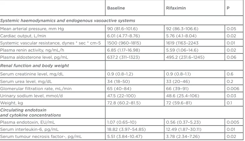
In an uncontrolled study, rifaximin has been shown to improve systemic haemodynamics and renal function in patients with alcohol-related cirrhosis and ascites.10 In an open label study, 15 patients with ascites and Child-Pugh B or C cirrhosis received 1200 mg of rifaximin daily for 4 weeks. Haemodynamic parameters, renal function tests and measurement of inlammation markers were measured at baseline and at day 28. The results showed that rifaximin had a positive efect on the systemic haemodynamic parameters
(Table 1). This study indicates that rifaximin has an efect on surrogate markers by decreases in cytokine levels, (which are the signals of bacterial translocation), and on haemodynamics.
None of the studies of the efects of gut decontamination in cirrhosis are double-blind or placebo controlled. However, it appears from the evidence available that gut decontamination prevents relapse of HE, improves systemic haemodynamics by an increase in BP and a decrease in cardiac output, decreases immune
activation, and may improve renal function. The efect of gut decontamination on splanchnic haemodynamics is yet to be proven.
A randomised, double-blind, placebo controlled study is in progress. This study is investigating intestinal decontamination with rifaximin in cirrhotic patients with ascites, and assessing the efects on haemodynamic and inlammatory factors.11 The aim of the study is to stop progression of liver disease by inhibiting bacterial translocation via bowel decontamination. The outcome measures are to reduce portal hypertension, diminish vasodilation, increase glomerular iltration rate, normalise inlammation and decrease risk of infection (Figure 5).
There is a need for a large scale, randomised, double-blind, placebo controlled study, addressing gut decontamination in decompensated cirrhosis. The clinical endpoints should include mortality, the risks of variceal bleeding, infections (especially spontaneous bacterial peritonitis), and hepatorenal syndrome.
Note: Data are expressed as the median and range.
Kalambokis GN et al.10
Table 1. Systemic haemodynamics, endogenous vasoactive systems, renal function, body weight, and inlammatory markers pre and post treatment with rifaximin.
Baseline Rifaximin P
Systemic haemodynamics and endogenous vasoactive systems
Mean arterial pressure, mm Hg 90 (81.6–101.6) 92 (86.3–106.6) 0.05
Cardiac output, L/min 6.01 (4.77–8.76) 5.76 (4.1–8.04) 0.02
Systemic vascular resistance, dynes * sec * cm-5 1500 (960–1815) 1619 (1163–2243 0.01
Plasma renin activity, ng/mL/h 6.85 (1.17–16.98) 5.59 (1.06–14.6) 0.02
Plasma aldosterone level, pg/mL 637.2 (311–1323) 495.2 (231.6–1245) 0.06
Renal function and body weight
Serum creatinine level, mg/dL 0.9 (0.8–1.2) 0.9 (0.8–1.1) 0.6
Serum urea level, mg/dL 34 (18–50) 33 (20–46) 0.2
Glomerular iltration rate, mL/min 65 (40–84) 66 (39–91) 0.006
Urinary sodium level, mmol/d 47.5 (22–100) 48.6 (25.4–106) 0.03
Weight, kg 72.8 (60.2–81.5) 72 (59.6–81) 0.1
Circulating endotoxin and cytokine concentrations
Plasma endotoxin, EU/mL 1.07 (0.65–10) 0.56 (0.37–5.23) 0.005
Serum interleukin-6, pg/mL 18.82 (3.97–54.85) 12.49 (1.87–30.11) 0.01
Treatment of Hepatic Encephalopathy:
Targeting the Gut-Liver-Brain Axis. Gut
Decontamination and HE. The More
Things Change, the More They Remain
the Same
Professor Rajiv Jalan
There have been signiicant advances in the treatment of HE, predominantly involving targeting the gut-liver-brain axis and gut decontamination. The current treatment concepts are illustrated by the following case study.
A 53 year-old male Afro-Caribbean lawyer was transferred from a neurology hospital where he had been an inpatient for 2 months. The patient had a history of haematemesis and melaena, spastic paraparesis, and a previous history of alcohol abuse and mildly abnormal liver function tests. On admission to the Intensive Care Unit (ICU) the patient had luctuating levels of consciousness with a Glasgow Coma Scale (GCS)
of 4-5. Immediate management included resuscitation, transfusion and an upper gastrointestinal endoscopy. This showed Grade 3 oesophageal varices which were treated with banding and achieved good control of the bleeding. The patient was treated with terlipressin for 3 days, antibiotics, thiamine and lactulose. The initial diagnosis was cirrhosis and oesophageal varices (a complication of the disease). The patient had been previously it and well, and had a lourishing law practice until 2006. He retired prematurely in July 2009 due to progressive leg weakness that started in 2006, and became wheelchair bound in 2011. The patient had undergone extensive investigations at the specialist neurology hospital and a working diagnosis of spastic paraplegia of unknown origin was made.
Following admission to the ICU, neurological examination on day 4 revealed that he was still unresponsive with a GCS of 4-5, had Grade 3-4 HE and had very little power in both legs. Abdominal examination showed mild
Figure 5. Outcome measures in an ongoing study of rifaximin in cirrhotic patients with ascites. Outcome
measures
Reduce portal hypertension
Hepatic venous cathertisation
CO, BP, SVR, vasoactive hormones
Chrome EDTA clearance
TNF-
α
, IL-6, IL-8, hs-CRPBacterial DNA and LPS-B Diminish
vasodilation
Increase GFR
Normalise inlammation
hepatosplenomegaly, no ascites and no peripheral stigmata of liver disease. Despite the very large ‘hit’ the patient had experienced, his chest was clear and lung function was normal, heart rate was 78 beats/minute, BP 143/78 mmHg, and kidney function was acceptable (creatinine 78
μmol/L). Liver investigations showed mildly
elevated bilirubin (37μ
m/L which equates to 2 mg/dL), and his creatinine was normal (65 μm/L which equates to between 0.7 and 0.8 mg/dL). The model for end-stage liver disease (MELD) score was 10; indicating very mild early chronic liver disease with portal hypertension. All the viral serology results were negative, except Hep B sAG and Hep B eAg (due to previous exposure to Hepatitis B), but HBV was negative. These indings suggested underlying alcoholic cirrhosis which was well compensated. Liver ultrasound demonstrated a normal liver, splenomegaly (14.5 cm) and a trace of pelvic ascites. MRI of the liver showed no evidence of chronic liver disease; the biliary system was normal and a normal total liver volume of 1,382 mL. Transjugular liver biopsy demonstrated evidence of alcoholic cirrhosis. Consequently, a diagnosis of well compensated alcoholic cirrhosis with portal hypertension was made. The patient had a neurological deicit; he was comatose and had spastic paraplegia which was initially thought to be unrelated. On day 5, the results of an ammonia test were 145 μmol/L (normal is <40), this indicated that the patient’s condition was due to HE. Therefore, lactulose was increased to 15 mL three times a day (tds) and treatment with rifaximin 550 mg twice a day (bd) was commenced. GCS remained at 4−5 for a further 5 days and the patient developed diarrhoea (culture and sensitivity was negative) which led to rifaximin and lactulose being stopped.A CT scan of the abdomen showed a large spontaneous portacaval shunt emanating from the left renal vein. Brain MRI showed an increased pallidal hyperintensity on T1 imaging, and MR spectroscopy showed an elevated glutamine level. The patient showed evidence of brain dysfunction with hyperammonemia, increased brain glutamine, spastic paraparesis, and pallidal hyperintensity indicating that the syndrome may be a result of brain dysfunction precipitated by cirrhosis. A diagnosis of severe HE was made and embolisation of the patient’s shunt was considered. Embolisation is a useful treatment for HE and a MELD score of
≤
11 predicts a goodresponse to therapy.12 However, this patient had portal hypertension and a recent variceal bleed and if the shunt were to be blocked, the portal hypertension would become worse. Consequently, the treatment strategy was to insert a small diameter Transjugular Intrahepatic PortoSystemic Shunt (TIPSS) and through the TIPSS, embolise the shunt and control the shunt. The patient remained in ICU and gradually woke up over the next 6 days.
On the ward between days 18 and 28 it became apparent that the patient had persistent Grade 2 HE and remained signiicantly hyperammonemic (75−90
μ
mol/L). Treatment with lactulose 15 mL tds and rifaximin 550 mg bd was restarted. Recommencement of rifaximin was in line with indings by Bass et al.13 that showed that patients discharged from hospital who had been treated with rifaximin had fewer HE relapses and hospital admissions. The study (Figure 6) found a 50% relative risk reduction and a 9% absolute risk reduction in patients treated with rifaximin (hazard ratio 0.50 [95% CI, 0.29−0.87] p=0.01).The patient was discharged from hospital to a rehabilitation centre on day 42. His ammonia levels had improved (35−42
μ
mol/L), neurologically the GCS was 15, and he remained mildly encephalopathic (Grade 1). The patient was followed-up closely; the treatment strategy was to continue lactulose and rifaximin, and if no improvement was seen liver transplantation would be considered. Weissenborn et al.14 showed that patients who have large portacaval shunts can develop HE and hepatic myelopathy, which may be reversible with liver transplantation. In this patient, liver transplantation was indicated for hepatic myelopathy because it was thought that the spastic paraparesis may be related to HE.Knowledge of the pathogenesis of cirrhosis may not have changed. The role of the gut was irst described in 1893 by Necki et al.15-17 who made the following observations: ammonia was coming from the gut and portal ammonia was greater than arterial ammonia (the current treatment for this is lactulose and rifaximin); ammonia concentration is increased in the muscle and kidney (the proposed treatment today is ornithine phenylacetate); ammonia concentration is raised
in the gastric mucosa even in a fasting state (now there may be a role for H. Pylori eradication therapy); ammonia was raised in the kidney of the portacaval shunted dog, and urinary ammonia excretion increased after a meat-meal or ammonia administration (the current treatments are volume expansion, ornithine phenylacetate, gut lavage, and rifaximin). These observations raise the question of how far we have actually progressed; the gut-liver axis was really described more than 120 years ago.
Figure 6. Time to irst HE-related hospitalisation (secondary endpoint). Hazard ratio with rifaximin, 0.50 (95% Cl, 0.29-0.87) p=0.01.
Bass et al.13
100
80
60
40
20
0
0 28 56 84 112 140 168
P
a
tients (%)
Hazard ratio with rifaximin, 0.50 (95% CI, 0.29-0.87) p=0.01
Rifaximin
Placebo
Panel Discussion:
The Role of Non-Absorbed Antibiotics in Gut Decontamination
GBT plays an important role in HE. Key mediators in GBT that drive this include the presence of bacteria in the circulation or tissues, as well as bacterial products that stimulate an inlammatory reaction (e.g. bacterial DNA). Furthermore, it is not the response itself that should be targeted but the location where it originated – the gut-liver axis.
There is a need for more controlled trials on GBT and gut microbiota, with patients with clear signs of bacterial translocation as the focus. Parameters that prove that bacterial translocation is present (e.g. an increased LBP or a high level of cytokines) should be measured. Targeting GBT requires the use of a very broad spectrum, non-absorbable antibiotic, such as rifaximin, which should be tested in this context. Rifaximin has shown eicacy in bowel decontamination with very few side-efects.
not be suicient to eradicate the bacteria and overgrowth in the intestine. Consequently, a drug is required to target both Gram-positive and Gram-negative bacteria: rifaximin has a non-selective, broad antibiotic spectrum and acts on both Gram-positive and Gram-negative bacteria.
The case study demonstrated that the pathology of hepatic myelopathy is a loss of the anterior horn cells; producing a very severe form of HE. There are two types of hepatic myelopathy: a reversible form, and an irreversible form. It is diicult to distinguish the types of hepatic myelopathy in a patient, but both types result from large, long-standing porto-systemic shunt. Liver transplantation is a treatment option for these patients but not all respond and the type of hepatic myelopathy present in responders is unknown.
In the treatment of HE, liver transplantation may not be the irst treatment choice – particularly if the chance of recovery is not evident. There are several diferent treatment options and indicators that can be considered. In comatose patients with liver disease who have clinical signs of HE, ammonia is a useful indicator of the severity of encephalopathy. Accurate measurement can indicate a target for intervention, and in the context of acute liver failure, ammonia provides a measurement to follow as an endpoint to intervention. However, the measurement of ammonia is diicult and must be performed using a validated technique.
The insertion of TIPSS in patients with long-term HE has been suggested to be contraindicated. The case study patient illustrated a treatment strategy when limited options are available. The patient had experienced a variceal bleed and there was a high chance of re-bleeding if the spontaneous large portosystemic shunt was occluded for the treatment of HE. Therefore, a small 8 mm shunt was inserted that could be stretched if the patient bled, providing an opportunity to rescue the patient. Small shunts are at risk of closure therefore a low level of anticoagulation is required.
Faecal transplantation is a therapeutic option that has been suggested in patients with Clostridium diicile infection. However, in patients who are immunosuppressed due to cirrhosis, this may not prevent bacterial overgrowth in the small intestine, and as a result is unlikely to beneit many patients.
Clinical experience demonstrates the important role of non-absorbable antibiotics in gut decontamination in patients with HE.
REFERENCES
1. Leise MD et al. Impact of rifaximin treatment on survival in patients with end-stage liver disease. Hepatology. 2010;52:331A.
2. Albillos A et al. Increased lipopolysaccharide binding protein in cirrhotic patients with marked immune and hemodynamic derangement. Hepatology. 2003;37:208-17.
3. Pérez-Páramo M et al. Efect of propranolol on the factors promoting bacterial translocation in cirrhotic rats with ascites. Hepatology. 2000;31:43-8. 4. Cirera I et al. Bacterial translocation of enteric organisms in patients with cirrhosis. J Hepatol. 2001;34:32-7.
5. Hooper LV et al. Interactions between the microbiota and the immune system. Science. 2012;336:1268-73.
6. Muñoz L et al. Interaction between intestinal dendritic cells and bacteria translocated from the gut in rats with cirrhosis. Hepatology. 2012;56:1861-9. 7. Krag A et al. The window hypothesis:
haemodynamic and non-haemodynamic efects of β-blockers improve survival of patients with cirrhosis during a window in the disease. Gut. 2012;61:967-9.
8. Rasaratnam B et al. The efect of selective intestinal decontamination on the hyperdynamic circulatory state in cirrhosis: a randomized trial. Ann Intern Med. 2003;139(3):186-193.
9. Vlachogiannakos J, et al. Intestinal decontamination improves liver haemodynamics in patients with alcohol-reduced decompensated cirrhosis. Aliment Pharmacol Ther. 2009;29(9):992-9.
10. Kalambokis GN et al. Rifaximin improves systemic hemodynamics and renal function in patients with alcohol-related cirrhosis and ascites. Clin Gastroenterol Hepatol. 2012;10(7):815.
11. ClinicalTrials.gov Identiier: NCT01769040
12. Laleman W. et al. Embolization of large spontaneous portosystemic shunts for refractory hepatic encephalopathy: a
multicenter survey on safety and eicacy. Hepatology.2013;57(6):2448-57.
13. Bass NM et al. Rifaximin treatment in hepatic encephalopathy. N Engl J Med. 2010;362:1071–81.
14. Weissenborn K et al. Liver transplantation improves hepatic myelopathy: evidence by three cases. Gastroenterology. 2003;124(2):346-51. 15. Hahn M et al. Die Eck’sche istel zwischen der unteren hohlvene und der pfortader und ihre folgen fur den organismus. Arch Exp Pathol Pharm. 1893;32:161-210.
16. Nencki et al. Ueber den ammoniakgehalt des blutes under der organe und die harnstofbildung bei den saugethieren. Archiv Exp Pathologie Pharm. 1896;37:26-51.