Submitted15 December 2015
Accepted 13 April 2016
Published24 May 2016
Corresponding author
Abhaya M. Dandekar, [email protected]
Academic editor
Abhishek Kumar
Additional Information and Declarations can be found on page 17
DOI10.7717/peerj.2007
Copyright
2016 Chakraborty et al.
Distributed under
Creative Commons CC-BY 4.0
OPEN ACCESS
Sequence/structural analysis of xylem
proteome emphasizes
pathogenesis-related proteins, chitinases and
β-1,
3-glucanases as key players in grapevine
defense against
Xylella fastidiosa
Sandeep Chakraborty1,*, Rafael Nascimento1,2,*, Paulo A. Zaini2,*,
Hossein Gouran1, Basuthkar J. Rao3, Luiz R. Goulart2,4and Abhaya M. Dandekar1
1Department of Plant Sciences, University of California, Davis (UC Davis), CA,
United States of America
2Institute of Genetics and Biochemistry, Federal University of Uberlândia, Campus Umuarama, Uberlândia
Minas Gerais, Brazil
3Department of Biological Sciences, Tata Institute of Fundamental Research, Mumbai, Maharashtra,
India
4Department of Medical Microbiology and Immunology, University of California, Davis (UC Davis), CA,
United States of America
*These authors contributed equally to this work.
ABSTRACT
Background.Xylella fastidiosa, the causative agent of various plant diseases including Pierce’s disease in the US, and Citrus Variegated Chlorosis in Brazil, remains a continual source of concern and economic losses, especially since almost all commercial varieties are sensitive to this Gammaproteobacteria. Differential expression of proteins in infected tissue is an established methodology to identify key elements involved in plant defense pathways.
Methods. In the current work, we developed a methodology named CHURNER that emphasizes relevant protein functions from proteomic data, based on identification of proteins with similar structures that do not necessarily have sequence homology. Such clustering emphasizes protein functions which have multiple copies that are up/down-regulated, and highlights similar proteins which are differentially regulated. As a working example we present proteomic data enumerating differentially expressed proteins in xylem sap from grapevines that were infected withX. fastidiosa.
Results. Analysis of this data by CHURNER highlighted pathogenesis related PR-1 proteins, reinforcing this as the foremost protein function in xylem sap involved in the grapevine defense response toX. fastidiosa.β-1, 3-glucanase, which has both anti-microbial and anti-fungal activities, is also up-regulated. Simultaneously, chitinases are found to be both up and down-regulated by CHURNER, and thus the net gain of this protein function loses its significance in the defense response.
SubjectsBiochemistry, Bioinformatics, Genomics, Plant Science
Keywords Plant-pathogen interaction, Proteome, Secreted effectors, Plant defense, Functional enrichment
INTRODUCTION
Xylella fastidiosa(X. fastidiosa) is a xylem-limited pathogen associated with diseases in many economically important plants, including Pierce’s Disease of grape (PD) and Citrus Varie-gated Chlorosis (CVC) (Chatterjee, Almeida & Lindow, 2008).X. fastidiosalives within the host’s water-conducting xylem vessels, where it forms biofilms believed to be responsible for reduced hydraulic conductance caused by clogging of the vessels, and not increased cavitation and embolism of xylem elements (McElrone, Sherald & Forseth, 2003).
The xylem is composed mainly of lignified vessels that are used for the transportation of water, mineral nutrients and metabolites throughout the vascular system, and in long-distance signaling in response to biotic and abiotic stresses (De Bernonville et al., 2014). Xylem sap contains small molecular weight inorganic compounds, organic substances ( Met-zner et al., 2010), amino acids and proteins (Biles & Abeles, 1991). Recent improvements in genomic and proteomic technologies are accelerating the characterization of these proteins. The xylem sap proteome has been characterized in different plants, which has been shown to contain several protein families such as metabolic enzymes, stress-related proteins and signal transduction proteins (Buhtz et al., 2004; Dafoe & Constabel, 2009; Djordjevic et al., 2007; Kehr, Buhtz & Giavalisco, 2005; Ligat et al., 2011; Rep et al., 2002; Zhang et al., 2015b). These include glycoside hydrolases, peroxidases, chitinases, lipid transfer proteins, proteases, lectins, pathogenesis-related proteins and cell wall structural proteins. The differential accumulation of proteins in xylem sap and apoplast fluid following pathogen infection has been investigated in some pathosystems, clearly indicating that protein composition changes during plant-pathogen interactions, both by the response of the host and by secreted effectors from the pathogen (Floerl et al., 2008;Gawehns et al., 2015;Houterman et al., 2007;Pu et al., 2016;Rep et al., 2002;Subramanian et al., 2009).
Vitis viniferacv. Chardonnay xylem sap protein composition was previously analyzed by two-dimensional gel electrophoresis, which identified only ten proteins (Agüero et al., 2008). While the role played by xylem proteins in defense against biotic stress has been established in other plant species, the only information available about grapevine xylem sap proteins and their importance to plant response duringX. fastidiosapathogenesis came from the pioneering work and Yang and collaborators(2011)and a recent contribution by
Moreover, the comparison of the xylem sap proteome of PD-tolerant and PD-susceptible grapevine species revealed the presence of few proteins that might be directly involved with plant defense againstX. fastidiosa(Basha, Mazhar & Vasanthaiah, 2010). These studies however rely on protein sequence-based approaches for peptide mapping and identification (Altschul et al., 1997; Fenyo & Beavis, 2003), which limits exploring the wealth of information generated in proteomic analysis. Proteins with no sequence homology often possess similar enzymatic capabilities due to convergent evolution (Gherardini et al., 2007) and promiscuity (Chakraborty & Rao, 2012;Copley, 2003;Jensen, 1976); two well-studied phenomena analyzed by considering structural features. As structural data analysis can focus on several properties of target proteins rather than the one-dimensional alignments inherent to sequence-based methods, a structure-based data analysis approach is not well established for proteomics. We present a simple method for classifying protein sets using metrics derived from protein fold which can suggest putative functions to uncharacterized proteins by structural similarity. Our pipeline also performs a more localized perspective and analyzes specific active site residues to determine functional equivalence (Chakraborty et al., 2011;Kleywegt, 1999). This approach was applied here to better understand the molecular basis of the interaction between this xylem-colonizing bacterium and grapevines, on data generated by comparing the composition of the xylem sap proteome of infected plants with that of healthy plants. Our analysis pipeline (CHURNER) was able to confirm previous stud-ies cited above and identify novel proteins not previously detected or yet uncharacterized, and is freely available to be used with other proteomic data sets.
MATERIALS & METHODS
Xylem sap collection and protein precipitation
Xylem sap was collected from six 3-year-old grapevines (Vitis viniferacv. ‘Thompson Seed-less’) located at the University of California Davis (Armstrong field). Three of these plants were mechanically inoculated withXylella fastidiosaTemecula1 12 months prior to sap collection. The presence ofX. fastidiosain the xylem sap of infected plants was confirmed using anti-X. fastidiosaantibodies in a Double Antibody Sandwich ELISA (Agdia, USA) following manufacturer’s instructions (Fig. S1). Xylem sap (30–50 mL per plant) was collected overnight in the second week of spring by drip of the cut stem ofX. fastidiosa -infected and non--infected plants. To initiate sap collection, an apical segment of approx-imately 10 cm was cut from the stem and the vine terminal introduced into a collection tube sealed with parafilm. Xylem sap was lyophilized followed by protein precipitation using TCA/Acetone (Gorg et al., 2000). The pellets were resuspended in 300µL of PBS (pH 7.4)
and total protein was quantified using BCA Protein Assay Kit (Thermo Fisher Scientific) following manufacturer’s instructions for subsequent SDS-PAGE and LC-MS/MS analysis.
Protein preparation, mass spectrometry analysis and NMR imaging
Proteins were precipitated using ProteoExtractTMProtein Precipitation kit (Calbiochem)
followed by dehydration overnight in a sterile fume hood. The protein pellet was then resuspended in 50 mM AmBic (pH 8.0) and 100 µg subjected to an in-solution tryptic
(Thermo Fisher Scientific) coupled with an Easy-LC (Thermo Fisher Scientific) and a nanospray ionization source. One microgram of digested peptides were loaded onto a trap (100 micron, C18 100◦A 5U) and desalted online before separation using a reverse phased column (75 micron, C18 200◦ A 3U). The gradient duration for separation of peptides was 60 min using 0.1% formic acid and 100% acetonitrile as solvents A and B, respectively. Raw data was analyzed using X!Tandem (Fenyo & Beavis, 2003) and visualized using Scaffold version 4.4.1 (Proteome Software, OR). Samples were searched against UniProt databases appended with the cRAP database, which recognizes common laboratory contaminants. Reverse decoy databases were also applied to the database prior to the X!Tandem searches. Peptide identifications were accepted if they could be established at greater than 95% probability by the Peptide Prophet algorithm (Keller et al., 2002;
Nesvizhskii et al., 2003) with Scaffold delta-mass correction. Protein identifications were accepted if they could be established at greater than 99% probability and contained at least 2 identified peptides (seeFile S1for raw data of identified proteins. All supplemental material is available athttp://dx.doi.org/10.5281/zenodo.50672). Proteins that contained similar peptides and could not be differentiated based on MS/MS analysis alone were grouped to satisfy the principles of parsimony. For relative protein quantification of xylem sap from infected and non-infected plants, the QSpec statistical framework (Version 2,
https://sourceforge.net/projects/qprot/) was used to assign significance to differentially regulated proteins, using a Bayes factor>10 (Choi, Fermin & Nesvizhskii, 2008).
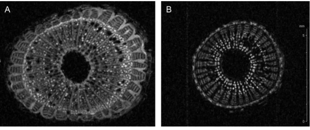
Nuclear magnetic resonance imaging (1H-MRI) was done in an Avance 400 spectrometer
equipped with Bruker DRX console microimaging accessory according toDandekar et al. (2012). Stem transverse sections of all non-infected and infected plants were collected between internodes located at the top (apical), middle and bottom of the central stem (three cuts per plant) and subjected to MRI.Fig. 2shows representative results.
DownstreamIn silicomethods
between proteins was performed with CLASP (Chakraborty et al., 2011). Adaptive Poisson– Boltzmann Solver (APBS) and PDB2PQR packages were used to calculate the electrostatic potentials of all the atoms in the protein (Baker et al., 2001;Dolinsky et al., 2004). The APBS parameters were set as described previously inChakraborty et al. (2011). APBS writes out the electrostatic potential in dimensionless units of kT/e where k is Boltzmann’s constant, T is the temperature in K and e is the charge of an electron, used to calculate the pairwise potential differences.
RESULTS & DISCUSSION
The CHURNER workflow for proteome analysis
CHURNER implements tandem analysis of sequence and structure of proteins to highlight potential functional similarities not obvious from simple sequence alignments. It uses simple Perl scripts to obtain the pairwise sequence and structural homology scores from BLAST and ProBiS, respectively (Fig. 1). The structural homology is then checked with FATCAT by the FCTSIG significance test (Ye & Godzik, 2004). We used these well established methods with different algorithms to detect structural similarity. This helps us corroborate the results between them (PZ scores). FATCAT treats the protein as flexible, allowing twists in the reference protein (akin to a real protein) and minimizes the number of rigid-body movements for the best structural alignment. ProBiS relies on common surface structural patches rather than global conservation in finding structural similarity, with the reasoning that the surface residues are more critical since they determine ligand or protein-protein interactions. The source code, working directory and a README script for the current example is available asSupplemental Information(see Methods). As a working example we used proteins identified by LC-MS/MS from xylem sap of grapevines infected withX. fastidiosaas described below. By using CHURNER we were able to highlight the Pathogenesis-Related proteins (PR-1) as the main proteins of grapevine defense againstX. fastidiosato be secreted in xylem sap and also to unravel structural similarity betweenβ -D-glucan exo-hydrolase and chitinases, which has no known reference in existing literature. To the best of our knowledge, this is the first attempt to analyze proteomic data for differentially expressed proteins based on structural features. A walkthrough of the steps implemented by CHURNER will be demonstrated next.
Our working example: xylem sap proteins identification by LC-MS/MS
In this study, proteins from xylem sap ofX. fastidiosa-infected and non-infected grapevines were lyophilized, precipitated by TCA/acetone and analyzed by SDS-PAGE (Fig. S2). A total of 91 proteins (Table 1) were identified herein by LC-MS/MS with at least two peptides sequenced per protein. SignalP and TargetP were used to predict the presence of signal peptides and sub-cellular localization, respectively, in all protein sequences. Signal peptides were found in 70 proteins (77%) of which 67 were predicted to be secreted while the others are directed towards an undetermined sub-cellular location.
Differentially expressed proteins in xylem sap of infected plants
Figure 1 The CHURNER workflow.After differentially expressed proteins are selected and grouped by functional analysis, individual protein sequences are used to retrieve UniProt and PDB identificators. All sequences and structures are compared pairwise, and significant structural alignments are then use to re-inforce protein functions that are significantly altered in the experiment. Yellow boxes indicate manual steps.
Table 1 Proteins identified in grapevine xylem sap ofXylella fastidiosa-infected and healthy plants by LC-MS/MS.a
UniProt ID Protein function Arabidopsisbest match MWb MPc Cov. (%)d SignalP TargetPe
ID E-value
Pathogenesis-related proteins
D7TXF5 Pathogenesis-related 4 AT3G04720.1 9E−51 15 6 62 21|22 Sec
F6HVL6 Pathogenesis-related 4 AT3G04720.1 2E−59 20 2 14 21|22 Sec
F6HBN7 Basic pathogenesis-related 1 AT2G14580.1 1E−47 18 4 37 25|26 Sec
A5BNW5 Pathogenesis-related 1 AT2G14610.1 2E−53 17 2 23 19|20 Sec
A5AWT7 Pathogenesis-related thaumatin AT1G20030.2 8E−83 45 5 17 26|27 Sec
A5AWT9 Osmotin 34 AT4G11650.1 1E−79 24 13 50 24|25 Sec
A5B4P9 Osmotin 34 AT4G11650.1 2E−82 24 6 47 24|25 Sec
Q9M4G7 Osmotin 34 AT4G11650.1 4E−72 20 5 42 – –
Q9M4G6 Osmotin 34 AT4G11650.1 7E−83 24 7 45 24|25 Sec
A5B2B6 Osmotin 34 AT4G11650.1 8E−80 24 3 35 24|25 Sec
Proteases
A5AZU5 Aspartyl protease AT5G07030.1 1E−134 40 5 15 – –
D7T5Q6 Aspartyl protease AT4G35880.1 1E−82 33 2 17 – –
F6H3K5 Aspartyl protease AT5G10770.1 1E−116 46 3 9 – –
F6H3K6 Aspartyl protease AT5G10770.1 1E−152 51 4 11 25|26 Sec
E0CQB3 Subtilisin AT1G20160.1 0E+00 79 19 36 – –
F6HNS0 Subtilisin AT5G59100.1 1E−157 69 9 24 – –
F6H4J9 Subtilisin AT5G67090.1 1E−157 79 2 3 – –
F6HSV1 Subtilase AT5G67360.1 0E+00 81 18 46 24|25 Sec
F6I357 Subtilase AT1G01900.1 0E+00 81 18 38 20|21 Sec
A5B179 Subtilase AT1G01900.1 1E−163 75 6 14 20|21 Sec
A5AIJ0 Serine carboxypeptidase 20 AT4G12910.1 0E+00 54 6 15 26|27 Sec
A5C816 Serine carboxypeptidase 51 AT2G27920.1 1E−163 51 4 16 21|22 Sec
Carbohydrate-active enzymes
F6GZC4 Basic chitinase AT3G12500.1 3E−89 35 15 68 21|22 Sec
Q9ZTK4 Basic chitinase AT3G12500.1 2E−85 35 11 52 21|22 Sec
A5BK69 Chitinase A AT5G24090.1 1E−109 32 12 48 25|26 Sec
F6H6H7 Chitinase A AT5G24090.1 1E−108 32 9 37 25|26 Sec
F6HB09 Carrot EP3-3 chitinase AT3G54420.1 1E−106 29 3 18 29|30 Sec
O24530 Carrot EP3-3 chitinase AT3G54420.1 5E−90 27 2 16 20|21 Sec
F6HQS7 Alpha-L-arabinofuranosidase AT3G10740.1 0E+00 84 20 26 28|29 Sec
F6HLL8 Beta-1, 3-glucanase 3 AT3G57240.1 1E−109 38 23 69 33|34 Sec
F6HLL9 Beta-1, 3-glucanase 3 AT3G57240.1 1E−110 37 4 22 32|33 Sec
A7PQW3 Beta-1, 3-glucanase 3 AT3G57240.1 3E−89 37 2 8 29|30 Sec
F6I6R4 Beta-D-xylosidase 4 AT5G64570.1 0E+00 83 23 42 33|34 Sec
D7TXW6 Alpha-galactosidase 2 AT5G08370.1 1E−169 45 11 31 24|25 Sec
F6HGW2 Beta galactosidase 1 AT3G13750.1 0E+00 92 2 2 24|25 Sec
D7SKW9 Beta-galactosidase 8 AT2G28470.2 0E+00 92 2 4 23|24 Sec
D7TPI6 Alpha-amylase-like AT4G25000.1 1E−161 47 6 20 22|23 Sec
Table 1(continued)
UniProt ID Protein function Arabidopsisbest match MWb MPc Cov. (%)d SignalP TargetPe
ID E-value
A5C7G0 Glucuronidase 3 AT5G34940.2 0E+00 71 3 8 18|19 Sec
F6GU88 Glycosyl hydrolase AT5G12950.1 0E+00 97 5 8 24|25 Sec
A5AZM8 Glycosyl hydrolase AT2G27500.1 1E−132 50 2 5 – –
F6H158 Glycosyl hydrolase AT1G58370.1 0E+00 105 5 7 – –
D7SVH6 Glycosyl hydrolase AT3G26720.1 0E+00 114 2 3 19|20 Sec
D7TQ09 O-Glycosyl hydrolases AT4G34480.1 1E−171 52 5 11 24|25 Sec
E0CQB9 O-Glycosyl hydrolases AT4G34480.1 1E−178 50 3 12 22|23 Sec
D7T828 O-Glycosyl hydrolases AT5G55180.1 0E+00 50 4 17 20|21 Sec
F6HCL5 Glycosyl hydrolases AT4G19810.1 1E−107 40 5 24 25|26 Sec
D7T548 Glycosyl hydrolases AT4G19810.1 1E−118 40 5 21 25|26 Sec
A7PZL3 Pectin lyase-like AT3G61490.3 0E+00 53 2 10 – –
F6HUM8 Pectin lyase-like AT3G61490.3 0E+00 52 7 24 – –
A5AZD0 Callose-binding protein 3 AT1G18650.1 3E−35 20 3 20 19|20 Sec
D7SI17 Callose-binding protein 3 AT1G18650.1 3E−39 21 2 20 19|20 Sec
A5C594 Expansin-like AT4G17030.1 2E−40 23 6 42 24|25 Sec
Receptor-like kinases (RLKs)
F6HIL5 Receptor-like kinase-related AT3G22060.1 8E−69 27 16 64 24|25 Sec
A5AID0 Receptor-like kinase-related AT5G48540.1 2E−74 45 3 11 25|26 Sec
D7TPF3 Leucine-rich repeat (LRR) SHV3-like 2
AT4G06744.1 1E−108 49 2 4 28|29 –
Peroxidases
F6GUF3 Peroxidase 2 AT5G06720.1 1E−109 36 2 7 23|24 Sec
F6GUE9 Peroxidase AT5G19890.1 1E−104 29 15 65 – –
F6HD61 Peroxidase AT1G49570.1 1E−110 36 11 35 25|26 Sec
A5BJV9 Peroxidase AT5G58390.1 3E−77 28 9 49 – –
F6H776 Peroxidase AT1G05260.1 8E−77 74 11 23 21|22 Sec
F6HIK4 Peroxidase AT1G05260.1 1E−135 76 2 3 26|27 Sec
D7TQI6 Peroxidase AT2G37130.1 1E−137 37 7 29 – –
F6H3X3 Peroxidase AT5G14130.1 8E−98 34 5 22 34|35 –
D7SVP1 Peroxidase AT5G14130.1 2E−20 10 2 43 – –
F6GXY7 Peroxidase AT5G05340.1 7E−96 28 2 13 – –
D7SR21 Peroxidase AT5G05340.1 3E−98 28 3 19 – –
F6H0Z1 Peroxidase AT5G05340.1 1E−113 34 2 13 22|23 Sec
A5B8V0 Peroxidase AT2G41480.1 6E−91 30 2 12 – –
F6HH88 Peroxidase AT2G41480.1 1E−120 70 2 7 24|25 Sec
F6HSU5 Peroxidase AT5G67400.1 1E−138 36 3 17 27|28 Sec
Others
E0CQL6 Basic blue protein-like AT2G02850.1 8E−38 19 8 49 – –
D7TML8 Inhibitor/LTP/seed storage AT3G53980.2 2E−33 12 11 67 27|28 Sec
D7SLG6 Inhibitor/LTP/seed storage AT2G44290.1 1E−35 19 3 26 – –
F6H7X9 Inhibitor/LTP/seed storage AT4G33550.2 1E−07 12 3 39 29|30 Sec
A5C9S3 Inhibitor/LTP/seed storage AT4G33550.2 4E−05 12 2 25 21|22 Sec
Table 1(continued)
UniProt ID Protein function Arabidopsisbest match MWb MPc Cov. (%)d SignalP TargetPe
ID E-value
F6I0G4 Pectin methylesterase inhibitor AT5G09760.1 0E+00 61 16 33 21|22 Sec
A5BS35 Basic seretory protein AT2G15220.1 1E−83 25 10 53 23|24 Sec
F6HS61 Glycine-rich protein AT4G30460.1 7E−03 13 7 67 22|23 Sec
A5AIZ1 Glycine-rich protein AT4G30460.1 3E−03 13 4 67 22|23 Sec
D7TY88 Protease inhibitor AT1G17860.1 6E−58 23 5 23 27|28 Sec
D7T293 Cupredoxin AT4G12420.2 0E+00 66 2 5 23|24 Sec
A5BMY7 Cupredoxin AT1G72230.1 7E−23 19 2 18 22|23 Sec
D7UBD5 Cupredoxin AT3G27200.1 4E−40 18 4 41 23|24 Sec
A5BZS1 FAD-binding Berberine AT4G20840.1 1E−179 59 5 10 30|31 Sec
A5B2E1 Cystatin/Monellin AT5G47550.1 2E−29 13 2 15 24|25 Sec
A5BH21 PLC-like phosphodiesterase AT1G66970.1 0E+00 70 3 5 21|22 Sec
D7SVW5 PI-PLC-like AT4G36945.1 1E−143 45 2 9 27|28 Sec
A5BB66 Fasciclin-like AT3G60900.1 1E−117 43 3 15 20|21 –
A5B7N6 Fasciclin-like AT4G12730.1 1E−115 44 2 8 26|27 Sec
D7SXH0 Lamin-like AT5G15350.1 3E−37 18 2 15 – –
A5AIY9 Unknown protein – – 15 4 59 23|24 Sec
Notes.
aA total of 91 proteins with at least two peptides sequenced per protein were identified and are displayed grouped by functional category. bPredicted molecular weight of proteins, in kDa.
cMatched peptides.
dPercentage of coverage.
eTargetP output: Sec, secreted protein; –, undefined.
mandarin infected withX. fastidiosa(Rodrigues et al., 2013), were not affected in our data, despite LRR-RLKs being detected in our proteomic analysis (Table 1). Plant receptor-like kinases (RLKs) are a large gene family (∼600 members inArabidopsis) (Shiu & Bleecker, 2001), consisting of an accelerated evolutionary domain implicated in signal reception through leucine-rich repeat (LRRs) (Afzal, Wood & Lightfoot, 2008). Resistance (R) genes have evolved to counter pathogens that bypass the pathogen-associated molecular patterns (PAMP) mechanism in plants (Nicaise, Roux & Zipfel, 2009). Most R genes encode proteins comprising of a nucleotide-binding site (NBS) and leucine-rich repeats (LRRs), and recognize and neutralize specialized pathogen avirulence (Avr) proteins, providing plants with resistance (Borhan et al., 2004;Chakraborty et al., 2016; Ernst et al., 2002;Hayashi et al., 2010;Zhang et al., 2010).
Up-regulated proteins
The up-regulated protein F6HLL8 listed in Table 2 is a β-1, 3-glucanase (GNS), a well-established pathogenesis related protein (Balasubramanian et al., 2012;Shinshi et al., 1988). GNS has strong anti-microbial (Xie et al., 2015) and anti-fungal activity (Su et al., 2013). Expectedly, it is a target of pathogen toxins in the ensuing evolutionary battle (Sanchez-Rangel, Sanchez-Nieto & Plasencia, 2012;Zhang et al., 2015b). GNS, along with chitinase, has been shown to inhibit fungal growth (Mauch, Mauch-Mani & Boller, 1988;
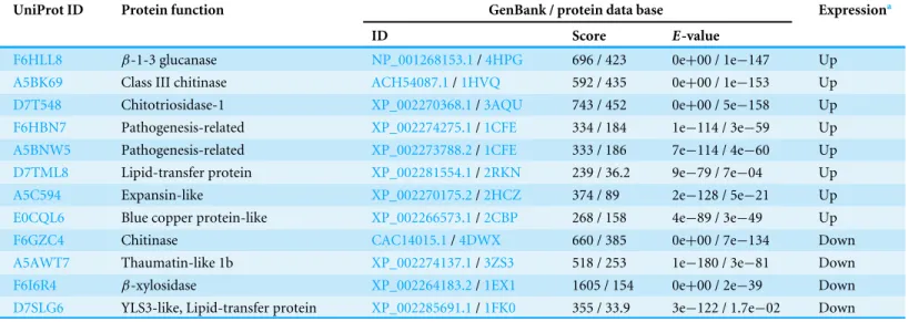
Table 2 Differentially expressed proteins and their sequence and structural similarities with reference proteins.
UniProt ID Protein function GenBank / protein data base Expressiona
ID Score E-value
F6HLL8 β-1-3 glucanase NP_001268153.1/4HPG 696 / 423 0e+00 / 1e−147 Up
A5BK69 Class III chitinase ACH54087.1/1HVQ 592 / 435 0e+00 / 1e−153 Up
D7T548 Chitotriosidase-1 XP_002270368.1/3AQU 743 / 452 0e+00 / 5e−158 Up
F6HBN7 Pathogenesis-related XP_002274275.1/1CFE 334 / 184 1e−114 / 3e−59 Up
A5BNW5 Pathogenesis-related XP_002273788.2/1CFE 333 / 186 7e−114 / 4e−60 Up
D7TML8 Lipid-transfer protein XP_002281554.1/2RKN 239 / 36.2 9e−79 / 7e−04 Up
A5C594 Expansin-like XP_002270175.2/2HCZ 374 / 89 2e−128 / 5e−21 Up
E0CQL6 Blue copper protein-like XP_002266573.1/2CBP 268 / 158 4e−89 / 3e−49 Up
F6GZC4 Chitinase CAC14015.1/4DWX 660 / 385 0e+00 / 7e−134 Down
A5AWT7 Thaumatin-like 1b XP_002274137.1/3ZS3 518 / 253 1e−180 / 3e−81 Down
F6I6R4 β-xylosidase XP_002264183.2/1EX1 1605 / 154 0e+00 / 2e−39 Down
D7SLG6 YLS3-like, Lipid-transfer protein XP_002285691.1/1FK0 355 / 33.9 3e−122 / 1.7e−02 Down
Notes.
aVariation of protein level detected in sap from infected grapevine compared to uninfected control.
Figure 2 Cell wall thickening of infected grapevines.Magnetic resonance imaging (1H-MRI) of stems
ofXylella fastidiosa(A) infected and (B) non-infected grapevines. Note the brighter contrast (denser
ma-terial) of secondary xylem and phloem vessels on the infected vine. Both images have the same magnifica-tion and scale bar is 5 mm. Images representative of transversal stem cuts obtained near 10 cm from top central stem. Similar thickening of cell walls were also observed in transversal cuts obtained along the vine until the base near the soil.
been demonstrated that citrus infected withX. fastidiosadisplay a thickening of secondary cell-walls (Niza et al., 2015).
Down-regulated proteins
As mentioned previously the GH19 sub-family chitinase (UID: F6GZC4) is down-regulated. A similar suppression of a chitinase gene in response to mycorrhizal fungus Glomus intraradicesinfection of tobacco roots has been noted previously (David et al., 1998). The GH19 sub-family has members that are sugar-binding proteins but without catalytic activity (Martinez-Caballero et al., 2014). Further experimentation is necessary to verify whether this is the case inVitis vinifera, as it can be part of the cell-wall remodeling in response to the pathogen, as previously suggested by other works (Lin et al., 2007;Rodrigues et al., 2013). A thaumatin-like protein (TLP) (UID: A5AWT7) is also found to be down-regulated upon
X. fastidiosainfection. TLP’s are found in most eukaryotes, and involved in host defense and several developmental processes (Liu, Sturrock & Ekramoddoullah, 2010). While TLP over expression has been shown to enhance resistance toAlternaria alternatain tobacco (Safavi, Zareie & Tabatabaei, 2012), these proteins can also be down-regulated in some cases, as shown in the compilation of transcriptomes of poplar leaf rust infections (Petre et al., 2011). Aβ-D-xilosidase 4 ortholog (UID: F6I6R4) was also down-regulated in infected vines, again reinforcing the drastic effect on cell-wall remodeling enzymes upon pathogen infection. The other down-regulated protein inTable 2is a LTP similar to YLS3 (yellow-leaf-specific) which is a marker of leaf senescence, being induced in earlier stages and repressed in later stages inArabidopsis(Yoshida et al., 2001).
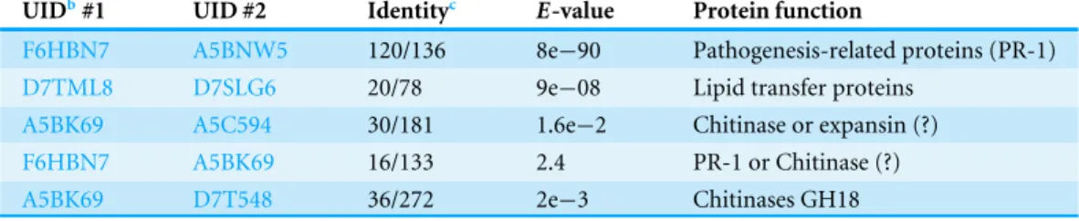
Sequence homology
Table 3 Pairwise BLASTaresults of example proteins analyzed with CHURNER.
UIDb#1 UID #2 Identityc E-value Protein function
F6HBN7 A5BNW5 120/136 8e−90 Pathogenesis-related proteins (PR-1)
D7TML8 D7SLG6 20/78 9e−08 Lipid transfer proteins
A5BK69 A5C594 30/181 1.6e−2 Chitinase or expansin (?)
F6HBN7 A5BK69 16/133 2.4 PR-1 or Chitinase (?)
A5BK69 D7T548 36/272 2e−3 Chitinases GH18
Notes.
aSequences aligned with bl2seq from NCBI. bUniProt identificator.
cTotal number of identical amino acid residues considering the best alignment between the two proteins.
homologous proteins difficult. Even in the current case, it is difficult to identify D7SLG6 as a lipid transfer protein (LTP) from the BLAST automated annotation (Table 2, value marked in italic). Thus, as the next step in CHURNER, we implemented a pairwise BLAST of all mature proteins (devoid of signal sequences).Table 3shows the pairwise sequence homology with anE-value cutoff of 0.005. There are several interesting aspects that emerge from this comparison. As expected, the two PR-1 proteins are found to be significantly homologous. The ‘YLS3-like’ protein (UID: D7SLG6) is found to be quite similar to another LTP (UID: D7TML8). Furthermore, we observe sequence homology between chitinases and expansin (E-value=4e−04), and much less between chitinases and PR-1 proteins
(E-value=0.002). Interestingly, these similarities are greater than that between the two known chitinases from sub-family GH18 (E-value =0.003). This raises the interesting question whether these proteins (chitinases/expansin/PR-1) have promiscuous functions (Chakraborty & Rao, 2012;Khersonsky & Tawfik, 2010), and underlines the problem of depending only on annotation of individual protein sequences, thus providing a more rational alternative to identify proteins with similar functions. Since the structure of a protein is intrinsically related to its function, we implemented structural annotation as the next step in CHURNER.
Structural annotation
CHURNER implements an automated search for homologous proteins with known PDB structures (Table 2). As expected, proteins with high similarity and alignment at the sequence level map to the same PDB structure, such as the PR-1 proteins (PDBid: 1CFE, PR-14a protein). Interestingly, this protein was identified as a possible replacement of the human neutrophil elastase component of the chimeric protein (Chakraborty, 2012;
Chakraborty et al., 2013) that provided enhanced grapevine resistance toX. fastidiosa
(Dandekar et al., 2012). Moreover, apart from the LTP (UID: D7SLG6,E-value=0.017), all matches are very significant to their PDB closest model.
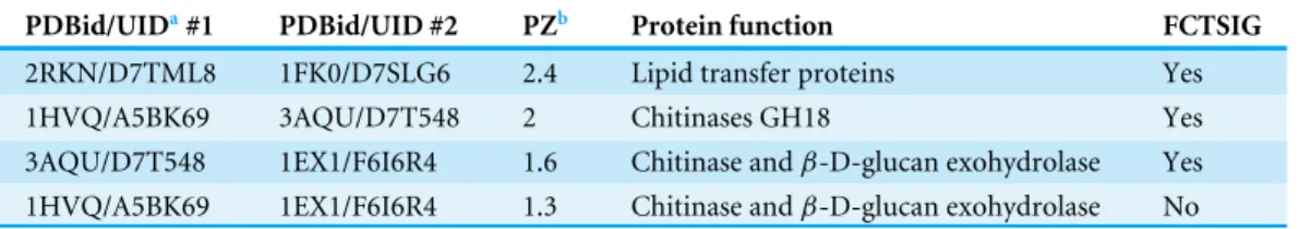
Structural homology
Table 4 Pairwise superimposition of the PDB structures using ProBiS.
PDBid/UIDa#1 PDBid/UID #2 PZb Protein function FCTSIG
2RKN/D7TML8 1FK0/D7SLG6 2.4 Lipid transfer proteins Yes
1HVQ/A5BK69 3AQU/D7T548 2 Chitinases GH18 Yes
3AQU/D7T548 1EX1/F6I6R4 1.6 Chitinase andβ-D-glucan exohydrolase Yes
1HVQ/A5BK69 1EX1/F6I6R4 1.3 Chitinase andβ-D-glucan exohydrolase No
Notes.
aUID: UniProt identificator.
bThe results are sorted based on the ProBiS ZScore (PZ). Significance of structural alignment was verified using FATCAT
(FCTSIG). Although the two chitinases have no sequence homology, their structural features are conserved. Similarly, we see signifiant structural similarity between a chitinase (UID: D7T548) and aβ-D-Glucan Exohydrolase (UID: F6I6R4).
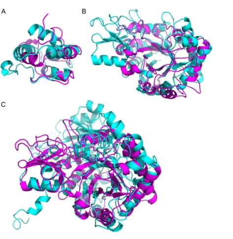
proteins which have the same PDB structure) computed using ProBiS (Konc & Janezic, 2010). We subsequently verified the alignment significance using the FATCAT server (Ye & Godzik, 2004). The ProBiS Z-score (PZ inTable 4) are standardized alignment scores (Konc & Janezic, 2010) which provide statistical and structural significance of local structural alignments. Z-scores>2 are considered highly significant (PDBs: 2RKN/1FK0 and 1HVQ/3AQU), although a Z-score of 1.6 is also significant (PDBs: 3AQU/1EX1), as confirmed by FATCAT (which looks at the global structure). The LTPs (with a sequence homologyE-value=1e−06) are structurally homologous, as expected (Fig. 3A). Noteworthy, the chitinases from the GH18 family with a low sequence homology (E -value =0.003) are structurally homologous (Fig. 3B). Finally, in spite of a much lower sequence homology (E-value=0.44), the chitinase (UID: D7T548) and theβ-D-glucan
exohydrolase (UID: F6I6R4) are structurally homologous (Fig. 3C). These observations highlight the necessity of structural comparison in annotating and grouping proteins based on functionality in proteomic analysis, and points to alternative protein functions that can be tested in subsequent studies.
Environmental stimuli (Yang et al., 2012), pathogens (Moy et al., 2004) or disease (Ng et al., 2009) induce differential expression of specific genes. Rapid technological advances have helped us identify these genes, and define their roles in defense or pathogenesis. While quantifying transcripts through high-throughput sequencing techniques have revolutionized these efforts (Wang, Gerstein & Snyder, 2009), the correlation between transcriptional and protein abundance remains suspect (Gygi et al., 1999) due to the complexity of the regulatory factors modulating translation (Zhu et al., 2012). Thus, identifying proteins through techniques like mass spectrometry (Witzel et al., 2009), and measuring their relative amounts (Hu, Rampitsch & Bykova, 2015), provides a true picture of the genes involved in pathogenesis and defense response, rather than measuring their RNA abundance (Moy et al., 2004). Nevertheless, the value of transcriptomic analysis should not be underestimated as it has proved to be an effective approach to discover genes responsive to infection, as exemplified by sequencing of expressed sequence tags fromX. fastidiosa-infected grapevines (Lin et al., 2007) and citrus (Rodrigues et al., 2013).
Figure 3 Superimposition of proteins that have significant structural homology.Structural homol-ogy has been detected using ProBiS, and confirmed using FATCAT. (A) Lipid transfer proteins: PDBid: 2RKNA (in magenta) and PDBid: 1FK0A (in cyan). (B) Chitinase GH18 proteins: PDBid: 1HVQA (in magenta) and PDBid: 3AQUA (in cyan). Note, that these proteins have low sequence homology (BLAST E-value=0.003). (C) Chitinase (PDBid: 3AQUA, in magenta) andβ-D-Glucan Exohydrolase (PDBid: 1EX1A, in cyan). These proteins have low sequence homology (E-value=0.44).
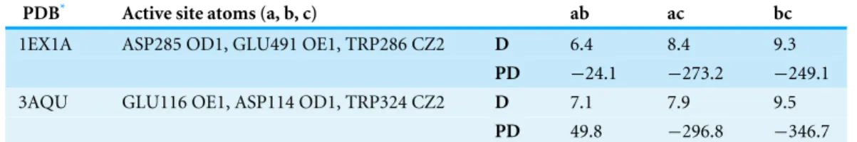
Table 5 Potential and spatial congruence of the active site residues in proteins chitinase andβ -D-glucan exohydrolase detected using CLASP.
PDB* Active site atoms (a, b, c) ab ac bc
1EX1A ASP285 OD1, GLU491 OE1, TRP286 CZ2 D 6.4 8.4 9.3
PD −24.1 −273.2 −249.1
3AQU GLU116 OE1, ASP114 OD1, TRP324 CZ2 D 7.1 7.9 9.5
PD 49.8 −296.8 −346.7
Notes.
*Chitinase: 1EX1A,β-D-glucan exohydrolase: 3AQU. The ability of CLASP to select stereo-chemically equivalent residues (Asp and Glu, both negatively charged residues) is critical to find the homologous active site. D, Pairwise distance in Å. PD, Pairwise potential difference. See Methods section for units of potential.
sequences with little sequence homology. For example, the two chitinase homologs have low pairwise sequence homology (E-value=0.003). However, their significant homologous counterparts in the PDB database are PDBid: 4DWXA (Secale cereale, rye) and PDBid: 3AQU (Arabidopsis thaliana) have significant structural homology, as computed using ProBiS (Konc & Janezic, 2010) and FATCAT (Ye & Godzik, 2004).
Corroborating the promiscuity of the chitinase andβ-D-glucan exohydrolase by active site structural homology
The β-D-glucan exohydrolase (PDBid: 1EX1) is critical for hydrolysis of cell walls, containing high levels of 1, 3-β-D-glucans, during wall degradation in germinated grain and during wall loosening in elongating coleoptiles (Varghese, Hrmova & Fincher, 1999). Interestingly, the fungal wall is composed of chitin, 1, 3-β- and 1, 6-β-glucan, mannan
and proteins (Adams, 2004). Thus, the up-regulation of both chitinases andβ-D-glucan exohydrolases is possibly an anti-fungal defense response that has been triggered by
X. fastidiosa. We have seen that the chitinase and theβ-D-glucan exohydrolase have no
sequence homology (Table 3), but partial structural homology (Table 4). A detailed analysis of their catalytic residues further strengthens credence of their functional similarity. For
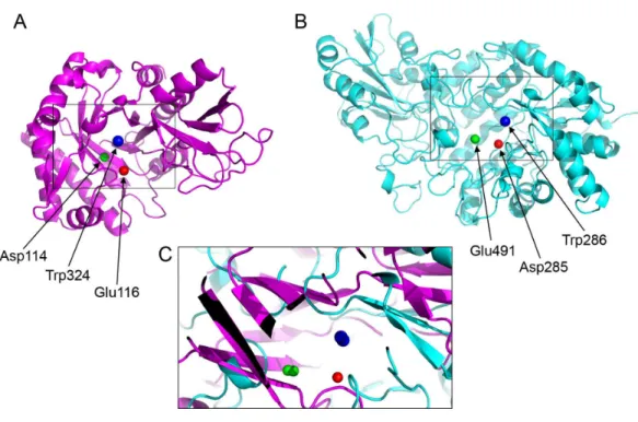
β-D-glucan exohydrolase, Asp285 and Glu491 are involved in catalysis (Varghese, Hrmova & Fincher, 1999). In chitinases, the Asp114 and Glu116 are a part of the conserved motif (DXXDXDXE) (Hamid et al., 2013). The active site residues of these proteins demonstrate significant spatial (Fig. 4) and electrostatic congruence (Table 5) determined using CLASP. The absence of sequence linearity indicates that this homology arose from convergent evolution. Remarkably, based on the catalytic triad of the barleyβ-D-glucan exohydrolase (PDBid: 1EX1A) provided to CLASP, it was able to pick up the catalytic triad from the
Arabidopsis chitinase (PDBid: 3AQU) despite the lack of sequence homology. We later realized this prediction was correct consulting the work from Ohnuma and collaborators (2011) on the crystallographic studies of this type V chitinase.
CONCLUSIONS
Figure 4 Active site residues in the chitinase andβ-D-glucan exohydrolase.(A) Glu116 (red), Asp114 (green), Trp324 (blue) in chitinase (PDBid:3AQU). (B) Asp285 (red), Glu491 (green), Trp286 (blue) in β-D-glucan exohydrolase (PDBid:1EX1A). (C) Superimposition of the chitinase (in magenta) andβ -D-glucan exohydrolase (in cyan).
viniferacv. Chardonnay vines (Agüero et al., 2008), and mass spectrometric analysis ofX. fastidiosa-infected grapevines (Katam et al., 2015;Yang et al., 2011). Thus, here we link the expression pattern of well-studied proteins in grapevines to the pathogen perception response, and present a methodology for assessing their significance by taking into account both their sequence and structural information. Although our data came from a single round of infection containing three plants (and three non-infected control plants), our findings are consistent among the different samples and to previous proteomic studies of
X. fastidiosainfected grapevines. In conclusion, CHURNER enhances our ability to find functionally-relevant protein candidates that have little or no sequence similarity, and thus would be considered as separate components of a data set. The name ‘‘CHURNER’’ was inspired in the mixing tool used to reach the ‘‘cream.’’ We intend to offer a tool to enable for detection of ‘‘cream’’ protein functions, not obvious from simple amino acid sequence alignments. The reduced data set employed in this work was used as a proof of concept, and we encourage readers to use complex data sets with thousands of proteins to find many putative functional relations among proteins that are yet unexplored.
ACKNOWLEDGEMENTS
ADDITIONAL INFORMATION AND DECLARATIONS
Funding
AMD received grant support from the California Department of Food and Agriculture PD/GWSS Board. BJR received financial support from Tata Institute of Fundamental Research (Department of Atomic Energy). Additionally, BJR received the JC Bose Award Grant from the Department of Science and Technology. RN, LRG, and PAZ received funding from the Brazilian Ministry of Science (CNPq) and Education (CAPES). The funders had no role in study design, data collection and analysis, decision to publish, or preparation of the manuscript.
Grant Disclosures
The following grant information was disclosed by the authors: California Department of Food and Agriculture PD/GWSS Board. Tata Institute of Fundamental Research (Department of Atomic Energy). JC Bose Award Grant from the Department of Science and Technology. Brazilian Ministry of Science (CNPq) and Education (CAPES).
Competing Interests
The authors declare there are no competing interests.
Author Contributions
• Sandeep Chakraborty conceived and designed the experiments, performed the experiments, analyzed the data, wrote the paper, prepared figures and/or tables, reviewed drafts of the paper.
• Rafael Nascimento and Hossein Gouran performed the experiments, analyzed the data, wrote the paper, prepared figures and/or tables, reviewed drafts of the paper.
• Paulo A. Zaini analyzed the data, wrote the paper, prepared figures and/or tables, reviewed drafts of the paper.
• Basuthkar J. Rao and Luiz R. Goulart contributed reagents/materials/analysis tools, reviewed drafts of the paper.
• Abhaya M. Dandekar conceived and designed the experiments, analyzed the data, contributed reagents/materials/analysis tools, wrote the paper, reviewed drafts of the paper.
Data Availability
The following information was supplied regarding data availability: Data can be found at Zenodo:http://dx.doi.org/10.5281/zenodo.50672.
Supplemental Information
REFERENCES
Adams DJ. 2004.Fungal cell wall chitinases and glucanases.Microbiology150:2029–2035
DOI 10.1099/mic.0.26980-0.
Afzal AJ, Wood AJ, Lightfoot DA. 2008.Plant receptor-like serine threonine kinases: roles in signaling and plant defense.Molecular Plant-Microbe Interactions 21:507–517DOI 10.1094/MPMI-21-5-0507.
Agüero CB, Thorne ET, Ibáñez AM, Gubler WD, Dandekar AM. 2008.Xylem sap proteins from Vitis vinifera L. Chardonnay.American Journal of Enology and Viticulture 59:306–311.
Altschul SF, Madden TL, Schaffer AA, Zhang J, Zhang Z, Miller W, Lipman DJ. 1997.
Gapped BLAST and PSI-BLAST: a new generation of protein database search programs.Nucleic Acids Research25:3389–3402DOI 10.1093/nar/25.17.3389.
Antoniw JF, White RF. 1980.The effects of aspirin and polyacrylic acid on soluble leaf proteins and resistance to virus infection in five cultivars of tobacco.Journal of Phytopathology98:331–341 DOI 10.1111/j.1439-0434.1980.tb03748.x.
Baker NA, Sept D, Joseph S, Holst MJ, McCammon JA. 2001.Electrostatics of nanosystems: application to microtubules and the ribosome.Proceedings of the National Academy of Sciences of the United States of America98:10037–10041
DOI 10.1073/pnas.181342398.
Balasubramanian V, Vashisht D, Cletus J, Sakthivel N. 2012.Plant beta-1, 3-glucanases: their biological functions and transgenic expression against phytopathogenic fungi.
Biotechnology Letters34:1983–1990DOI 10.1007/s10529-012-1012-6.
Basha SM, Mazhar H, Vasanthaiah HK. 2010.Proteomics approach to identify unique xylem sap proteins in Pierce’s disease-tolerant Vitis species.Applied Biochemistry and Biotechnology160:932–944DOI 10.1007/s12010-009-8620-1.
Bendtsen JD, Nielsen H, Von Heijne G, Brunak S. 2004.Improved prediction of signal peptides: SignalP 3.0.Journal of Molecular Biology 340:783–795
DOI 10.1016/j.jmb.2004.05.028.
Biles CL, Abeles FB. 1991.Xylem sap proteins.Plant Physiology96:597–601
DOI 10.1104/pp.96.2.597.
Borhan MH, Holub EB, Beynon JL, Rozwadowski K, Rimmer SR. 2004.TheArabidopsis
TIR-NB-LRR gene RAC1 confers resistance to Albugo candida (white rust) and is dependent on EDS1 but not PAD4.Molecular Plant-Microbe Interactions17:711–719
DOI 10.1094/MPMI.2004.17.7.711.
Buhtz A, Kolasa A, Arlt K, Walz C, Kehr J. 2004.Xylem sap protein composition is conserved among different plant species.Planta219(4):610–618.
Camacho C. 2008.BLASTR Command Line Applications User Manual. Bethesda:
National Center for Biotechnology Information. Available athttp:// www.ncbi.nlm. nih.gov/ books/ NBK279690/.
Cao J, Li X, Lv Y, Ding L. 2015.Comparative analysis of the phytocyanin gene family in 10 plant species: a focus on Zea mays.Frontiers in Plant Science6:515
Chakraborty S. 2012.An automated flow for directed evolution based on detection of promiscuous scaffolds using spatial and electrostatic properties of catalytic residues.
PLoS ONE7:e40408DOI 10.1371/journal.pone.0040408.
Chakraborty S, Britton M, Martinez-Garcia PJ, Dandekar AM. 2016.Deep RNA-Seq profile reveals biodiversity, plant–microbe interactions and a large family of NBS-LRR resistance genes in walnut (Juglans regia) tissues.AMB Express6:12
DOI 10.1186/s13568-016-0182-3.
Chakraborty S, Minda R, Salaye L, Bhattacharjee SK, Rao BJ. 2011.Active site detection by spatial conformity and electrostatic analysis–unravelling a proteolytic function in shrimp alkaline phosphatase.PLoS ONE6:e28470
DOI 10.1371/journal.pone.0028470.
Chakraborty S, Minda R, Salaye L, Dandekar AM, Bhattacharjee SK, Rao BJ. 2013.
Promiscuity-based enzyme selection for rational directed evolution experiments.
Methods in Molecular Biology 978:205–216DOI 10.1007/978-1-62703-293-3_15.
Chakraborty S, Rao BJ. 2012.A measure of the promiscuity of proteins and character-istics of residues in the vicinity of the catalytic site that regulate promiscuity.PLoS ONE7:e32011DOI 10.1371/journal.pone.0032011.
Champigny MJ, Isaacs M, Carella P, Faubert J, Fobert PR, Cameron RK. 2013.Long distance movement of DIR1 and investigation of the role of DIR1-like during systemic acquired resistance inArabidopsis.Frontiers in Plant Science4:230
DOI 10.3389/fpls.2013.00230.
Chatterjee S, Almeida RP, Lindow S. 2008.Living in two worlds: the plant and in-sect lifestyles ofXylella fastidiosa.Annual Review of Phytopathology46:243–271
DOI 10.1146/annurev.phyto.45.062806.094342.
Choi H, Fermin D, Nesvizhskii AI. 2008.Significance analysis of spectral count data in label-free shotgun proteomics.Molecular & Cellular Proteomics7:2373–2385
DOI 10.1074/mcp.M800203-MCP200.
Copley SD. 2003.Enzymes with extra talents: moonlighting functions and catalytic promiscuity.Current Opinion in Chemical Biology7:265–272
DOI 10.1016/S1367-5931(03)00032-2.
Dafoe NJ, Constabel CP. 2009.Proteomic analysis of hybrid poplar xylem sap. Phyto-chemistry70(7):856–863DOI 10.1016/j.phytochem.2009.04.016.
Dandekar AM, Gouran H, Ibanez AM, Uratsu SL, Aguero CB, McFarland S, Borhani Y, Feldstein PA, Bruening G, Nascimento R, Goulart LR, Pardington PE, Chaudhary A, Norvell M, Civerolo E, Gupta G. 2012.An engineered innate immune defense protects grapevines from Pierce disease.Proceedings of the National Academy of Sci-ences of the United States of America109:3721–3725DOI 10.1073/pnas.1116027109.
De Bernonville TD, Albenne C, Arlat M, Hoffmann L, Lauber L, Jamet E. 2014.Xylem sap proteomics.Methods in Molecular Biology 1072:391–405
DOI 10.1007/978-1-62703-631-3_28.
Djordjevic MA, Oakes M, Li DX, Hwang CH, Hocart CH, Gresshoff PM. 2007.The glycine max xylem sap and apoplast proteome.Journal of Proteome Research 6(9):3771–3779.
Dolinsky TJ, Nielsen JE, McCammon JA, Baker NA. 2004.PDB2PQR: an automated pipeline for the setup of Poisson–Boltzmann electrostatics calculations.Nucleic Acids Research32:W665–W667DOI 10.1093/nar/gkh381.
Emanuelsson O, Brunak S, Von Heijne G, Nielsen H. 2007.Locating proteins in the cell using TargetP, SignalP and related tools.Nature Protocols2:953–971
DOI 10.1038/nprot.2007.131.
Ernst K, Kumar A, Kriseleit D, Kloos DU, Phillips MS, Ganal MW. 2002.The broad-spectrum potato cyst nematode resistance gene (Hero) from tomato is the only member of a large gene family of NBS-LRR genes with an unusual amino acid repeat in the LRR region.The Plant Journal31:127–136
DOI 10.1046/j.1365-313X.2002.01341.x.
Fenyo D, Beavis RC. 2003.A method for assessing the statistical significance of mass spectrometry-based protein identifications using general scoring schemes.Analytical Chemistry75:768–774 DOI 10.1021/ac0258709.
Floerl S, Druebert C, Majcherczyk A, Karlovsky P, Kues U, Polle A. 2008.Defence reactions in the apoplastic proteome of oilseed rape (Brassica napus var. napus) attenuate Verticillium longisporum growth but not disease symptoms.BMC Plant Biology8:129DOI 10.1186/1471-2229-8-129.
Funkhouser JD, Aronson Jr NN. 2007.Chitinase family GH18: evolutionary insights from the genomic history of a diverse protein family.BMC Evolutionary Biology7:96
DOI 10.1186/1471-2148-7-96.
Gawehns F, Ma L, Bruning O, Houterman PM, Boeren S, Cornelissen BJC, Rep M, Takken FLW. 2015.The effector repertoire of Fusarium oxysporum determines the tomato xylem proteome composition following infection.Frontiers in Plant Science 6:967 DOI 10.3389/fpls.2015.00967.
Gherardini PF, Wass MN, Helmer-Citterich M, Sternberg MJ. 2007.Convergent evolution of enzyme active sites is not a rare phenomenon.Journal of Molecular Biology372:817–845DOI 10.1016/j.jmb.2007.06.017.
Gorg A, Obermaier C, Boguth G, Harder A, Scheibe B, Wildgruber R, Weiss W. 2000.
The current state of two-dimensional electrophoresis with immobilized pH gradi-ents.Electrophoresis21:1037–1053
DOI 10.1002/(SICI)1522-2683(20000401)21:6<1037::AID-ELPS1037>3.0.CO;2-V.
Gygi SP, Rochon Y, Franza BR, Aebersold R. 1999.Correlation between protein and mRNA abundance in yeast.Molecular and Cellular Biology 19:1720–1730
Hamid R, Khan MA, Ahmad M, Ahmad MM, Abdin MZ, Musarrat J, Javed S. 2013.Chitinases: an update.Journal of Pharmacy and Bioallied Sciences5:21–29
DOI 10.4103/0975-7406.106559.
Han Y, Chen Y, Yin S, Zhang M, Wang W. 2014.Over-expression of TaEXPB23, a wheat expansin gene, improves oxidative stress tolerance in transgenic tobacco plants.
Journal of Plant Physiology 173C:62–71DOI 10.1016/j.jplph.2014.09.007.
Hayashi N, Inoue H, Kato T, Funao T, Shirota M, Shimizu T, Kanamori H, Yamane H, Hayano-Saito Y, Matsumoto T, Yano M, Takatsuji H. 2010.Durable panicle blast-resistance gene Pb1 encodes an atypical CC-NBS-LRR protein and was generated by acquiring a promoter through local genome duplication.The Plant Journal 64:498–510DOI 10.1111/j.1365-313X.2010.04348.x.
Hilaire E, Young SA, Willard LH, McGee JD, Sweat T, Chittoor JM, Guikema JA, Leach JE. 2001.Vascular defense responses in rice: peroxidase accumulation in xylem parenchyma cells and xylem wall thickening.Molecular Plant-Microbe Interactions 14:1411–1419DOI 10.1094/MPMI.2001.14.12.1411.
Houterman PM, Speijer D, Dekker HL, CG DEK, Cornelissen BJ, Rep M. 2007.
The mixed xylem sap proteome of Fusarium oxysporum-infected tomato plants.
Molecular Plant Pathology 8:215–221DOI 10.1111/j.1364-3703.2007.00384.x.
Hu J, Rampitsch C, Bykova NV. 2015.Advances in plant proteomics toward improve-ment of crop productivity and stress resistance.Frontiers in Plant Science6:209
DOI 10.3389/fpls.2015.00209.
Jensen RA. 1976.Enzyme recruitment in evolution of new function.Annual Review of Microbiology30:409–425DOI 10.1146/annurev.mi.30.100176.002205.
Katam R, Chibanguza K, Latinwo LM, Smith D. 2015.Proteome biomarkers in xylem reveal pierce’s disease tolerance in grape.Journal of Proteomics & Bioinformatics 8:217–224.
Kehr J, Buhtz A, Giavalisco P. 2005.Analysis of xylem sap proteins from Brassica napus.
BMC Plant Biology5:11.
Keller A, Nesvizhskii AI, Kolker E, Aebersold R. 2002.Empirical statistical model to estimate the accuracy of peptide identifications made by MS/MS and database search.Analytical Chemistry74:5383–5392DOI 10.1021/ac025747h.
Khersonsky O, Tawfik DS. 2010.Enzyme promiscuity: a mechanistic and evolutionary perspective.Annual Review of Biochemistry79:471–505
DOI 10.1146/annurev-biochem-030409-143718.
Kleywegt GJ. 1999.Recognition of spatial motifs in protein structures.Journal of Molecular Biology 285:1887–1897DOI 10.1006/jmbi.1998.2393.
Konc J, Janezic D. 2010.ProBiS algorithm for detection of structurally similar protein binding sites by local structural alignment.Bioinformatics26:1160–1168
DOI 10.1093/bioinformatics/btq100.
Ligat L, Lauber E, Albenne C, San Clemente H, Valot B, Zivy M, Pont-Lezica R, Arlat M, Jamet E. 2011.Analysis of the xylem sap proteome of Brassica oler-acea reveals a high content in secreted proteins.Proteomics11(9):1798–1813
Lin H, Doddapaneni H, Takahashi Y, Walker MA. 2007.Comparative analysis of ESTs involved in grape responses toXylella fastidiosainfection.BMC Plant Biology7:8
DOI 10.1186/1471-2229-7-8.
Liu JJ, Sturrock R, Ekramoddoullah AK. 2010.The superfamily of thaumatin-like proteins: its origin, evolution, and expression towards biological function.Plant Cell Reports29:419–436 DOI 10.1007/s00299-010-0826-8.
Lu S, Faris JD, Sherwood R, Edwards MC. 2013.Dimerization and protease resistance: new insight into the function of PR-1.Journal of Plant Physiology170:105–110
DOI 10.1016/j.jplph.2012.08.006.
Martinez-Caballero S, Cano-Sanchez P, Mares-Mejia I, Diaz-Sanchez AG, Macias-Rubalcava ML, Hermoso JA, Rodriguez-Romero A. 2014.Comparative study of two GH19 chitinase-like proteins from Hevea brasiliensis, one exhibiting a novel carbohydrate-binding domain.FEBS Journal281:4535–4554
DOI 10.1111/febs.12962.
Mauch F, Mauch-Mani B, Boller T. 1988.Antifungal Hydrolases in Pea Tissue : II. Inhibition of Fungal Growth by Combinations of Chitinase and beta-1, 3-Glucanase.
Plant Physiology88:936–942DOI 10.1104/pp.88.3.936.
McElrone AJ, Sherald JL, Forseth IN. 2003.Interactive effects of water stress and xylem-limited bacterial infection on the water relations of a host vine.Journal of Experimental Botany54:419–430DOI 10.1093/jxb/erg046.
Metzner R, Schneider HU, Breuer U, Thorpe MR, Schurr U, Schroeder WH. 2010.
Tracing cationic nutrients from xylem into stem tissue of French bean by stable isotope tracers and cryo-secondary ion mass spectrometry.Plant Physiology 152:1030–1043DOI 10.1104/pp.109.143776.
Mi H, Muruganujan A, Casagrande JT, Thomas PD. 2013.Large-scale gene function analysis with the PANTHER classification system.Nature Protocols8:1551–1566
DOI 10.1038/nprot.2013.092.
Moy P, Qutob D, Chapman BP, Atkinson I, Gijzen M. 2004.Patterns of gene expression upon infection of soybean plants by Phytophthora sojae.Molecular Plant-Microbe Interactions17:1051–1062DOI 10.1094/MPMI.2004.17.10.1051.
Nanjo Y, Nakamura T, Komatsu S. 2013.Identification of indicator proteins associated with flooding injury in soybean seedlings using label-free quantitative proteomics.
Journal of Proteome Research12:4785–4798DOI 10.1021/pr4002349.
Nersissian AM, Immoos C, Hill MG, Hart PJ, Williams G, Herrmann RG, Valentine JS. 1998.Uclacyanins, stellacyanins, and plantacyanins are distinct subfamilies of phytocyanins: plant-specific mononuclear blue copper proteins.Protein Science 7:1915–1929DOI 10.1002/pro.5560070907.
Nesvizhskii AI, Keller A, Kolker E, Aebersold R. 2003.A statistical model for identifying proteins by tandem mass spectrometry.Analytical Chemistry75:4646–4658
Ng EK, Chong WW, Jin H, Lam EK, Shin VY, Yu J, Poon TC, Ng SS, Sung JJ. 2009.
Differential expression of microRNAs in plasma of patients with colorectal cancer: a potential marker for colorectal cancer screening.Gut58:1375–1381
DOI 10.1136/gut.2008.167817.
Nicaise V, Roux M, Zipfel C. 2009.Recent advances in PAMP-triggered immunity against bacteria: pattern recognition receptors watch over and raise the alarm.Plant Physiology150:1638–1647DOI 10.1104/pp.109.139709.
Niza B, Coletta-Filho HD, Merfa MV, Takita MA, De Souza AA. 2015.Differential colonization patterns ofXylella fastidiosainfecting citrus genotypes.Plant Pathology 64:1259–1269DOI 10.1111/ppa.12381.
Ohnuma T, Numata T, Osawa T, Mizuhara M, Lampela O, Juffer AH, Skriver K, Fukamizo T. 2011.A class V chitinase fromArabidopsisthaliana: gene re-sponses, enzymatic properties, and crystallographic analysis.Planta234:123–137
DOI 10.1007/s00425-011-1390-3.
Okushima Y, Koizumi N, Kusano T, Sano H. 2000.Secreted proteins of tobacco cultured BY2 cells: identification of a new member of pathogenesis-related proteins.Plant Molecular Biology 42:479–488DOI 10.1023/A:1006393326985.
Petre B, Major I, Rouhier N, Duplessis S. 2011.Genome-wide analysis of eukaryote thaumatin-like proteins (TLPs) with an emphasis on poplar.BMC Plant Biology 11:33DOI 10.1186/1471-2229-11-33.
Pu Z, Ino Y, Kimura Y, Tago A, Shimizu M, Natsume S, Sano Y, Fujimoto R, Kaneko K, Shea DJ, Fukai E, Fuji S-I, Hirano H, Okazaki K. 2016.Changes in the proteome of xylem sap in Brassica oleracea in response to Fusarium oxysporum stress.Frontiers in Plant Science7:31DOI 10.3389/fpls.2016.00031.
Rep M, Dekker HL, Vossen JH, De Boer AD, Houterman PM, Speijer D, Back JW, De Koster CG, Cornelissen BJ. 2002.Mass spectrometric identification of isoforms of PR proteins in xylem sap of fungus-infected tomato.Plant Physiology130:904–917
DOI 10.1104/pp.007427.
Rice P, Longden I, Bleasby A. 2000.EMBOSS: the European Molecular Biology Open Software Suite.Trends in Genetics16:276–277DOI 10.1016/S0168-9525(00)02024-2.
Rodrigues CM, De Souza AA, Takita MA, Kishi LT, Machado MA. 2013. RNA-Seq analysis of Citrus reticulata in the early stages ofXylella fastidiosa infec-tion reveals auxin-related genes as a defense response.BMC Genomics14:676
DOI 10.1186/1471-2164-14-676.
Safavi K, Zareie R, Tabatabaei BES. 2012.Constitutive expression of thaumatin-like protein (TLP-3) in transgenic tobacco plants leads to enhance resistance to Alternaria alternata.Archives of Phytopathology and Plant Protection45:161–169
DOI 10.1080/03235408.2010.507947.
Sela-Buurlage MB, Ponstein AS, Bres-Vloemans SA, Melchers LS, Van Den Elzen P, Cornelissen B. 1993.Only specific tobacco (Nicotiana tabacum) chitinases and [beta]-1, 3-glucanases exhibit antifungal activity.Plant Physiology101:857–863.
Shi X, Bi J, Morse JG, Toscano NC, Cooksey DA. 2010.Differential expression of genes ofXylella fastidiosain xylem fluid of citrus and grapevine.FEMS Microbiology Letters 304:82–88DOI 10.1111/j.1574-6968.2009.01885.x.
Shinshi H, Wenzler H, Neuhaus JM, Felix G, Hofsteenge J, Meins F. 1988.Evidence for N- and C-terminal processing of a plant defense-related enzyme: Primary structure of tobacco prepro-beta-1, 3-glucanase.Proceedings of the National Academy of Sciences of the United States of America85:5541–5545DOI 10.1073/pnas.85.15.5541.
Shiu SH, Bleecker AB. 2001.Receptor-like kinases fromArabidopsisform a mono-phyletic gene family related to animal receptor kinases.Proceedings of the National Academy of Sciences of the United States of America98:10763–10768
DOI 10.1073/pnas.181141598.
Stajich JE, Block D, Boulez K, Brenner SE, Chervitz SA, Dagdigian C, Fuellen G, Gilbert JG, Korf I, Lapp H, Lehvaslaiho H, Matsalla C, Mungall CJ, Osborne BI, Pocock MR, Schattner P, Senger M, Stein LD, Stupka E, Wilkinson MD, Birney E. 2002.The Bioperl toolkit: perl modules for the life sciences.Genome Research 12:1611–1618DOI 10.1101/gr.361602.
Subramanian S, Cho UH, Keyes C, Yu O. 2009.Distinct changes in soybean xylem sap proteome in response to pathogenic and symbiotic microbe interactions.BMC Plant Biology9:119DOI 10.1186/1471-2229-9-119.
Sudisha J, Sharathchandra R, Amruthesh K, Kumar A, Shetty HS. 2012. Pathogenesis related proteins in plant defense response. In:Plant defence: biological control. New York: Springer, 379–403.
Su YC, Xu LP, Xue BT, Wu QB, Guo JL, Wu LG, Que YX. 2013.Molecular cloning and characterization of two pathogenesis-related beta-1, 3-glucanase genes ScGluA1 and ScGluD1 from sugarcane infected by Sporisorium scitamineum.Plant Cell Reports 32:1503–1519DOI 10.1007/s00299-013-1463-9.
Van Loon LC, Rep M, Pieterse CM. 2006.Significance of inducible defense-related proteins in infected plants.Annual Review of Phytopathology44:135–162
DOI 10.1146/annurev.phyto.44.070505.143425.
Varghese JN, Hrmova M, Fincher GB. 1999.Three-dimensional structure of a barley beta-D-glucan exohydrolase, a family 3 glycosyl hydrolase.Structure7:179–190
DOI 10.1016/S0969-2126(99)80024-0.
Wallis CM, Chen J. 2012.Grapevine phenolic compounds in xylem sap and tissues are significantly altered during infection byXylella fastidiosa.Phytopathology 102:816–826DOI 10.1094/PHYTO-04-12-0074-R.
Wang Z, Gerstein M, Snyder M. 2009.RNA-Seq: a revolutionary tool for transcrip-tomics.Nature Reviews Genetics10:57–63DOI 10.1038/nrg2484.
Xie YR, Raruang Y, Chen ZY, Brown RL, Cleveland TE. 2015.ZmGns, a maize class I beta-1, 3-glucanase, is induced by biotic stresses and possesses strong antimicrobial activity.Journal of Integrative Plant Biology 57:271–283DOI 10.1111/jipb.12286.
Yang Y, He M, Zhu Z, Li S, Xu Y, Zhang C, Singer SD, Wang Y. 2012.Identification of the dehydrin gene family from grapevine species and analysis of their respon-siveness to various forms of abiotic and biotic stress.BMC Plant Biology12:140
DOI 10.1186/1471-2229-12-140.
Yang L, Lin H, Takahashi Y, Chen F, Walker MA, Civerolo EL. 2011.Proteomic analysis of grapevine stem in response toXylella fastidiosainoculation.Physiological and Molecular Plant Pathology 75:90–99DOI 10.1016/j.pmpp.2010.11.002.
Ye Y, Godzik A. 2004.FATCAT: a web server for flexible structure comparison and structure similarity searching.Nucleic Acids Research32:W582–W585
DOI 10.1093/nar/gkh430.
Yoshida S, Ito M, Nishida I, Watanabe A. 2001.Isolation and RNA gel blot analysis of genes that could serve as potential molecular markers for leaf senescence in
Arabidopsisthaliana.Plant and Cell Physiology42:170–178DOI 10.1093/pcp/pce021.
Zhang J, Li W, Xiang T, Liu Z, Laluk K, Ding X, Zou Y, Gao M, Zhang X, Chen S, Mengiste T, Zhang Y, Zhou JM. 2010.Receptor-like cytoplasmic kinases integrate signaling from multiple plant immune receptors and are targeted by a Pseudomonas syringae effector.Cell Host Microbe7:290–301DOI 10.1016/j.chom.2010.03.007.
Zhang Z, Xin W, Wang S, Zhang X, Dai H, Sun R, Frazier T, Zhang B, Wang Q. 2015b.
Xylem sap in cotton contains proteins that contribute to environmental stress response and cell wall development.Functional and Integrative Genomics15:17–26
DOI 10.1007/s10142-014-0395-y.
Zhang W, Yan H, Chen W, Liu J, Jiang C, Jiang H, Zhu S, Cheng B. 2014.Genome-wide identification and characterization of maize expansin genes expressed in endosperm.
Molecular Genetics and Genomics289:1061–1074DOI 10.1007/s00438-014-0867-8.
Zhang YT, Zhang YL, Chen SX, Yin GH, Yang ZZ, Lee S, Liu CG, Zhao DD, Ma YK, Song FQ, Bennett JW, Yang FS. 2015a.Proteomics of methyl jasmonate induced defense response in maize leaves against Asian corn borer.BMC Genomics16:224
DOI 10.1186/s12864-015-1363-1.