UNIVERSIDADE FEDERAL DO CEARÁ
FACULDADE DE FARMÁCIA, ODONTOLOGIA E ENFERMAGEM PROGRAMA DE PÓS-GRADUAÇÃO EM ODONTOLOGIA
TÁCIO PINHEIRO BEZERRA
AVALIAÇÃO DA FRAGILIDADE MANDIBULAR AO TRAUMA DEVIDO À
PRESENÇA DE TERCEIROS MOLARES: UMA ANÁLISE TRIDIMENSIONAL
COM ELEMENTOS FINITOS
TÁCIO PINHEIRO BEZERRA
AVALIAÇÃO DA FRAGILIDADE MANDIBULAR AO TRAUMA DEVIDO À PRESENÇA DE TERCEIROS MOLARES: UMA ANÁLISE TRIDIMENSIONAL COM
ELEMENTOS FINITOS
Tese submetida ao Programa de Pós-Graduação em Odontologia, da Universidade Federal do Ceará, como requisito parcial para a obtenção do grau de Doutor em Odontologia.
Área de Concentração: Clínica Odontológica. Orientador: Prof. Dr. Eduardo Costa Studart Soares Coorientador: Prof. Dr. Francisco Ilson da Silva Júnior
Dados Internacionais de Catalogação na Publicação Universidade Federal do Ceará
Biblioteca de Ciências da Saúde
B469a Bezerra, Tácio Pinheiro
Avaliação da fragilidade mandibular ao trauma devido à presença de terceiros molares: uma análise tridimensional com elementos finitos/ Tácio Pinheiro Bezerra. – 2012.
34 f.
Tese (Doutorado) - Universidade Federal do Ceará, Faculdade de Farmácia, Odontologia e Enfermagem, Programa de Pós-Graduação em Odontologia, Fortaleza, 2012.
Área de concentração: Clinica Odontológica Orientação: Prof. Dr. Eduardo Costa Studart Soares Coorientação: Prof. Dr. Francisco Ilson da Silva Júnior
TÁCIO PINHEIRO BEZERRA
AVALIAÇÃO DA FRAGILIDADE MANDIBULAR AO TRAUMA DEVIDO À PRESENÇA DE TERCEIROS MOLARES: UMA ANÁLISE TRIDIMENSIONAL COM
ELEMENTOS FINITOS
Tese submetida ao Programa de Pós-Graduação em Odontologia, da Universidade Federal do Ceará, como requisito parcial para a obtenção do grau de Doutor em Odontologia
Aprovada em 17 de maio de 2012
BANCA EXAMINADORA
___________________________________________ Prof. Dr. Eduardo Costa Studart Soares (Orientador)
Universidade Federal do Ceará-UFC
___________________________________________ Prof. Dr. Francisco Ilson da Silva Junior
Universidade Federal do Ceará-UFC
___________________________________________ Prof. Dr. Henrique Clasen Scaparo
Universidade Federal do Ceará - UFC
___________________________________________ Prof. Dr. Francisco Wagner Vasconcelos Freire Filho
Universidade de Fortaleza - UNIFOR
___________________________________________ Prof. Dr. Luis Raimundo Serra Rabelo
AGRADECIMENTOS ESPECIAIS
Em primeiro lugar à Deus, que é a luz que ilumina os meus caminhos, que graças a Ele são prósperos. Por ter me dado a família que me orgulho, a esposa que amo e os amigos que mereço.
Ao meu pai Bezerra e minha mãe Celina, fontes de amor inesgotável, exemplos discretos, presentes, constantes, espontâneos e fundamentais para o que sou hoje. Devo tudo o que sou ao que vocês me ensinaram. Agradeço a Deus por todos os sonhos que deixaram de realizar e todas as privações que tiveram para poder dar a mim e à minha irmã a educação que nos deram. Hoje eu tenho certeza de que eu fui abençoado por ter sido gerado nessa família.
À minha esposa Denise exemplo de mulher, amorosa e presente, ainda me lembro do dia que te disse: do inesperado nasce algo inevitável que aos poucos se torna inseparável. Obrigado por ter me acompanhado em boa parte da minha formação, por ter me dado apoio nos momentos difíceis, suportar os momentos de dificuldades, impaciências ausências e presenças ausentes, dividir comigo os problemas, as alegrias, as conquistas mútuas e a nossa profissão.
À minha irmã, exemplo de determinação e simplicidade. Seus atos objetivos e focados foram muito importantes para a minha formação como pessoa. As minhas dificuldades se tornavam simples quando eu me dava conta dos obstáculos que eu ví você passar para chegar onde está hoje. Obrigado!
Minhas sobrinhas Maria Cláudia e Maria Clara, que conseguem nos fazer sorrir até mesmo quando as coisas não estão bem. Com certeza, são enviadas de Deus para nos ajudar a vencer cada dia.
Aos meus cunhados José Claudio, Samara e Cláudio Milério, muito obrigado por compartilharem comigo muitos momentos especiais da minha vida, viagens, brincadeiras, farras, conversas, jantares... Podem ter certeza que vocês são como irmãos.
À toda minha família que se fez presente em algum momento da minha vida trazendo certamente alguma contribuição.
muito bem orientou meus passos de formação. Por fim, passou a ser um grande amigo que hoje faz parte da minha pequena e seleta roda de confiança.
Ao Prof. Dr. Francisco Ilson, um desconhecido que surpreendeu todas as melhores expectativas. Pessoa, simples, atenciosa, capacitada e sem a qual esta pesquisa ainda estaria nas fases de simulação computacional. Muito obrigado por ter realizado uma importante parte deste trabalho. Espero que o grupo de trabalho tenha sido valoroso e que não se encerre com a defesa desta tese. Muito mais há por produzir. Obrigado!
Aos amigos de vida e de especialidade Ivo Pita, Fábio Wildson, Marcelo Ferraro, Alexandre Nogueira, Romulo Medeiros, Ernest Pouchain, Diego Esses, Carlos Diego, Bárbara Carneiro, Rafael Linard, Rodrygo Tavares, Saulo Elery, Sormani Queiroz, Patrício, Nelson Studart, Ricardo Bessa, Abrahão Cavalcante, Patrício Oliveira, Saulo Batista e todos os outros, pela forma sempre unida como lidamos a nossa relação profissional e de amigos. Ao amigo Carlos Bruno Pinheiro pela colaboração com a elaboração e realização deste trabalho.
A todos os colegas de graduação, residência, mestrado e doutorado por todas as trocas de experiência, brincadeiras, farras, brigas e conquistas que tivemos. Saibam que cada um tem uma parcela de participação nas minhas conquistas.
Aos colegas de trabalho na Faculdade Católica Rainha do Sertão, na Perícia Forense do Ceará e no Hospital Geral Dr. Waldemar Alcântara por possibilitarem um ambiente de trabalho de muito respeito e aprendizado mútuo.
AGRADECIMENTOS
À Universidade Federal do Ceará (UFC) e ao Programa de Pós-Graduação em Odontologia, pela oportunidade de realizar a minha formação em odontologia, graduação, mestrado e doutorado. É uma grande honra ser filho desta escola.
Ao Conselho Nacional de Desenvolvimento Científico e Tecnológico (CNPq) pelo suporte financeiro a este estudo.
À coordenadora do Programa de Pós-Graduação em Odontologia da Universidade Federal do Ceará, Profa. Dra. Lidiany Rodrigues, em nome de quem agradeço a todos os professores do programa. Obrigado por terem dedicado o seu tempo e conhecimento em prol da nossa formação.
À todos os meus professores de graduação, residência, especialização, mestrado e doutorado que contribuíram para o meu desenvolvimento pessoal, profissional e científico.
À professora Dra. Andrea Walter por estar sempre me estimulando a trilhar um caminho embasado na ciência e nos valores humanos.
À professora Dra. Cristiane Fonteles por suas valorosas e importantes sugestões e por ser um exemplo de caráter e competência.
Ao professor Dr. Henrique Scarparo pelos importantes ensinamentos profissionais e pelos momentos divididos em várias cirurgias desde o tempo de professor substituto.
Ao professor Dr. Wagner Freire pela sua grande determinação em prol da nossa especialidade e por ter aceito o convite para contribuir com seus conhecimentos na avaliação do presente trabalho.
Ao professor Dr. Luis Rabelo pelo exemplo de docente presente e atencioso desde o tempo de residência e por ter aceito o convite para se fazer presente na avaliação desta tese.
À professora Marivan Ferraro pela revisão gramatical do presente trabalho.
Às secretarias do Programa de Pós-Graduação em Odontologia pela atenção e apoio sempre prestados.
RESUMO
O ângulo mandibular tem sido descrito como uma área frágil e, acredita-se que, com a presença do terceiro molar, a mandíbula perde parte de sua estrutura para abrigar tecidos que não contribuem para sua resistência estrutural. Neste contexto, o artigo produzido nessa tese teve como objetivo identificar áreas de concentração e distribuição de estresse na região do ângulo em uma mandíbula portando os dois terceiros molares erupcionados, outra portando apenas um terceiro molar erupcionado e outra sem terceiros molares após a aplicação de um trauma mentual por meio de simulações computacionais por elementos finitos. Foi reconstruída uma mandíbula através da discretização de máscaras por um processo de identificação digital e atribuição de propriedades de acordo com a densidade tomográfica para o osso cortical e medular, ligamento periodontal, cemento, dentina, esmalte e polpa. Para diferenciar as estruturas, a mandíbula portando os terceiros molares recebeu uma substituição digital das propriedades correspondentes a esses tecidos dentários por máscaras correspondente aos tecidos ósseos, assim produzindo as duas outras estruturas do estudo. De modo a reproduzir a condição anatomia real, os nós da vertente posterior dos côndilos foram deixados imóveis e foram criados elementos para reproduzir a ação da musculatura mastigatória. Cada um dos corpos recebeu um impacto mentual perpendicular ao plano frontal de 250 quilograma-força. Os resultados foram avaliados por análise descritiva do diagrama cromático da dispersão de estresses de Von Misses. Um modelo detalhado, relacionado ao paciente, de alta resolução foi produzido com uma alta densidade de elementos finitos (914.952 elementos para mandíbula 01, 867.183 para a 02 e 831.897 para a 03). Devido ao método detalhado de obtenção dos corpos e à metodologia do estudo foi possível obter resultados adequados para a resposta ao impacto. De acordo com o diagrama de dispersão de tensões, sempre que o terceiro molar esteve presente, seja bilateral ou unilateralmente, houve uma maior concentração de tensões ao redor da porção cervical do alvéolo desse dente. No entanto, quando o dente estava ausente, houve uma concentração de energia maior no colo do côndilo. O presente trabalho apresentou um modelo experimental que reproduz adequadamente a dinâmica mandibular e que evidenciou o comportamento da estrutura mandibular frente à um impacto mentual. Neste caso, o terceiro molar é responsável pela concentração de tensões na região da linha oblíqua externa e trígono retro-molar, justificando que esse dente contribui para uma maior fragilidade do ângulo mandibular.
ABSTRACT
The mandibular angle has been described as fragile area, the presence of the third molar has been suggested to contribute to increased mandibular fragility because the mandible loses parts of its bone structure to harbor an organ that does not contribute to its strength. In this context, the article produced on this thesis had the objective of identify areas of tension concentration and its distribution on the mandibular angle if both third molar were present, if only one was present, and without third molars. Each mandible was submitted to blunt mentual trauma and evaluated by a finite element methodology. A mandible was reconstructed through the discretization of masks by a digital process of identification of structures considering the tomographic density to the cortical and medullar bone, periodontal ligament, cement, dentin, enamel and pulp. To differentiate the structures, the first mandible with both third molars was submitted to a digital replacement of the mask from the third molar structures to the masks of the cortical and medullar bone. Therefore, producing the two other structures of the study. To reproduce the normal anatomic situation, the external nodes of the most posterior and superior part of the mandibular condyle were fixated in all degrees of freedom bilaterally, and elements were created to reproduce the actions of the masticatory muscles. Each structure was submitted to a blunt mentual trauma, perpendicularly to the frontal plane, with 250 kilograms of magnitude. The results were evaluated by the description of the chromatic stress distribution diagram of Von Misses. A highly detailed, patient-specific, custom-made, high-resolution model of the mandible could be generated with a very dense volume mesh (914.952 elements for the mandible 01, 867.183 for 02, and 831.897 for 03). Due to the detailed method of body prove attainment and to the study methodology it was possible to obtain adequate results to the dynamic of the impact. According to the diagram of the dispersion of tensions, whenever the third molar was present, unilateral or bilateral, there was a greater concentration of tensions around the cervical part of the alveolus. However, when absent the stress concentration was more significant on the condylar neck. The present study showed an experimental model that reproduces the mandibular dynamics and the behavior of the mandibular structure to a mentual trauma. As a conclusion, the third molar is responsible to a tension concentration on the region of the external oblique ridge and retromolar area justifying that these teeth contribute to the mandibular angle fragility.
LISTA DE ABREVIATURAS
EVM Escala de Von Misses
MEF Método de elementos finitos
SUMÁRIO
1 INTRODUÇÃO GERAL... 11 2 PROPOSIÇÃO... 15 3 CAPITULOS ... 16
3.1 CAPITULO 1
Do third molars weaken the mandibular angle in a mentual trauma? A finite element study...
16
11
1 INTRODUÇÃO GERAL
A mandíbula é uma estrutura óssea especializada, com músculos, articulações e dentes em complexa sinergia. A forma e função mandibular são altamente adaptadas para o desenvolvimento do sistema mastigatório (VOLLMER et al., 2000). Devido sua projeção no esqueleto facial, a mandíbula é um dos ossos mais suscetíveis ao trauma na face, com uma frequência estimada de 11,5 casos por 100.000 indivíduos por ano (INAOKA et al., 2009; MA’AITA; ALWRIKAT; JORDAN, 2000).
O ângulo mandibular tem sido descrito como uma área frágil e a presença do terceiro molar sugere um fator de aumento no risco de fraturas de ângulo (IIDA et al., 2005; UGBOKO; OGINNI; OWOTADE, 2000). Acredita-se que, com a presença desse dente, a mandíbula perde parte de sua estrutura óssea ao abrigar tecidos que não contribuem para sua resistência (MEISAMI et al., 2002). Um estudo experimental em macacos demonstrou que mandíbulas contendo terceiros molares não erupcionados fraturaram com aproximadamente 60% da força requerida para fraturar mandíbulas com esses dentes erupcionados (REITZIK et al., 1978).
Vários estudos reportam aumentos de duas a três vezes de risco de fraturas de ângulo mandibular quando da presença de um terceiro molar (HALMOS; ELLIS III; DODSON, 2004; MEISAMI et al., 2002; FUSELIER; ELLIS; DODSON, 2002; LEE; DODSON, 2000). Bezerra et al. (2010) relataram que a presença do terceiro molar inferior acarreta um risco 1,94 vezes maior de fratura do ângulo mandibular.
12
fraturas de ângulo. No entanto, quando o dente não estava presente, houve um aumento estatisticamente significante na prevalência de fraturas de côndilo (DUAN; ZANG 2008).
A precisa avaliação do comportamento biomecânico da mandíbula é fundamental para diversas áreas da Odontologia, em especial para a Cirurgia e Traumatologia Buco-Maxilo-Faciais, uma vez que esta deverá avaliar a biofuncionalidade dos sistemas atualmente disponíveis para a fixação interna rígida de fraturas faciais (MEYER et al., 2000), bem como indicar ou não a remoção profilática de terceiros molares (GOMES et al., 2004).
Em 1956, Turner e cols., trabalhando em um projeto de aeronaves para a Boeing, propuseram um método de análise estrutural computacional, mais tarde, denominado pelos autores como Método dos Elementos Finitos (MEF). A simulação computacional por elementos finitos possibilita a modelagem matemática de estruturas complexas com geometrias irregulares de tecidos naturais e artificiais, como os dentes e ossos maxilares, além de permitir a modificação dos parâmetros de sua geometria. Com isso, torna-se possível a aplicação de um sistema de forças em qualquer ponto e/ou direção, promovendo, assim, informações sobre o deslocamento e o grau de tensão provocado por essas cargas ao tecido analisado. Neste método, o modelo computacional é realizado por meio da determinação de elementos de tamanhos e propriedades físicas finitas, o que o torna adaptável a estruturas reais e possibilita a avaliação da deformação e distribuição de tensões sob estresse na estrutura estudada (MAURER et al., 2002).
Segundo Wong et al. (2011), o método de elementos finitos é uma ferramenta poderosa que permite a análise de sistemas geométricos complexos de uma maneira viável. Os modelos são feitos por meio da divisão do objeto em um grande número de partes menores (elementos), através de uma malha unida por pontos nodais. Em seguida, executa-se então a aplicação de cargas e são avaliadas as consequências, como o comportamento de deformação da estrutura, de acordo com as propriedades e condições de limite dos elementos dos materiais individuais. O processamento do modelo envolve certificar-se de que todas as mudanças em todos os elementos são consistentes entre si, que no caso do modelo, é convertido a uma solução. A modelagem computacional permite que o trabalho seja realizado num ambiente virtual, no qual modelos numéricos complexos podem ser trabalhados com relativa facilidade (WONG et al., 2011).
13
vitro, com aquela obtida por meio de elementos finitos, empregando a mesma mandíbula. Concluíram que o estresse gerado e a distribuição de forças obtida pelos dois métodos obedeceram a uma correlação de 99,2%, ratificando o uso de tal método como uma potente ferramenta computacional para a compreensão da biomecânica mandibular.
Wroe et al. (2010) realizou um estudo com metodologia de elementos finitos procurando esclarecer a evolução do processo mastigatório desde os ancestrais hominídios até o homem moderno (Homo sapiens) para esclarecer o motivo de mudança evolutiva da dieta. Os autores conseguiram reproduzir os crânios com metodologia de elementos finitos ao ponto de afirmarem que a mudança do padrão alimentar ocorreu devido à uma modificação evolutiva de músculos e do crânio. Desta forma, seus resultados evidenciaram que o método é passível de ser utilizado para predizer comportamentos mandibulares, e assim ser empregado em odontologia.
Gallas-Torreira e Fernandez (2004) aplicaram o método de elementos finitos a um modelo mandibular simulado para avaliar a repercussão de traumas aplicados sobre a sínfise e corpo mandibular. Os autores observaram que há uma concentração de tensões na região alveolar do ângulo mandibular. No entanto, os autores não consideraram as propriedades dos dentes e do ligamento periodontal, fazendo com que haja uma situação que não reproduz adequadamente o real.
Takada et al. (2006) avaliaram, por meio do método de elementos finitos produzidos por microtomografias, a interferência de terceiros molares sobre a distribuição de tensões aplicadas perpendicularmente ao ângulo mandibular. Concluíram haver uma concentração de tensões na região apical e cervical do terceiro molar, ao passo que a tensão se distribui para o corpo e ramo nas mandíbulas sem tais dentes.
14
15
2 PROPOSIÇÃO
O presente trabalho tem como objetivo geral avaliar o impacto da presença do terceiro molar na fragilidade mandibular, tendo como objetivos específicos:
- Identificar áreas de concentração e distribuição de tensão na região do ângulo mandibular, em mandíbula portando os dois terceiros molares erupcionados, após a aplicação de um trauma mentual.
- Identificar áreas de concentração e distribuição de tensão na região do ângulo mandibular, em mandíbula portando apenas um terceiro molar erupcionado, após a aplicação de um trauma mentual.
16
3 CAPÍTULOS
Esta tese está baseada no Artigo 46, do Regimento Interno do Programa de Pós-Graduação em Odontologia da Universidade Federal do Ceará, que regulamenta o formato alternativo para teses de Doutorado e permite a inserção de artigos científicos de autoria ou co-autoria do candidato.
Por se tratar de pesquisa envolvendo seres humanos, os protocolos utilizados neste trabalho foram submetidos à apreciação e foram devidamente aprovados pelo Comitê de Ética em Pesquisa do Hospital Universitário Walter Cantídio (Anexo).
Dessa forma, a presente tese é composta por um artigo científico redigido de acordo com a revista científica escolhida.
3.1 Capítulo 1
“Do third molars weaken the mandibular angle in a mentual trauma? A three-dimentional finite element study.” Este artigo seguiu normas de publicação do periódico
17
Do third molars weaken the mandibular angle in a mentual trauma? A three-dimensional finite element study.
TITLE PAGE
Do third molars weaken the mandibular angle in a mentual trauma? A three-dimensional finite element study.
Authors:
Tácio Pinheiro BEZERRA – DDS, MS, PhD Francisco Ilson SILVA-JUNIOR – MS, PhD Henrique Clasen SCARPARO – DDS, MS, PhD Fábio Wildson Gurgel COSTA – DDS, MS
Eduardo Costa STUDART-SOARES – DDS, MS, PhD Intitutional Affiliation:
Post-Graduation Program in Dentistry of the Federal University of Ceará Address: St. Monsenhor Furtado, without number. Rodolfo Teófilo 60.430-350 – Fortaleza – Ceará – Brazil
Corresponding Author: Tácio Pinheiro Bezerra
Address: St. Tomaz Pompeu, 340, apto 802. Meireles. 60.160-080 – Fortaleza – Ceará – Brazil
Phone: 55-85-88995998 Fax: 55-85-32412855
e-mail: taciopb@uol.com.br drtpb@hotmail.com
Sources of support:
This research received the financial support of Grant # 478819/2010-2 from Conselho Nacional de Desenvolvimento Científico e Tecnológico (MTC/CNPq nº 14/2010).
Keywords:
Finite Element Analysis; Molar, third; Mandible
Running title:
18
ABSTRACT
19
INTRODUCTION
The mandible is one of the bones most susceptible to trauma in the facial region due to its more projected position in the facial skeleton (1-3). This prevalence is influenced by a series of factors such as sex, age, socioeconomic condition and the characteristics of trauma (4). An experimental study on monkeys has shown that mandibles containing unerupted third molars fractured at approximately 60% of the force required to fracture mandibles with erupted third molars (5). Bezerra et al (6) reported a 1.94-fold higher risk of mandibular angle fractures when the third molar is present.
Force applied directly in the symphisis region in axial plane is distributed along the arch of the mandible. Because the condylar heads are free to rotate within the glenoid fossa to a certain degree, tension develops along the lateral aspect of the condylar neck and mandibular body regions, as well as along the lingual aspect of the symphysis. This leads to a bilateral condylar fracture and a symphysis fracture, unless a fragility factor exists (7).
The reason for the increased prevalence of mandibular angle fractures is not well established (1). Presence of third molars has been suggested to contribute to an increased mandibular fragility because the mandible loses parts of its bone structure to harbor an organ that does not contribute to its strength (4, 8, 9). In addition, some authors support that completely unerupted teeth are more associated with mandibular fragility because they compromise more extensively the bone structure. However, the effect of partially erupted teeth on the support structures of the mandibular framework (external oblique line) should be taken into account (3, 4, 5, 10). A retrospective data files review indicated that the prevalence and relative risk of mandibular angle fracture are both significantly higher in subjects with fully erupted third molars than in individual lacking those teeth (11).
For obvious ethical reasons, there is no human study design that would permit the elucidation of this interference, since it would be impossible to submit experimental and control groups to injuries likely to fracture the mandible, in order to evaluate the resistance of this bone and the effect of the third molar on mandibular fractures.
20
accuracy of FEA to describe the biomechanical behavior of bone specimens has been shown by different authors (13, 14, 15, 16).
In the present study, a three-dimensional computed tomographic-based finite element reconstruction of three human mandibles with or without third molars was performed to evaluate these mechanical properties. It aimed to evaluate the impact of the erupted third molar presence on the mandibular angle stiffness when submitted to a mentual trauma.
MATERIALS AND METHODS
The ethics committee of the local institution approved the protocol for this study. An informed written consent was obtained from a 30 year old, male patient who had a computer tomography selected for the study, based on the fact that he had all the lower teeth without any structural mandibular changes (osseous callus/fracture, pathologic entities, previous orthodontic treatment maxilla-mandibular discrepancy or periodontal illness).
The images were obtained by a cone beam CT, and were imported by the ScanIP software (Simpleware ® Ltd., Exeter, UK) in which was defined the tomographic density window applicable to the object in study (2240 X 550 UH) and pixel size to be used (0.55 mm).
Modeling
To produce the virtual structure, the three-dimensional mesh and all the steps to the finite element analysis, it was done an adaptation of what was written by Silva et al (17).
21
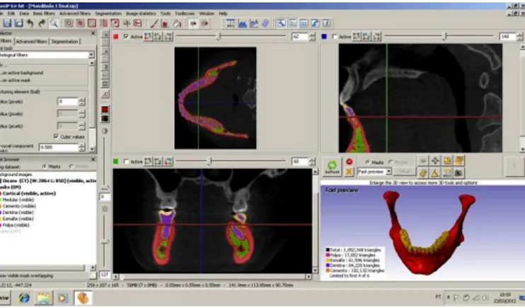
Figure 01 – ScanIP interface. Discretization process on three spacial plans and a 3D visualization.
After the production of the final model in all slices, the software generated a 3D structure maintaining in position each mask discretized. For greater smoothness on the surface of the structure, a software tool was used to fill small gaps and round angles. The result was a very detailed 3D mesh.
Virtual Sample Differentiation
To create the three meshes to be included in this study, the initial mandible structure (mandible 01) was submitted to a digital mask substitution. On the software interface the pixels of the third molar were changed from the initial tooth structure masks to those from cortical and medullar bone in each CT slice in accordance to an anatomic aspect. Therefore, it was possible to create a second structure without the right third molar (mandible 02), and third one without third molars (mandible 03) (figure 02). But the rest of the structure remained the same.
Figure 02 – The three structures developed to the study. Mandible 01 with both third molars; Mandible 02 without the left third molar; Mandible 03 without both third molars.
Meshing
22
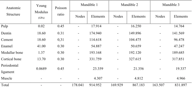
ANSYS® (SIMULIA, Providence, RI, USA), version 13.0, for structural analysis of the mechanical tests. The homogeneity of the structures, linear elastic deformation pattern, and the standardization of the isotropic mechanical properties were ensured for each discretized mask (Table 01). The values of Young modulus and Poison ratio were based on the Lotti et al (18) reference. Anatomic Structure Young Modulus (GPa) Poisson ratio
Mandible 1 Mandible 2 Mandible 3
Nodes Elements Nodes Elements Nodes Elements
Pulp 0.02 0.45 - 17.914 - 16.250 - 14.764
Dentin 18.60 0.31 - 174.940 - 149.896 - 141.569
Cement 18.60 0,31 - 114.618 - 104.475 - 96.478
Enamel 41.00 0.30 - 54.887 - 50.659 - 47.247
Medullar bone 1.37 0.30 - 193.168 - 192.120 - 189.685
Cortical bone 13.70 0.30 - 331.759 - 327.615 - 317.851
Periodontal
ligament 0.0689 0.45 - 23.359 - 21.356 - 19.337
Muscle - - - 4.307 - 4.812 - 4.966
Total - - 178.041 914.952 169.929 867.183 163.507 831.897
Table 01. Mechanical properties, references of number of nodes and elements in each mask reconstructed.
Boundary Conditions
To simulate an anatomically normal mandibular function, the external nodes of the most posterior and superior part of the mandibular condyle were fixated in all degrees of freedom bilaterally (figure 03). The actions of the masticatory muscles were reproduced by the creation of spring resistance elements with vectors as described by Bujtár et al (19). And the rigidity based on an estimation of deformation of the muscles.
23
Load Application
A blunt trauma, perpendicularly to the frontal plane, with a magnitude of 250 kgf, was applied, to a 1-cm diameter circular area (center on the pogonium), in the midline of the symphysis, perpendicularly to the coronal plane. This was a simulation representative of a frontal aggression (punch).
The results were evaluated by a descriptive analysis of the chromatic Von Misses stresses distribution of the tension after the impact (EVM).
RESULTS
A highly detailed, patient-specific, custom-made, high-resolution yet simplified model of the mandible could be generated with a very dense volume mesh of 914.952 finite elements for the mandible 01, 867.183 for 02, and 831.897 for 03. Based on this method the details of the mandible could be emphasized and successfully included into an analysis of the dynamics of a response to an impact.
The maximum stresses were located at the symphysis (point of impact), at the retromolar area and both condyles on the three experimental models.
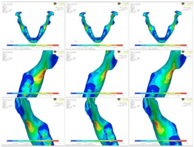
The third molar presence resulted in a difference of the tension distribution on the three meshes studied. Whenever the third molar was present there was a greater concentration of tensions around the cervical part of the alveolus (figure 04).
24
It was noticeable that the impact resulted on a concentration of tension on the external oblique ridge, and when the third molar was present this concentration extended to the alveolar process (figure 5).
Figure 05 – The Von Misses chromatic stress distribution showing the concentration of stress on the external oblique ridge in all models. On mandible 01 it can be seen that the concentration goes to the cervical alveolar area.
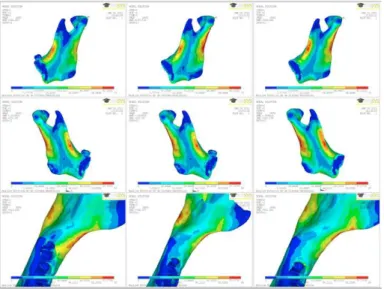
However, on the mandibles 02 and 03, the structural reinforcement provided by the bone on the retromolar area without third molar made the tension concentrate more on the condilar region on the side of the removal (figure 06).
25
Approximated Von Mises equivalent stress in the third molar region was 107.035 MPa in the mandible with a third molar and 64.6948 MPa in the mandible without a third molar. Whereas in the condilar region the Von Misses equivalent stress was 151.65 MPa when the third molar was present and 184.496 MPa when absent.
DISCUSSION
Finite element analysis has been developed into a branch of applied mathematics for numeric modeling of physical systems that is used in many engineering disciplines. In its simplest mathematical terms, this numeric technique is used to find approximate solutions for partial differential equations and integral equations through the generation of meshes of a continuous domain for a set of discrete subdomains or “elements.” Numeric methods are then used to predict the behavior of the object in question in various situations, for example, under conditions of loading (20). The external forces and the mechanical properties/geometry are used to calculate the nodal displacements; the differentiation of the displacement field yields the strain distribution; and the stress distribution is determined mathematically (21).
The FEA is being developed to overcome the experimental models to biomechanics studies. It is extremely difficult to create an experimental model of the mandible; the geometry, internal structures and the function cannot be grossly simplified. Moreover, the muscular action cannot be reproduced as a unique vector of force attached to a single point (22). The muscle tension is needed to cause an angle fracture (23). This reduction of detail leads to a simplified model of the item's behavior; it may often lead to incomplete or incorrect models of the mechanics of the structure (22). Avoiding these significant variables, the experimental computational model developed in this study approached the real mandibular behavior. It’s geometric shape was reconstructed based on a real mandibular structure, the masticatory muscles were attached to the mandible as in an anatomic body, and the physical and mechanical properties were reproduced similarly to the normal body.
In addition, to allow the comparison between the structures in order to evaluate the influence of the third molar presence, the only difference was on the mechanical properties of the third molars pixels. The rest of the structures remained exactly similar to diminish structural bias.
26
tension than compression (24). In undertaking any therapy that affects the skeleton, it is important to understand the potential problem of excessive loading of bone (25).
Huelk and Harger (24) described that once a force is applied to an anterior mandibular region the energy dispersion will occur along the body toward the condyles, causing tension on the lateral aspect of the angle and condyle. Furthermore, the force seeks out the weakest point in the arch and causes extreme bending and tensile failure at that point. These aspects could be verified in the present study, characterized by stress concentration on the external oblique ridge near the third molar, and in the neck region of the condyle on it’s buccal and posterior sides.
The knowledge of local or systemic factors, which weaken the jaw structure, is of great relevance to maxillofacial trauma. The third molar can be related to this fragility, since their presence significantly alters the biomechanics of the mandibular angle. Retrospective studies, case series or literature reviews show that the presence of third molar relates to the increased prevalence of mandibular angle fracture (6). However, there is not enough studies that show, in a biomechanics perspective, the true impact of it’s presence and the occurrence of fractures, considering that reproducing the real model of the mandible is extremely difficult.
The FEA is a valid and non-invasive method that provides useful results to predict different parameters of the complex biomechanical behavior of human mandibles (14).
With these suitable digital models, this study proved that the mandibular angle becomes more fragile when the third molar is present. Gallas-Torreira and Fernandez (15) was the only study with a similar methodology and results. But the authors highlighted that clinical extrapolations from mathematical models may not give absolute values. The reason to that is due to a not adequately recreation of the computational model, considering that they did not apply differential mechanical properties to the teeth parts, allowing then to function as a part of the mandibular structure. Moreover, Vollmer et al (14) added the necessity of attribution the boundary conditions of the condyles and the distribution of the masticatory muscles to obtain as adequate computational model. With the accurate methodology used, it can be assumed that the results of the present work approached to a real situation.
27
the fragility of mandibular angle. This is an important information to know for decision making of third molar removal.
In a retrospective study, Inaoka et al (1) reported that the percentage of impacted third molar was greater in angle fractures than in condylar fractures. Duan and Zang (23) stated that when the mandible is submitted to a low force trauma, the third molar presence prone the bone to fracture on the mandibular angle. But, in an overall evaluation of the sample reviewed, the data revealed that patients without third molars had a significantly higher risk of sustaining condylar fractures than those with third molars. This information can be confirmed by the results of this essay based on the increased equivalent Von Misses stress on the third molar region when the tooth was present and on the condilar region when it was absent. Comparing the sub-condilar region between the three mandibles it can be seen that when the third molar was substituted by normal bone, the energy concentrates more on the sub-condilar region. That’s why a mandible with third molar tends to fail on the angle region, while the one without those teeth tend to fail on the condilar neck. The surgeon must think carefully to indicate the third molar removal to reduce the mandibular angle fracture risk, considering that it may predispose to condylar fracture, a significantly more morbid injury with an increased risk of complications (11).
Duan and Zang (23) considered that a low force trauma was the one able to fracture the mandible in one site, the moderate in two, and the high in three or more. With respect to prophylactic third molar extraction, it appears that the impacted ones in patients with a high risk of suffering low trauma forces, as in contact sports, should be extracted, whereas patients more often subjected to moderate or high trauma forces might not benefit from prophylactic third molar extraction. After analyzing the relationship between multiple mandibular fractures and the presence of lower third molars, Choi et al (11) found that, in mandibles with embedded lower third molars, the mandibular symphysis is the most common site of comorbid fracture in individuals with a mandibular angle fracture. The studies say that the third molar presence prone the mandible to fracture on the third molar region when the trauma is of moderate intensity and can cause two fractures, one in the place of the impact (symphysis) and the other on the angle. The images obtained in the present study support this information as far as the energy concentrates on the impact point and on the retro-molar area of the third molar.
28
with a real body simulation in vivo and can be extrapolated to computational models. To overcome this limitation a larger sample would be need. However, it would be impossible in a real situation to submit a great number of subjects to mandibular impacts and in the computational simulation it would be a laborious and time-consuming task.
The present study aimed to evaluate the influence of the third molar presence on the mandibular angle weakness through a finite element model methodology. Under the conditions of this study it can be said that this experimental model produced a reliable reproduction of a real situation found in “in vivo” facial traumas. More over, the comparative analysis showed a tension concentration on the vestibular aspect of the mandibular angle when the third molar was present, and when it was absent the concentration was on the condilar neck. These findings must be considered in the decision making to prophylactically remove third molars in subjects prone to receive facial trauma. Future simplifications of this method and its evolution into a more user-friendly modality for dentistry may facilitate the use of finite element analysis in the preoperative analysis of specific surgical sites (25).
Funding:
This research was funded by Grant # 478819/2010-2 from CNPq. Competing interests - None declared.
Ethical approval
This study was submitted and approved by the local committee on human research of Walter Cantídio University Hospital, registered under protocol 0.43.04.11.
REFERENCES
1. Inaoka SD, Carneiro SC, Vasconcelos BC, Leal J, Porto GG. Relationship between mandibular fracture and impacted lower third molar. Med Oral Patol Oral Cir Bucal 2009: 14: 349-54.
2. Libersa P, Roze D, Cachart T, Libersa JC. Immediate and late mandibular fractures after third molar removal. J Oral Maxillofac Surg 2002: 60: 163-5.
3. Ma’aita J, Alwrikat A, Jordan A. Is the mandibular third molar a risk for mandibular fracture? Oral Surg Oral Med Oral Pathol Oral Radiol Endod 2000: 89: 143-6.
4. Meisami T, Sojat A, Sandor GKB, Lawrence HP, Clokie CML. Impacted third molars and risk of angle fractures. Int J Oral Maxillofac Surg 2002: 31: 140-4.
29
6. Bezerra TP, Studart-Soares EC, Pita-Neto IC, Costa FWG, Batista SHB. Do third molars weaken the mandibular angle? Medicina oral, patología oral y cirugía bucal 2009: 16: e657-e663. doi: 10.4317/medoral.16970.
7. Chacon GE; Larsen PE. Principles of management of mandibular fractures. In: Miloro M. In: Miloro, M, ed,: Peterson´s Principles Of Oral and Maxillofacial Surgery. London: BC Decker, 2004: 401-433.
8. Iida S, Hassfeld S, Reuther T, Nomura K, Muhling J. Relationship between the risk of mandibular angle fractures and the status of incompletely erupted mandibular third molar. J Craniomaxillofac Surg 2005: 33: 158-63.
9. Ugboko VI, Oginni FO, Owotade FJ. An investigation into the relationship between mandibular third molars and angle fractures in Nigerians. Br J Oral Maxillofac Surg 2000: 38: 427-9.
10. Halmos DR, Ellis EE 3rd, Dodson TB. Mandibular third molars and angle fractures. J Oral Maxillofac Surg 2004: 62: 1076-81.
11. Choi BJ, Park S, Lee DW, Ohe JY, Kwon YD. Effect of Lower Third Molars on the Incidence of Mandibular Angle and Condylar Fractures. J Craniomaxillofac Surg 2011: 22(4): 1521-25.
12. Maurer P, Holweg S, Knoll WD, Schubert J. Study by finite element method of the mechanical stress of selected biodegradable osteosynthesis screws in sagittal ramus osteotomy. Br J Oral Maxillofac Surg 2000: 40: 76–83.
13. Groning F, Liu J, Fagan MJ, O’Higgins P. Validating a voxel-based finite element model of a human mandible using digital speckle pattern interferometry. J Biomechan 2009: 42: 1224-29.
14. Vollmer D, Meyer U, Joos U, Vegh A, Piffko J: Experimental and finite element study of a human mandible. J Cranio Maxillofac Surg 28: 91-96, 2000.
15. Gallas-Torreira M, Fernandez JR. A three-dimensional computer model of the human mandible in two simulated standard trauma situations. J Craniomaxillofac Surg 2004: 32: 303–307.
16. Liao SH, Tong RF, Dong JX. Anisotropic finite element modeling for patient-specific mandible. Comput methods programs biomed 2007: 88: 197-209.
30
18. Lotti RS, Machado AW, Mazzieiro ET, Landre Júnior J. Aplicabilidade científica do método dos elementos finitos. R Dental Press Ortodon Ortop Facial 2006: 11(2): 35-43.
19. Bujtár, P, Sandor, G K B, Bojtos A, Szucs, A, Barabas, J. Finite element analysis of the human mandible at 3 different stages of life. Oral Surg Oral Med Oral Pathol Oral Radiol Endod 2010;110:301-309.
20. Strang G, Fix G. An analysis of the finite element method. Englewood Cliffs (NJ): Prentice-Hall; 1973.
21. Wong RCW, Tideman H, Merkx MAW, Jansen J, Goh SM, Liao K. Review of biomechanical models used in studying the biomechanics of reconstructed mandibles. Int J Oral Maxillofac Surg 2011: 40: 393-400.
22. Rudderman RH, Mullen RL. Biomechanics of the facial skeleton. Clin Plast Surg 1992: 19: 11.
23. Duan DH, Zang Y. Does the presence of mandibular third molars increase the risk of angle fracture and simultaneously decrease the risk of condylar fracture? Int J Oral Maxillofac Surg 2008: 37: 25-28.
24. Huelke DF, Harger JH. Maxillofacial injuries: their nature and mechanisms of production. J Oral Surg 1969: 27: 451–60.
31
4 CONCLUSÃO GERAL
Os resultados deste estudo mostraram que há uma concentração de tensões ao redor do terceiro molar erupcionado, quando a mandíbula é submetida a um trauma na região mentual. Além disso, a ausência do referido dente leva a uma menor concentração de tensões da região do trígono retro-molar. No entanto, há um direcionamento destas tensões para o colo do côndilo mandibular.
32
REFERÊNCIAS
BEZERRA, T.P.; STUDART-SOARES, E.C.; PITA-NETO, I.C.; COSTA, F.W.; BATISTA, S.H. Do third molars weaken the mandibular angle? Med. Oral Patol. Cir. Bucal, v. 16, n. 5, p. e657-e663, 2010.
CHOI, B.J.; PARK, S.; LEE, D.W.; OHE, J.Y.; KWON, Y.D. Effect of Lower Third Molars on the Incidence of Mandibular Angle and Condylar Fractures. J. Craniomaxillofac. Surg.,
v. 22, n. 4, p. 1521-1525, 2011.
DUAN, D.H.; ZANG, Y. Does the presence of mandibular third molars increase the risk of angle fracture and simultaneously decrease the risk of condylar fracture? Int. J. Oral Maxillofac. Surg., v. 37, p. 25-28, 2008.
FUSELIER, J.C.; ELLIS, E.; DODSON, T.B. Do mandibular third molars alter the risk of angle fracture? J. Oral Maxillofac. Surg., v. 60, p. 514-518, 2002.
GALLAS-TORREIRA, M.; FERNANDEZ, J.R. A three-dimensional computer model of the human mandible in two simulated standard trauma situations. J. Craniomaxillofac. Surg., v. 32, p. 303–307, 2004.
GOMES, A. C. A.; DIAS, E. O. S.; BEZERRA, T. P.; PONTUAL, M. M.; VASCONCELOS, Z. R. Terceiros moleres: O que fazer? Rev. Cir. Traumatol. Buco-Max-Fac., v. 4, n. 2, p. 137-143, 2004.
HALMOS, D.R.; ELLIS, E.; DODSON, T.B. Mandibular third molars and angle fractures. J. Oral Maxillofac. Surg., v. 62, p. 1076-1081, 2004.
IIDA, S.; HASSFELD, S.; REUTHER, T.; NOMURA, K.; MUHLING, J. Relationship between the risk of mandibular angle fractures and the status of incompletely erupted mandibular third molar. J. Craniomaxillofac. Surg., v. 33, p. 158-163, 2005.
INAOKA, S.D.; CARNEIRO, S.C.; VASCONCELOS, B.C.; LEAL, J.; PORTO, G.G. Relationship between mandibular fracture and impacted lower third molar. Med. Oral Patol. Oral Cir. Bucal., v. 1, n. 14, p. 349-354, 2009.
LEE, J.T.; DODSON, T.B. The effect of mandibular third molar presence and position on the risk of an angle fracture. J. Oral Moxillofac. Surg., v. 58, p. 394-398, 2000.
MA’AITA, J.; ALWRIKAT, A. Is the mandibular third molar a risk factor for mandibular angle fracture? Oral Surg. Oral Med. Oral Pathol. Oral Radiol. Endod., v. 89, p. 143-146, 2000.
MAURER, P.; HOLWEG, S.; KNOLL, W.D.; SCHUBERT, J. Study by finite element method of the mechanical stress of selected biodegradable osteosynthesis screws in sagittal ramus osteotomy. Br. J. Oral Maxillofac. Surg., v. 40, p. 76–83, 2000.
33
MEYER, C. et al. Development of a static simulator of the mandible. J. Craniomaxillofac. Surg., v. 28, p. 278 – 286, 2000.
REITZIK, M.; LOWNIE, J.F.; CLEATON-JONES, P.; AUSTIN, J. Experimental fractures of monkey mandibles. Int. J. Oral Surg., v. 7, p. 100-103, 1978.
TAKADA, H.; ABE, S.; TAMATSU, Y.; MITARASHI, S.; SAKA, H.; IDE, Y. Three-dimensional bone microstructures of the mandibular angel using micro-CT and finite element analysis: relationship between partially impacted mandibular third molars and angle fractures.
Dent. Traumatol., v. 22, p. 18-24, 2006.
TURNER, M. J.; CLOUGH, R. W.; MARTIN, H. C.; TOPP, L. J. Stiffness and Deflection Analysis of Complex Structures. J. Aeron. Sci., v. 23, n. 9, p. 805-823, 1956.
UGBOKO, V.I.; OGINNI, F.O.; OWOTADE, F.J. An investigation into the relationship between mandibular third molars and angle fractures in Nigerians. Br. J. Oral Maxillofac. Surg., v. 38, p. 427-429, 2000.
VOLLMER, D. et al. Experimental and Finite element study of a human mandible. J. Cranio-Maxillofac. Surg., v. 28, p. 91-96, 2000.
WONG, R.C.W.; TIDEMAN, H.; MERKX, M.A.W.; JANSEN, J.; GOH, S.M.; LIAO, K. Review of biomechanical models used in studying the biomechanics of reconstructed mandibles. Int. J. Oral Maxillofac. Surg., v. 40, p. 393-400, 2011.
34