Reveals Three Classes of Methanogens
Iain Anderson1*, Luke E. Ulrich2, Boguslaw Lupa3, Dwi Susanti4,7, Iris Porat3¤, Sean D. Hooper1, Athanasios Lykidis1, Magdalena Sieprawska-Lupa3, Lakshmi Dharmarajan4,7, Eugene Goltsman1, Alla Lapidus1, Elizabeth Saunders8, Cliff Han8, Miriam Land9, Susan Lucas1, Biswarup Mukhopadhyay4,5,6,7, William B. Whitman3, Carl Woese10, James Bristow1, Nikos Kyrpides1
1Joint Genome Institute, Walnut Creek, California, United States of America,2Joint Institute for Computational Sciences, University of Tennessee – Oak Ridge National Laboratory, Oak Ridge, Tennessee, United States of America,3Department of Microbiology, University of Georgia, Athens, Georgia, United States of America,4Virginia Bioinformatics Institute, Bioinformatics and Computational Biology Graduate Program, Virginia Polytechnic Institute and State University, Blacksburg, Virginia, United States of America,5Department of Biochemistry, Bioinformatics and Computational Biology Graduate Program, Virginia Polytechnic Institute and State University, Blacksburg, Virginia, United States of America,6Department of Biological Sciences, Bioinformatics and Computational Biology Graduate Program, Virginia Polytechnic Institute and State University, Blacksburg, Virginia, United States of America,7Department of Genetics, Bioinformatics and Computational Biology Graduate Program, Virginia Polytechnic Institute and State University, Blacksburg, Virginia, United States of America,8Joint Genome Institute, Los Alamos National Laboratory, Bioscience Division, Los Alamos, New Mexico, United States of America,9Bioscience Division, Oak Ridge National Laboratory, Oak Ridge, Tennessee, United States of America, 10Department of Microbiology, University of Illinois, Urbana, Illinois, United States of America
Abstract
Background:Methanomicrobiales is the least studied order of methanogens. While these organisms appear to be more closely related to the Methanosarcinales in ribosomal-based phylogenetic analyses, they are metabolically more similar to Class I methanogens.
Methodology/Principal Findings:In order to improve our understanding of this lineage, we have completely sequenced
the genomes of two members of this order,Methanocorpusculum labreanum Z and Methanoculleus marisnigri JR1, and
compared them with the genome of a third, Methanospirillum hungatei JF-1. Similar to Class I methanogens,
Methanomicrobiales use a partial reductive citric acid cycle for 2-oxoglutarate biosynthesis, and they have the Eha energy-converting hydrogenase. In common with Methanosarcinales, Methanomicrobiales possess the Ech hydrogenase and at least some of them may couple formylmethanofuran formation and heterodisulfide reduction to transmembrane ion
gradients. Uniquely, M. labreanum and M. hungatei contain hydrogenases similar to the Pyrococcus furiosus Mbh
hydrogenase, and all three Methanomicrobiales have anti-sigma factor and anti-anti-sigma factor regulatory proteins not found in other methanogens. Phylogenetic analysis based on seven core proteins of methanogenesis and cofactor biosynthesis places the Methanomicrobiales equidistant from Class I methanogens and Methanosarcinales.
Conclusions/Significance:Our results indicate that Methanomicrobiales, rather than being similar to Class I methanogens or Methanomicrobiales, share some features of both and have some unique properties. We find that there are three distinct classes of methanogens: the Class I methanogens, the Methanomicrobiales (Class II), and the Methanosarcinales (Class III).
Citation: Anderson I, Ulrich LE, Lupa B, Susanti D, Porat I, et al. (2009) Genomic Characterization of Methanomicrobiales Reveals Three Classes of Methanogens. PLoS ONE 4(6): e5797. doi:10.1371/journal.pone.0005797
Editor:Niyaz Ahmed, University of Hyderabad, India
ReceivedMarch 30, 2009;AcceptedMay 7, 2009;PublishedJune 4, 2009
Copyright:ß2009 Anderson et al. This is an open-access article distributed under the terms of the Creative Commons Attribution License, which permits unrestricted use, distribution, and reproduction in any medium, provided the original author and source are credited.
Funding:This work was performed under the auspices of the US Department of Energy’s Office of Science, Biological and Environmental Research Program, and by the University of California, Lawrence Berkeley National Laboratory under Contract No. DE-AC02-05CH11231, Lawrence Livermore National Laboratory under Contract No. DE-AC52-07NA27344, and Los Alamos National Laboratory under Contract No. DE-AC02-06NA25396. L. E. U. was supported by grant number GM72285 from the National Institutes of Health. B. L., I. P., M. S.-L., and W. B. W. were supported by DOE contract number DE-FG02-97ER20269. D. S., L. D., and B. M. were supported by NASA Astrobiology, Exobiology, and Evolutionary Biology grant NNG05GP24G. M. L. was supported by the Department of Energy under contract DE-AC05-000R22725. The funders had no role in study design, data collection and analysis, decision to publish, or preparation of the manuscript. Department of Energy Office of Biological and Environmental Research: http://www.er.doe.gov/ober/ National Institutes of Health: http://www.nih.gov National Aeronautics and Space Administration http://www.nasa.gov
Competing Interests:The authors have declared that no competing interests exist.
* E-mail: [email protected]
¤ Current address: Biological and Environmental Sciences Directorate, Oak Ridge National Laboratory, Oak Ridge, Tennessee, United States of America
Introduction
The Archaea were discovered to form a distinct domain in 1977 [1] and subsequently were found to be comprised of two major
thermophiles, and thermoacidophiles. Recently a third kingdom, Thaumarchaeota, has been proposed that includes mesophilic organisms previously classified as Crenarchaeota [3].
Methanogens play a major role in the global carbon cycle [4] by carrying out the final steps in the anaerobic degradation of organic material. In the process, they are estimated to produce close to 400 million metric tons of methane per year. Much of the methane is converted back to carbon dioxide by methanotrophs, but some is released to the atmosphere where it is a potent greenhouse gas. As a result of human activities, the concentration of methane in the atmosphere has almost tripled in the last 200 years [5].
Methanogens are currently classified in five orders: Methano-bacteriales, Methanococcales, Methanopyrales, Methanomicro-biales, and Methanosarcinales.
It has been recognized that the methanogens can be divided into two major groups based on phylogenetic analysis [6,7]. The first group contains the orders Methanobacteriales, Methanococ-cales, and Methanopyrales, and has been named Class I methanogens by Bapteste et al. [7]. The second group, the Class II methanogens, includes Methanomicrobiales and Methanosarci-nales. However, the Methanomicrobiales are physiologically more similar to the Class I methanogens than to the Methanosarcinales, growing on H2/CO2 or formate, while members of the Methanosarcinales can produce methane from acetate, methanol, methylamines, and other C-1 compounds. Recently Thauer et al. [8] have argued that methanogens can be divided into two groups based on the presence or lack of cytochromes, with Methano-sarcinales alone possessing cytochromes. The Methanomicrobiales thus belong to the phylogenetic group of Class II methanogens, but to the physiological group of methanogens without cyto-chromes.
Members of the order Methanomicrobiales have few known unique properties. However their membrane lipid composition is distinctive, and they are unique in possessing aminopentanetetrols in their lipids (reviewed in [9]). In addition to growth on H2/CO2 or formate, some are capable of using secondary alcohols as electron donors [10]. Methanomicrobiales have been detected in marine environments, in landfills and wastewater reactors, and as symbionts of ciliates (reviewed in [9]).
This is the first publication to describe genomes from the order Methanomicrobiales.
We report here the genome of the marine methanogen Methanoculleus marisnigri JR1 [11] and that of Methanocorpusculum labreanumZ, a methanogen isolated from tar pit sediments [12]. We include comparisons of these two with the genome sequence of Methanospirillum hungateiJF-1, a spiral-shaped methanogen isolated from sewage sludge [13]. We also present a comparative analysis of Methanomicrobiales genomes with those of Methanosarcinales and Class I methanogens.
Results
General features
The genomes ofM. labreanum and M. marisnigriconsist of one chromosome and no plasmids (Table 1), and the same is true for M. hungatei. The size of the M. hungatei genome is substantially larger than those of the other two. M. marisnigri has only one ribosomal RNA (rRNA) operon, whileM. labreanumhas three and M. hungateihas four. In two of theM. hungateirRNA operons, there are two copies of the 5S rRNA.
Methanogenesis
As expected, the three Methanomicrobiales have all of the genes required for methanogenesis from hydrogen and carbon dioxide. All three species are capable of utilizing formate, and they have formate transporters as well as cytosolic formate dehydrogenases that probably reduce coenzyme F420. No homologs were found to C-1 compound:corrinoid methyltransferases, corrinoid proteins, and methylcobalamin:Coenzyme M methyltransferases involved in methanogenesis from methanol and methylamines (no BLAST hit to Methanosarcina acetivorans proteins with 1025 cutoff value). Some methanogens, includingM. marisnigri, can utilize secondary alcohols as electron donors for methanogenesis [10], whereasM. hungateiJF-1 can not [14], andM. labreanumhas not been tested. Alcohol dehydrogenases that oxidize secondary alcohols and use the electrons to reduce coenzyme F420 have been characterized [15] and the structure has been determined for one enzyme [16]. M. marisnigrihas a gene (Memar_0783) that is closely related to the
Table 1.General genome statistics.
M. labreanum M. marisnigri M. hungatei
Genome size (bp) 1,804,962 2,478,101 3,544,738
G+C content (bp) 902,600 (50.0%) 1,537,981 (62.1%) 1,600,415 (45.1%)
Number of genes 1828 2559 3305
RNA genes 63 (3.4%) 53 (2.1%) 66 (2.0%)
Protein-coding genes 1765 (96.6%) 2506 (97.9%) 3239 (98.0%)
Pseudogenes 26 (1.4%) 17 (0.7%) 99 (3.0%)
Genes in ortholog clusters 1676 (91.7%) 2294 (89.6%) 3031 (91.7%)
Genes assigned to COGs 1358 (74.3%) 1832 (71.6%) 2314 (70.0%)
Genes assigned to Pfam domains 1335 (73.0%) 1790 (69.9%) 2326 (70.4%)
Genes with signal peptides 406 (22.2%) 620 (24.2%) 771 (23.3%)
Genes with transmembrane helices 368 (20.1%) 595 (23.3%) 762 (23.1%)
Fusion genes 73 (4.0%) 104 (4.1%) 171 (5.2%)
Transposable elements 0 3 76
CRISPR-associated genes 8 1 21
CRISPR repeat arrays 1 0 6
F420-dependent secondary alcohol dehydrogenase from
Methano-culleus thermophilus, but M. labreanum and M. hungatei do not (no BLAST hit with cutoff of 10210).
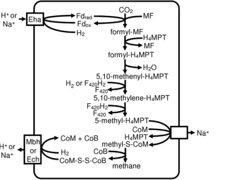
In Class I methanogens, F420-non-reducing hydrogenase provides electrons to heterodisulfide reductase, and its D subunit interfaces with heterodisulfide reductase [17]. In all three Methanomicrobiales the gene for the D subunit of the hydroge-nase (Mlab_0242, Memar_0622, Mhun_1839) is adjacent to the genes for heterodisulfide reductase, but phylogenetic analysis of hydrogenase alpha subunits (not shown) reveals that only M. marisnigri possesses the F420-non-reducing hydrogenase (Memar_1007–1008) (Table 2). Apparently,M. labreanumand M. hungateiuse a different source of electrons for their heterodisulfide reductase. Based on the lack of F420-nonreducing hydrogenase and the fact that the Eha hydrogenase is located adjacent to formylmethanofuran dehydrogenase (Fmd) (see below), we pro-pose that, in at least some Methanomicrobiales, Fmd and heterodisulfide reductase are linked to transmembrane proton or sodium ion transport (Figure 1) rather than flavin-based electron bifurcation as proposed by Thauer et al. [8].
M. labreanum has a hydrogen-forming methylene-tetrahydro-methanopterin dehydrogenase (COG4074), an enzyme previously
found only in Class I methanogens. The other two genomes lack genes assigned to COG4074 and thus are unlikely to have this enzyme. The enzyme functions under conditions of nickel limitation (reviewed in [18]), so this suggests that M. labreanum can tolerate lower environmental nickel concentrations. When this gene is found in a Class I methanogen genome, it is often accompanied by one or two paralogs of unknown function belonging to COG4007. However, M. labreanum lacks these paralogs.
Membrane-Bound Hydrogenases
Methanogens have several families of membrane-bound hydrogenases that participate in various processes including methanogenesis and biosynthesis (reviewed in [19]). These hydrogenases are encoded by a core of conserved genes that includes from six to more than 20 subunits. The three Methanomicrobiales genomes encode two to three membrane-bound hydrogenases (Table 2). All three possess the genes for a membrane-bound hydrogenase similar to that encoded by the Methanothermobacter thermautotrophicus eha operon (Memar_1172– 1185, Mlab_0561–0573, Mhun_2094–2106). Their genes for the enzyme subunits are in the same order as those in the M.
Figure 1. Proposed pathway for methanogenesis in Methanomicrobiales.Methanomicrobiales are predicted to couple formylmethanofuran formation and CoM-CoB heterodisulfide reduction to ion gradients. Fd: ferredoxin; MF: methanofuran; H4MPT: tetrahydromethanopterin. doi:10.1371/journal.pone.0005797.g001
Table 2.Hydrogenases in methanogen genomes.
Frh Mvh Eha Ehb Ech Mbh
Class I methanogens All All all except Msp all except Mka
Methanosarcinales Mac, Mba, Mmz Mba, Mmz
Methanomicrobiales All Mmar All All Mlab, Mhun
Frh: F420-reducing hydrogenase; Mvh: F420-non-reducing hydrogenase; Eha: converting hydrogenase A; Ehb: converting hydrogenase B; Ech: energy-converting hydrogenase; Mbh: membrane-bound hydrogenase; Msp:Methanosphaera stadtmanae; Mka:Methanopyrus kandleri; Mac:Methanosarcina acetivorans; Mba:
thermautotrophicus eha operon. However, some of the smaller subunits have diverged so extensively that homology can not be detected, and subunits A and M are absent. Adjacent to the hydrogenase operon are genes for the subunits of formylmetha-nofuran dehydrogenase, suggesting that the Eha hydrogenase may reduce the ferredoxin used by this enzyme (Figure 1).
All three genomes also have a six-subunit membrane-bound hydrogenase operon similar to Ech hydrogenase (Mlab_1619– 1624, Memar_0359–0364, Mhun_1741–1746), which has multi-ple functions in Methanosarcina barkeri [20]. M. labreanumand M. hungatei,but notM. marisnigri, also have an operon very similar to the mbh operon of Pyrococcus furiosus. Since this hydrogenase is found in the two Methanomicrobiales genomes that lack F420 -nonreducing hydrogenase, the Mbh hydrogenase may be involved in heterodisulfide reduction (Figure 1). M. hungatei has another operon similar to membrane-bound hydrogenases (Mhun_1817– 1822). Homologous operons are absent from the other two Methanomicrobiales, but they are found in two Methanosarci-nales,Methanosarcina acetivoransandMethanosarcina mazei. However, the hydrogenase large subunits of these operons appear to lack the cysteine residues necessary for binding to the nickel-iron center, so these operons may not encode hydrogenases.
Metabolism and Transport
The Embden-Meyerhof pathway is present in many methan-ogens, where it is thought to play a role in the metabolism of stored glycogen. Although M. hungatei and M. marisnigri have putative glycogen phosphorylases (Mhun_1203, Memar_1262, Memar_ 2480), andM. marisnigrihas a putative glycogen branching enzyme (Memar_1265), none of the three Methanomicrobiales has an identifiable glycogen synthase.M. marisnigriandM. hungateiappear to encode a complete glycolysis pathway. This suggests that they may be able to utilize glucose from the environment (although they lack identifiable sugar transporters) or that they have a novel glycogen synthase. M. hungatei was previously reported to lack phosphofructokinase activity [21], but the genome contains two putative phosphofructokinase genes (Mhun_0556 and Mhun_ 1465). The Embden-Meyerhof pathway appears to be absent from M. labreanum as it lacks both phosphofructokinase and pyruvate kinase. A gluconeogenesis pathway is present in all three, as it is necessary for biosynthesis of pentoses and hexoses.
The pathway for 2-oxoglutarate production differs significantly between the Methanosarcinales and the Class I methanogens. Methanosarcinales generate 2-oxoglutarate through a partial oxidative TCA cycle with isocitrate as an intermediate, while Class I methanogens use a partial reductive TCA cycle with succinate as an intermediate (Figure 2). Methanomicrobiales appear to use the partial reductive TCA cycle, similar to the Class I methanogens, as they have genes for all of the necessary enzymes and they lack genes for citrate synthase and isocitrate dehydro-genase. They possess genes encoding the two subunits of the predicted archaeal aconitase [22], but this enzymatic activity has not been verified experimentally.
Sigma Factor Regulators
BothM. labreanumand M. marisnigricontain an anti-anti-sigma factor (Memar_02467, Mlab_1451), an anti-sigma factor (Memar_2469, Mlab_1452), and a serine phosphatase (Memar_2468, Mlab_1450) that are similar to the SpoIIAA/ SpoIIAB/SpoIIE components of the Bacillus subtilis sporulation pathway. Moreover, these SpoII-type proteins are also found inM. hungatei, but not outside the order Methanomicrobiales. This finding is intriguing given that nobona fidesigma factors have been identified in Archaea. Kyrpides and Ouzounis identified proteins
inM. jannaschii with similarity to conserved region 4 of bacterial sigma factors [23], and the Methanomicrobiales have homologs of three of these proteins (MJ0173, MJ0272, and MJ1243). However, the SpoIIAB anti-sigma factor binds to three separate regions of sigma F [24] corresponding to conserved regions 2, 3, and 4, and regions 2 and 3 are not present in the archaeal proteins. Therefore the targets of these archaeal anti-sigma factors can not be determined from the genome sequence.
Phylogenetic Analysis of Enzymes for Methanogenesis and Cofactor Biosynthesis
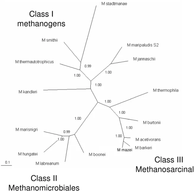
Bapteste et al. [7] determined the relationships among the various groups of methanogens by generating phylogenetic trees for enzymes of methanogenesis and cofactor biosynthesis. Their analysis found that methanogens could be divided into two groups: Class I and Class II methanogens. We present here an updated analysis that includes additional sequenced genomes. Further-more, the protein-coding genes that we used in the analysis (see Materials and Methods) are present in only one copy per genome. Inclusion of the additional genomes reveals that, surprisingly, Methanomicrobiales are equally distant from Class I methanogens and from the Methanosarcinales (Figure 3). Therefore there appear to be three distinct classes of methanogens: the Class I methanogens, the Methanomicrobiales (that we have termed Class II methanogens), and the Methanosarcinales (that we have termed Class III methanogens).
Comparative genomics of methanogens
Now that several sequenced genomes from the order Metha-nomicrobiales are available, it is possible to carry out comparative genomic analyses between this order and the other methanogens. We used a protein clustering method to identify and cluster related proteins from 15 species representing Class I, II, and III methanogens (see Materials and Methods for the list of organisms). We then searched for the signature clusters, i.e. clusters of homologous proteins that are present in all members of a phylogenetic group and absent from other groups. Of particular interest are the exclusive signature clusters, those whose member Figure 2. Alternate pathways for synthesis of 2-oxoglutarate from oxaloacetate. Class I methanogens and Methanomicrobiales use a partial reductive citric acid cycle while Methanosarcinales use a partial oxidative citric acid cycle.
proteins are found in all sequenced genomes from only one class. We also identified shared signature clusters (present in only two classes) and common signature clusters (present in all three).
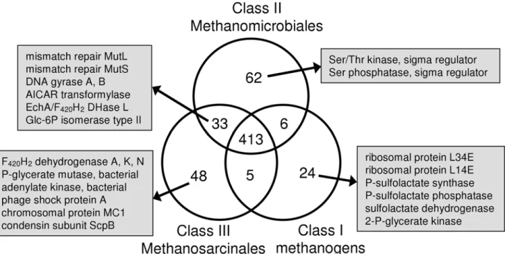
We found 413 common signature clusters (Figure 4, Supple-mentary Table S1). These proteins are involved primarily in core information processing and essential metabolic activities (i.e. transcription, translation, methanogenesis, etc.). We found 62 exclusive signature clusters for Methanomicrobiales, 24 for Class I methanogens, and 48 for Methanosarcinales. Given the relatively close phylogenetic relationship between Methanomicrobiales and Methanosarcinales in ribosomal RNA and ribosomal protein-based trees, it is surprising that they share only 33 clusters to the exclusion of the Class I methanogens. While this is more than either class shares with the Class I methanogens, it represents but a very small proportion of the genome. In the following sections we describe some of the signature proteins associated with each of the
three classes, as well as those shared by Methanomicrobiales and Methanosarcinales.
Class I. The Class I methanogen exclusive signature clusters include two LSU ribosomal proteins (L34E and L14E) and three enzymes of coenzyme M (CoM) biosynthesis (phosphosulfolactate synthase, phosphosulfolactate phosphatase, and sulfolactate dehydrogenase). This suggests that other methanogens possess either unrelated genes for these enzymes or a different pathway for CoM biosynthesis. Also present in only Class I methanogens is 2-phosphoglycerate kinase, an enzyme used in the synthesis of cyclic 2,3-diphosphoglycerate, which is thought to be a thermoprotectant. Its presence in mesophilic Class I methanogens suggests that it carries out a different function in these organisms. The second enzyme of the pathway, cyclic 2,3-diphosphoglycerate synthetase, is found only in a subset of Class I methanogens and is not part of the signature.
Figure 3. Phylogenetic tree of methanogens based on seven core enzymes of methanogenesis and cofactor biosynthesis. See Materials and Methods for a list of the proteins and organisms included. Protein sequences were concatenated and aligned with Clustal W. The tree was generated with MrBayes 3.1.2 and viewed with TreeView.
All Class I methanogens also have a homolog of seryl-tRNA(Sec) selenium transferase, used for the synthesis of selenocysteine in bacteria. However, this gene is likely to have a different function in archaea because not all of the Class I methanogens use selenocysteine [25], and those that do utilize a different pathway for selenocysteine synthesis, one that is shared with eukaryotes [26,27]. Experimental testing of this protein found that it did not catalyze selenocysteine formation [28].
Methanomicrobiales (Class II). Of the 62 exclusive
signature clusters for Methanomicrobiales, 26 are hypothetical proteins, reflecting the fact that this order has been less studied. A serine/threonine kinase and a serine phosphatase, both of which regulate sigma factors in bacteria (see the Sigma Factor Regulators section above), are part of the Methanomicrobiales signature. In addition to a full-length heterodisulfide reductase subunit A (HdrA), Methanomicrobiales also contain a homolog that is truncated at both the N- and C-terminus. Similarly, the A and G subunits of their tetrahydromethanopterin S-methyltransferase are fused. A separate A subunit was found, but no other G subunit is present.
Methanosarcinales (Class III). Among the exclusive
signature proteins found in Methanosarcinales are subunits A, K, and N of reduced coenzyme F420 (F420H2) dehydrogenase. Since only Methanosarcinales can use methyl compounds as a substrate for methanogenesis, it is not surprising that this enzyme, used for growth on methyl compounds, is not found in the other methanogens. All methanogens have the archaeal bisphosphoglycerate-independent phosphoglycerate mutase, but Methanosarcinales also have a bacterial version. Similarly, Methanosarcinales use the bacterial adenylate kinase while other methanogens have the archaeal enzyme.
Methanosarcinales exclusive signature proteins include phage shock protein A, a protein that functions in the repair of damaged
cell membranes. Likewise, they encode two proteins involved in DNA compaction: the non-histone chromosomal protein MC1 and a unique variant (,200 amino acids longer) of the ScpB subunit of the condensin complex. That these two chromosome condensation proteins are found only in Methanosarcinales may be related to the larger genome size of some members. The other two components of the condensin complex, ScpA and Smc, are present in most methanogens, including Methanosarcinales. In addition to these DNA condensation proteins, all methanogens have at least one histone gene. Most also have a copy of the gene encoding the Alba protein, but among Methanosarcinales it is present only inMethanosaeta thermophila.
Missing from Class I. There are 33 clusters shared by
Methanomicrobiales and Methanosarcinales that are absent from Class I methanogens. Among these are the DNA mismatch repair proteins MutL and MutS. MutH, however, is not present in any methanogen. This suggests that, if Class I methanogens have methyl-directed mismatch repair, they use a different system. Class I methanogens also lack DNA gyrase subunits A and B. This is unexpected as several Class I methanogens were found to be sensitive to coumarins that target bacterial DNA gyrase [29]. Furthermore, DNA gyrase is the only protein known to introduce negative supercoils into DNA, and these are required for many cellular processes including transcription and DNA replication [30]. Another enzyme missing from Class I methanogens is 5-amino-4-imidazolecarboxamide ribonucleotide (AICAR) transformylase in the pathway forde novopurine synthesis. Since most Class I methanogens are autotrophs, they must have this capability provided by a protein unrelated to the known enzyme. Another shared cluster is the one containing Ech hydrogenase subunit A. Although M. acetivorans lacks Ech, it does have the F420H2 dehydrogenase subunit L and a subunit of multisubunit sodium/proton antiporters, both of which cluster with EchA. Figure 4. Venn diagram of signature clusters.The clusters were generated using a spectral clustering procedure (see Materials and Methods section for details). Signature protein clusters were identified as clusters for which a member protein was present in every analyzed species from one or more classes of methanogens. The number of exclusive, shared, and common signature clusters associated with each methanogen class are shown. The functions of characterized proteins belonging to exclusive signature clusters and to clusters shared between the Methanomicrobiales and the Methanosarcinales are also noted.
Lastly, Methanomicrobiales and Methanosarcinales have one form of glucose-6-phosphate isomerase (COG2140), while most Class I methanogens use another (COG0166). Since a glucose-6-phosphate isomerase could not be identified inMethanopyrus kandleri or in Methanobacteriales, there is probably a third form of this enzyme.
Discussion
Phylogenetics
The sequencing of the genomes ofM. labreanumandM. marisnigri reported in this paper, combined with the previously sequenced genome ofM. hungatei, has enabled further characterization of the order Methanomicrobiales and clarification of its relationship to other methanogens. Our analyses including these species reveal that the order Methanomicrobiales is clearly distinct from other methanogens. The phylogenetic tree built for seven core methanogenesis and cofactor biosynthesis enzymes reveals three discrete groups of methanogens: the Class I methanogens, the Methanomicrobiales (termed here Class II), and the Methano-sarcinales (termed here Class III). This classification differs significantly from the previous study by Bapteste et al. [7] that divided the methanogens into two major groups. In that earlier study, the order Methanosarcinales was represented by only species from the genus Methanosarcina, whereas our study also included two genomes from other genera. Likewise, their analysis included only one representative of the Methanomicrobiales, while we included four species from this order. Because our study encompassed more species and greater diversity, our results may be a more accurate representation of the relationships among these groups. A relatively close relationship was previously seen between Methanosarcinales and Methanomicrobiales in 16S rRNA trees [3,31] and ribosomal protein trees [3,7]. In contrast, Methanomicrobiales are equally distant from Class I methanogens and Methanosarcinales in the tree built in this study from core methanogenesis proteins.
Genomic Analyses
The protein clustering results reported also suggest a significant distance between Methanomicrobiales and all other methanogens. They share only 6 signature clusters with Class I methanogens and 33 with Methanosarcinales. In addition, the number of exclusive signature clusters for the Methanomicrobiales is of the same magnitude as the signatures for the other two groups. The complement of membrane-bound hydrogenases also shows the uniqueness of Methanomicrobiales. They all have the Eha hydrogenase similar to Class I methanogens and the Ech hydrogenase found in Methanosarcinales, while some of them have hydrogenases similar to Mbh fromP. furiosusand a putative membrane-bound hydrogenase from Methanosarcinales.
Methanomicrobiales share some capabilities with Class I methanogens to the exclusion of Methanosarcinales. Both groups are capable of using only H2/CO2or formate for methanogenesis. The genomes show that they also share the pathway for 2-oxoglutarate synthesis. Both use a partial reductive TCA cycle, while Methanosarcinales use a partial oxidative TCA cycle. This could reflect the observations that Methanomicrobiales efficiently use low concentrations of H2, while the Methanosarcinales dominate in environments in which acetate is plentiful. The partial oxidative TCA cycle results in the loss of one carbon as CO2, therefore the use of the reductive cycle by Methanomicro-biales and Class I methanogens would be predicted to preserve more fixed carbon. On the other hand, we propose that, similar to Methanosarcinales, Methanomicrobiales link formylmethanofuran
synthesis and heterodisulfide reduction to membrane ion gradi-ents, even though they lack cytochromes and methanophenazine that are present in Methanosarcinales.
Hydrogenases
Methanomicrobiales encode from two to four membrane-bound hydrogenases. In all three genomes (M. labreanum,M. marisnigri, and M. hungatei), the genes for Eha hydrogenase are found adjacent to genes for formylmethanofuran dehydrogenase (Fmd), suggesting that the Eha hydrogenase may reduce a low potential ferredoxin that is required for the reduction of CO2to formylmethanofuran. In contrast, in the Class I methanogenMethanococcus maripaludis, the eha and fmd operons are not linked, and Eha hydrogenase presumably plays a role in carbon assimilation similar to Ehb and not methanogenesis [8,32,33].
All Methanomicrobiales also contain genes for the Ech hydrogenase that has been characterized in M. barkeri. Ech hydrogenase is involved in reduction of ferredoxin for the first step of methanogenesis from H2/CO2, in the reduction of ferredoxin for biosynthesis, and in the formation of H2 from ferredoxin during aceticlastic methanogenesis [20]. Since the Ech hydroge-nase is found in all three Methanomicrobiales, it is likely that its function is common to all three, e.g. the reduction of ferredoxin for 2-oxoglutarate synthesis. Another putative membrane-bound hydrogenase (Pmh) is found only in M. hungatei where it may perform a function that is unique to this organism, such as producing ferredoxin for acetyl-CoA decarbonylase/synthase, an enzyme that is absent from the other two.
Experimental evidence is needed to determine the functions of these hydrogenases. Nevertheless, their distribution within the Methanomicrobiales is clearly distinct from that in the Class I methanogens and the Methanosarcinales (Table 2), supporting the functional and evolutionary uniqueness of this group. Their distribution and other features of the operons suggest that their roles in energy conservation differ in Class I methanogens, Methanosarcinales, and Methanomicrobiales.
Materials and Methods
DNA Preparation
M. marisnigristrain JR1 was obtained from the ATCC (ATCC 35101). It was cultured at room temperature in modified McC medium [34] that contained 0.1 M NaCl, 3 g/L of sodium bicarbonate, 2 g/L of Trypticase (replacing yeast extract), and 0.17 g/L of Na2S?9H2O.M. labreanumstrain Z was obtained from the ATCC (ATCC 43576). It was cultured at 37uC in MS-OCM Base Medium with 2.5 g/L NaCl, 5 mM sodium acetate, 50 mM sodium formate, and 2.5% (v/v) of rumen fluid.
Genome Sequencing and Assembly
The genome of M. labreanum Z was sequenced at the Joint Genome Institute (JGI) using a combination of Sanger shotgun sequencing and 454 sequencing-by-synthesis technology. All general aspects of library construction and sequencing performed at the JGI can be found at http://www.jgi.doe.gov/sequencing/ protocols/prots_production.html. Draft assemblies were based on 26,432 Sanger shotgun and 390,106 pyrosequencing reads. The combined reads provided 346coverage of the genome. The Newbler assembly software (www.454.com) and the Paracel Genome Assembler (Paracel, Pasadena, CA) were used for fragment assembly, and the Consed finishing package (www. phrap.org) was used for quality assessment and editing. All mis-assemblies were corrected and all gaps between contigs were closed by custom primer walk using subclones or PCR products as templates. A total of 196 additional reactions were run to close gaps and to raise the quality of the finished sequence.
The genome ofM. marisnigri JR1 was sequenced at the Joint Genome Institute (JGI) using a combination of 3 kb, 7 kb and 36 kb (fosmid) DNA libraries. Draft assemblies were based on 29,769 total reads. The three libraries combined provided 116
coverage of the genome. The Phred/Phrap/Consed software package (www.phrap.com) was used for sequence assembly and quality assessment [35–37]. All mis-assemblies were corrected and all gaps between contigs were closed by custom primer walk using subclones or PCR products as templates. A total of 702 primer walk reactions, PCR end reads and 3 mini-libraries were required to close gaps and to raise the quality of the finished sequence.
Genome Analysis
Automatic genome annotation was performed at Oak Ridge National Laboratory. Genes were identified using a combination of Critica [38] and Glimmer [39]. In addition, predicted coding regions (CDSs) were manually curated using JGI’s Gene-PRIMP Quality Assurance pipeline (http://tunis.jgi-psf.org/geneprimp) (Pati et al., in preparation). Comparative genome analysis was performed within the Integrated Microbial Genomes (IMG) system [40]. CRISPR repeats were identified with the CRISPR Recognition Tool [41].
A phylogenetic tree was constructed using the concatenated sequences of seven core proteins found in all methanogens and involved in methanogenesis and cofactor biosynthesis. The genes included are F420-dependent methylenetetrahydromethanopterin dehydrogenase (mtd, COG1927), tetrahydromethanopterin:coen-zyme M methyltransferase subunits B (mtrB, COG4062), C (mtrC, COG4061), D (mtrD, COG4060) and E (mtrE, COG4059), FO synthase subunit 1 (cofG), and sulfopyruvate decarboxylase alpha subunit (comD). Protein sequences were downloaded from IMG [40]. The concatenated amino acid sequences were aligned with Clustal W [42], and the tree was generated with MrBayes 3.1.2 [43] with 1,000,000 generations sampled every 100 generations. The first 250,000 generations were discarded as burn-in. The tree was visualized with TreeView [44].
For protein clustering, methanogens were included from all three groups: six Class I methanogens, four Methanomicrobiales, and five Methanosarcinales. Class I included Methanocaldococcus
jannaschii,Methanothermobacter thermautotrophicus,Methanopyrus kandleri, Methanococcus maripaludisS2, Methanobrevibacter smithii, and Methano-sphaera stadtmanae. Methanomicrobiales includedMethanocorpusculum labreanum, Methanoculleus marisnigri, Methanospirillum hungatei, and Candidatus Methanoregula boonei. Methanosarcinales included Methanosarcina acetivorans,Methanosarcina mazei,Methanosarcina barkeri, Methanococcoides burtonii, and Methanosaeta thermophila. Protein sequences for these organisms were downloaded from IMG [40]. We applied a spectral clustering procedure [45,46] to cluster similar proteins based on the topology of their similarity graph, rather than using a fixed threshold value for sequence similarity. The proteins are represented as nodes in a connected undirected graph with edges that carry weights based on node-to-node similarity according to the protein identity. The clustering procedure is analogous to a random walk of a particle moving on this graph from one node to another. In each node the particle moves to another node based on the probabilities corresponding to the weights of the edges. The amount of time the particle spends in a given subgraph will determine whether this is indeed a cluster of its own or not.
The second eigenvalue of the transition matrix is a measure of how easily a graph (i.e. a cluster) can be partitioned. A cutoff value of 0.8 was applied; if the second eigenvalue exceeds 0.8, the cluster is further partitioned. This approach provides a relatively flexible partitioning that can reveal protein similarities despite sequence differences due to phylogenetic distance.
Signature protein clusters were identified as clusters for which a member protein was present in every analyzed species from one (or more) class of methanogens. Those clusters were binned into groups: exclusive signature clusters found in all members of only one class, shared signature clusters found in all members of a specified pair of classes, and common clusters found in all three classes. The resultant cluster distribution was visualized as a Venn diagram.
Accession Numbers
The genome sequences ofMethanoculleus marisnigri JR1, Metha-nocorpusculum labreanumZ, andMethanospirillum hungateiJF-1 can be accessed in GenBank (CP000562, CP000559, and CP000254, respectively). The Genomes OnLine Database accession numbers are Gc00512, Gc00506, and Gc00350, respectively.
Supporting Information
Table S1 Signature clusters of methanogens. List of signature clusters including exclusive clusters that are present in one class only, shared clusters that are present in two classes, and common clusters that are present in all three classes.
Found at: doi:10.1371/journal.pone.0005797.s001 (0.11 MB TXT)
Author Contributions
Conceived and designed the experiments: CW JB NCK. Performed the experiments: SDH AL EG AL ES CSH MLL SL. Analyzed the data: IA LEU BL DS IP LD BM WW NCK. Contributed reagents/materials/ analysis tools: MSL. Wrote the paper: IA LEU BL DS IP BM WW NCK.
References
1. Woese CR, Fox GE (1977) Phylogenetic structure of the prokaryotic domain: the primary kingdoms. Proc Natl Acad Sci U S A 74: 5088–5090.
2. Woese CR, Kandler O, Wheelis ML (1990) Towards a natural system of organisms: proposal for the domains Archaea, Bacteria, and Eucarya. Proc Natl Acad Sci U S A 87: 4576–4579.
3. Brochier-Armanet C, Boussau B, Gribaldo S, Forterre P (2008) Mesophilic crenarchaeota: proposal for a third archaeal phylum, the Thaumarchaeota. Nat Rev Microbiol 6: 245–252.
4. Ferry JG (1997) Methane: small molecule, big impact. Science 278: 1413– 1414.
5. Etheridge DM, Steele LP, Francey RJ, Langenfelds RL (1998) Atmospheric methane between 1000 A.D. and present: evidence of anthropogenic emissions and climatic variability. J Geophys Res 103: 15979–15993.
7. Bapteste E´ , Brochier C, Boucher Y (2005) Higher-level classification of the Archaea: evolution of methanogenesis and methanogens. Archaea 1: 353–363. 8. Thauer RK, Kaster A-K, Seedorf H, Buckel W, Hedderich R (2008) Methanogenic archaea: ecologically relevant differences in energy conservation. Nat Rev Microbiol 6: 579–591.
9. Garcia J-L, Ollivier B, Whitman WB (2006) The order Methanomicrobiales. Prokaryotes 3: 208–230.
10. Zellner G, Winter J (1987) Secondary alcohols as hydrogen donors for CO2-reduction by methanogens. FEMS Microbiol Lett 44: 323–328.
11. Romesser JA, Wolfe RS, Mayer F, Spiess E, Walther-Mauruschat A (1979)
Methanogenium, a new genus of marine methanogenic bacteria, and character-ization ofMethanogenium cariacisp. nov. andMethanogenium marisnigrisp. nov. Arch Microbiol 121: 147–153.
12. Zhao Y, Boone DR, Mah RA, Boone JE, Xun L (1989) Isolation and characterization ofMethanocorpusculum labreanumsp. nov. from the LaBrea tar pits. Int J Syst Bacteriol 39: 10–13.
13. Ferry JG, Smith PH, Wolfe RS (1974) Methanospirillum, a new genus of methanogenic bacteria, and characterization ofMethanospirillum hungatiisp. nov. Int J Syst Bacteriol 24: 465–469.
14. Widdel F, Rouvie`re PE, Wolfe RS (1988) Classification of secondary alcohol-utilizing methanogens including a new thermophilic isolate. Arch Microbiol 150: 477–481.
15. Bleicher K, Winter J (1991) Purification and properties of F420- and NADP+
-dependent alcohol dehydrogenases ofMethanogenium liminatansand Methanobacter-ium palustre, specific for secondary alcohols. Eur J Biochem 200: 43–51. 16. Aufhammer SW, Warkentin E, Berk H, Shima S, Thauer RK, et al. (2004)
Coenzyme binding in F420-dependent secondary alcohol dehydrogenase, a member of the bacterial luciferase family. Structure 12: 361–370.
17. Stojanowic A, Mander GJ, Duin EC, Hedderich R (2003) Physiological role of the F420-non-reducing hydrogenase (Mvh) fromMethanothermobacter marburgensis. Arch Microbiol 180: 194–203.
18. Shima S, Thauer RK (2007) A third type of hydrogenase catalyzing H2 activation. Chem Rec 7: 37–46.
19. Hedderich R, Forzi L (2005) Energy-converting [NiFe] hydrogenases: more than just H2activation. J Mol Microbiol Biotechnol 10: 92–104.
20. Meuer J, Kuettner HC, Zhang JK, Hedderich R, Metcalf WW (2002) Genetic analysis of the archaeonMethanosarcina barkeriFusaro reveals a central role for Ech hydrogenase and ferredoxin in methanogenesis and carbon fixation. Proc Natl Acad Sci U S A 99: 5632–5637.
21. Verhees CH, Tuininga JE, Kengen SWM, Stams AJM, van der Oost J, et al. (2001) ADP-dependent phosphofructokinases in mesophilic and thermophilic methanogenic archaea. J Bacteriol 183: 7145–7153.
22. Makarova KS, Koonin EV (2003) Filling a gap in the central metabolism of archaea: prediction of a novel aconitase by comparative-genomic analysis. FEMS Microbiol Lett 227: 17–23.
23. Kyrpides NC, Ouzounis CA (1997) Bacterial sigma 70 transcription factor DNA-binding domains in the archaeonMethanococcus jannaschii. J Mol Evol 45: 706–707.
24. Decatur AL, Losick R (1996) Three sites of contact between theBacillus subtilis
transcription factor sigma F and its antisigma factor SpoIIAB. Genes Dev 10: 2348–2358.
25. Zhang Y, Turanov AA, Hatfield DL, Gladyshev VN (2008)In silicoidentification of genes involved in selenium metabolism: evidence for a third selenium utilization trait. BMC Genomics 9: 251.
26. Yuan J, Palioura S, Salazar JC, Su D, O’Donoghue P, et al. (2006) RNA-dependent conversion of phosphoserine forms selenocysteine in eukaryotes and archaea. Proc Natl Acad Sci U S A 103: 18923–18927.
27. Xu X-M, Carlson BA, Mix H, Zhang Y, Saira K, et al. (2007) Biosynthesis of selenocysteine on its tRNA in eukaryotes. PLoS Biol 5: e4.
28. Kaiser JT, Gromadski K, Rother M, Engelhardt H, Rodnina MV, et al. (2005) Structural and functional investigation of a putative archaeal selenocysteine synthase. Biochemistry 44: 13315–13327.
29. Sioud M, Possot O, Elie C, Sibold L, Forterre P (1989) Coumarin and quinolone action in archaebacteria: evidence for the presence of a DNA gyrase-like enzyme. J Bacteriol 170: 946–953.
30. No¨llman M, Crisona NJ, Arimondo PB (2007) Thirty years ofEscherichia coli
DNA gyrase: Fromin vivofunction to single-molecule mechanism. Biochimie 89: 490–499.
31. Schleper C, Jurgens G, Jonuscheit M (2005) Genomic studies of uncultivated archaea. Nat Rev Microbiol 3: 479–488.
32. Porat I, Kim W, Hendrickson EL, Xia Q, Zhang Y, et al. (2006) Disruption of the operon encoding Ehb hydrogenase limits anabolic CO2assimilation in the archaeonMethanococcus maripaludis. J Bacteriol 188: 1373–1380.
33. Lupa B, Hendrickson EL, Leigh JA, Whitman WB (2008) Formate-dependent H2production by the mesophilic methanogenMethanococcus maripaludis. Appl Env Microbiol 74: 6584–6590.
34. Whitman WB, Shieh J-S, Sohn S-H, Caras DS, Premachandran U (1986) Isolation and characterization of 22 mesophilic methanococci. Syst Appl Microbiol 7: 235–240.
35. Ewing B, Green P (1998) Base-calling of automated sequencer traces using phred. II. Error probabilities. Genome Res 8: 186–194.
36. Ewing B, Hillier L, Wendl MC, Green P (1998) Base-calling of automated sequencer traces using phred. I. Accuracy assessment. Genome Res 8: 175–185. 37. Gordon D, Abajian C, Green P (1998) Consed: a graphical tool for sequence
finishing. Genome Res 8: 195–202.
38. Badger JH, Olsen GJ (1999) CRITICA: coding region identification tool invoking comparative analysis. Mol Biol Evol 16: 512–524.
39. Delcher AL, Harmon D, Kasif S, White O, Salzberg SL (1999) Improved microbial gene identification with GLIMMER. Nucleic Acids Res 27: 4636–4641.
40. Markowitz VM, Szeto E, Palaniappan K, Grechkin Y, Chu K, et al. (2008) The integrated microbial genomes (IMG) system in 2007: data content and analysis tool extensions. Nucleic Acids Res 36: D528–D533.
41. Bland C, Ramsey TL, Sabree F, Lowe M, Brown K, et al. (2007) CRISPR Recognition Tool (CRT): a tool for automatic detection of clustered regularly interspaced palindromic repeats. BMC Bioinformatics 8: 209.
42. Thompson JD, Higgins DG, Gibson TJ (1994) CLUSTAL W: improving the sensitivity of progressive multiple sequence alignment through sequence weighting, position-specific gap penalties and weight matrix choice. Nucleic Acids Res 22: 4673–4680.
43. Ronquist F, Huelsenbeck JP (2003) MrBayes 3: Bayesian phylogenetic inference under mixed models. Bioinformatics 19: 1572–1574.
44. Page RDM (1996) TREEVIEW: an application to display phylogenetic trees on personal computers. Comput Appl Biosci 12: 357–358.
45. Paccanaro A, Casbon JA, Saqi MAS (2006) Spectral clustering of protein sequences. Nucleic Acids Res 34: 1571–1580.