O R I G I N A L A R T I C L E
Carlos D. BrondinoÆ Mario C. G. Passeggi
Jorge CaldeiraÆMaria J. Almendra ÆMaria J. Feio Jose J. G. MouraÆ Isabel Moura
Incorporation of either molybdenum or tungsten into formate
dehydrogenase from
Desulfovibrio alaskensis
NCIMB 13491; EPR
assignment of the proximal iron-sulfur cluster to the pterin cofactor in
formate dehydrogenases from sulfate-reducing bacteria
Received: 3 June 2003 / Accepted: 24 October 2003 / Published online: 11 December 2003 SBIC 2003
AbstractWe report the characterization of the molecular properties and EPR studies of a new formate dehydro-genase (FDH) from the sulfate-reducing organism Des-ulfovibrio alaskensisNCIMB 13491. FDHs are enzymes that catalyze the two-electron oxidation of formate to carbon dioxide in several aerobic and anaerobic organ-isms.D.alaskensisFDH is a heterodimeric protein with a molecular weight of 126±2 kDa composed of two subunits, a=93±3 kDa and b=32±2 kDa, which contains 6±1 Fe/molecule, 0.4±0.1 Mo/molecule, 0.3±0.1 W/molecule, and 1.3±0.1 guanine monophos-phate nucleotides. The UV-vis absorption spectrum of D. alaskensis FDH is typical of an iron-sulfur protein with a broad band around 400 nm. Variable-tempera-ture EPR studies performed on reduced samples of D. alaskensis FDH showed the presence of signals
associated with the different paramagnetic centers of D. alaskensis FDH. Three rhombic signals having g-values and relaxation behavior characteristic of [4Fe-4S] clusters were observed in the 5–40 K temperature range. Two EPR signals with all the g-values less than two, which accounted for less than 0.1 spin/protein, typical of mononuclear Mo(V) and W(V), respectively, were observed. The signal associated with the W(V) ion has a larger deviation from the free electrong-value, as expected for tungsten in a d1 configuration, albeit with an unusual relaxation behavior. The EPR parameters of the Mo(V) signal are within the range of values typically found for the slow-type signal observed in several Mo-containing proteins belonging to the xanthine oxi-dase family of enzymes. Mo(V) resonances are split at temperatures below 50 K by magnetic coupling with one of the Fe/S clusters. The analysis of the inter-center magnetic interaction allowed us to assign the EPR-dis-tinguishable iron-sulfur clusters with those seen in the crystal structure of a homologous enzyme.
Keywords Electron paramagnetic resonance Æ Formate dehydrogenaseÆMagnetic interactions Æ Molybdenum-containing enzymesÆ
Tungsten-containing enzymes
Abbreviations AORaldehyde oxidoreductaseÆFDH formate dehydrogenaseÆNAPperiplasmic nitrate reductaseÆ SRBsulfate-reducing bacteria
Introduction
Molybdenum- and tungsten-containing enzymes are a wide group of proteins present in several living systems that participate in hydroxylation and oxo-transfer reactions. With the exception of nitrogenase, the metal ion is associated with either one or two pterin derivatives
DOI 10.1007/s00775-003-0506-z
C. D. BrondinoÆJ. CaldeiraÆM. J. Almendra J. J. G. MouraÆI. Moura (&)
REQUIMTE, Departamento de Quı´mica, Centro de Quı´mica Fisica e Biotecnologia, Faculdade de Cieˆncias e Tecnologia,
Universidade Nova de Lisboa, 2829-516 Caparica, Portugal E-mail: [email protected]
Tel.: +351-21-2948381 Fax: +351-21-2948550
C. D. BrondinoÆM. C. G. Passeggi Departamento de Fı´sica,
Facultad de Bioquı´mica y Ciencias Biologicas,
Universidad Nacional del Litoral, 3000 Santa Fe, Argentina
M. C. G. Passeggi
Instituto de Desarrollo Tecnologico (UNL-CONICET), 3000 Santa Fe, Argentina
M. J. Feio
School of Pharmacy and Biomedical Sciences, University of Portsmouth, St. MichaelÕs Building, White Swan Road, Portsmouth, PO1 2DT UK
Present address: M. J. Feio
Instituto de Bioquı´mica Vegetal y Fotosı´ntesis, Centro de Inves-tigaciones Cientı´ficas Isla de la Cartuja,
to form a metallic cofactor (hereafter called pterin co-factor) [1, 2, 3]. Most of the proteins studied so far contain molybdenum in their active sites, though in the last two years the number of proteins having tungsten has considerably increased. Tungstoproteins are usually found in thermophilic organisms that grow in extreme environments. However, the number of reports of tungstoproteins from other sources has considerably increased in recent years [4, 5].
We have reported the characterization of two formate dehydrogenases (FDHs) from the sulfate-reducing bac-teria (SRB)Desulfovibrio desulfuricans ATCC 27774 [6, 7] and Desulfovibrio gigas [4, 8, 9] that belong to the DMSO reductase family (DMSO=dimethyl sulfoxide). FDHs catalyze the two-electron oxidation of formate to carbon dioxide. D. desulfuricans FDH contains molyb-denum at the pterin cofactor whereas the one from D. gigascontains tungsten.D.desulfuricansFDH presents a heterotrimeric structure (a=88 kDa, b=29 kDa, and
c=16 kDa), whereas D. gigas FDH is heterodimeric (a=92 kDa andb=29 kDa). The crystal structure ofD. gigas FDH revealed that besides the pterin cofactor, four iron-sulfur clusters of the [4Fe-4S] type are present (Fe/S I, II, III, and IV), one of them being 14.4 A˚ from the pterin cofactor (proximal center). The larger subunit of this enzyme contains the pterin cofactor and the proximal iron-sulfur cluster. The smaller subunit con-tains the remaining three iron-sulfur clusters. The four iron-sulfur centers are along a ‘‘ziz-zag’’ line separated from each other by10 A˚. The larger subunit of theD. gigas FDH is homologous to the periplasmic nitrate reductase (NAP) from D. desulfuricans [10] and the FDH-H from Escherichia coli[11]. Furthermore, all the evidence obtained up to the present suggests that D. gigas FDH presents a high homology with the recently reported W-containing FDH-2 fromS. fumaroxidans[5] and with the two larger subunits present in D. desulfu-ricansFDH [6].
The presence of iron-sulfur clusters of the type [4Fe-4S] in bothD.gigasandD.desulfuricanshas been shown by EPR and Mo¨ssbauer spectroscopies [4, 6, 8]. EPR studies performed on both enzymes in the dithionite-reduced state showed two types of EPR signals. The Fe/S I EPR signal appears below 70 K and shows no broadening below 40 K. The other one, which is identified as Fe/S II, appears below 30 K and shows much broader resonance lines. It was suggested that the remaining iron-sulfur centers present both EPR and Mo¨ssbauer properties similar to Fe/S I and II, but additional work should be performed to confirm this hypothesis [8]. The EPR properties of the Fe/S I center evaluated in FDHs from SRB are similar to those observed in NAP [12] andE.coli FDH [13]. On this basis, this center should be the center present in the larger subunit of these enzymes, but no spectroscopic evidence has been shown so far.
We report herein the characterization of the molec-ular properties and EPR studies performed on a FDH obtained from D. alaskensis NCIMB 13491, a SRB in-volved in metal corrosion [14]. D. alaskensis FDH is
closely related to the W-containing D. gigas FDH. However, metal quantification and EPR studies indicate that the enzyme is a mixture of two forms, each con-taining either a mononuclear Mo site or a mononuclear W site. Furthermore, the Mo site is magnetically cou-pled to one of the iron-sulfur clusters. The results obtained for this protein are discussed in comparison with other W- and Mo-containing proteins.4
Materials and methods
Growth conditions, protein purification, and enzymatic activity
D. alaskensis NCIMB 13491 was grown at 37C in Postgate C medium [15] under anaerobic conditions in the Unite´ de Fermen-tation at LBC-CNRS in Marseille, France, and cells were harvested at the logarithmic/early stationary phase of bacterial growth. In general, the growth media used for biomass production of SRB intended for protein isolation includes a metal supplement. In this experiment a more basic medium was used for the cell growth. It contained a large amount of iron (140lM) but only trace amounts
of all other metals (added mainly as contaminants by the various medium components, in particular yeast extract). This medium was chosen because it mimics better the natural growth conditions experienced by SRB in their natural habitats, where nutrient sources are often scarce [14].
All extraction and purification procedures were performed aer-obically at 4C and pH 7.6 with reagent grade or higher. The pellet of cells (265 g wet weight) was suspended in 10 mM Tris-HCl buffer. Deoxyribonuclease and ribonuclease were added to lower the vis-cosity. A cell-free extract was obtained by centrifugation of the cel-lular suspension at 5000·gfor 40 min and the supernatant (300 mL), containing mostly the periplasmic proteins, was collected. To remove any membrane contamination, the supernatant was spun at 180,000·gfor 1 h. The periplasmic fraction (300 mL), initially with 381 total units (U), was loaded onto an anion exchange column (DEAE-52 cellulose, Whatman 6·32.5 cm) equilibrated with 10 mM Tris-HCl. The column was eluted with a linear gradient (0.01–0.60 M) of the same buffer with a total volume of 2 L. The enzyme, with 122 U and a specific activity of 0.13 U mg)1and a yield of 32%, was found in the fraction eluted at200 mM Tris-HCl. This
fraction was then dialyzed against Millipore water and applied to an anionic exchange column (Source 15, Pharmacia 2.7·20 cm). The proteins were eluted using a Tris-HCl buffer gradient (0.01–0.50 M); the fraction that mainly contained FDH activity was collected at
200 mM ionic strength and its specific activity was 1.63 U mg )1
(82 U and a yield of 22%). The ionic strength of this fraction was decreased adding Millipore water. The next and final step was per-formed on a Source-Q column, using a Tris-HCl buffer gradient (0.01–0.50 M). The enzyme was found to be pure with 38.1 U, a specific activity=3.51 U mg)1and a final yield of 10%.
The FDH activity assay was performed with formate as sub-strate and benzyl viologen as electron acceptor. A standard optical assay of FDH was performed at room temperature under an argon atmosphere, by monitoring the benzyl viologen reduction at 555 nm (=12 mM)1cm)1), as described elsewhere [6]. One unit of
FDH activity was defined as the amount of enzyme catalyzing the reduction of 2lmol of benzyl viologen/min.
Molecular mass determination
(232 kDa), and ferritin (440 kDa), from Pharmacia Biotech. Sub-unit composition was determined by SDS-PAGE at 12.5% using the Pharmacia low molecular mass kit as standard for calibration. Gels were stained with Coomassie brilliant blue G-250 (Merck).
Protein and metals quantification
Protein concentration was determined by the Lowry method using bovine serum albumin as standard [16]. The quantification of tungsten, molybdenum, and iron was performed by inductively coupled plasma emission analysis.
Analysis of pterin nucleotides
Nucleotides were extracted from D. alaskensis FDH by a previ-ously published procedure [17, 18]. Cofactor extraction was per-formed with sulfuric acid (3% v/v) for 10 min at 95C, with subsequent centrifugation in a microcentrifuge for 5 min at 10,000 rpm. Chromatography analysis was carried out using a Merck L-7100 HPLC equipped with a L-7100 UV detector and D-7000 computer interface. Hydrolyzed nucleotide peak areas were quantified at 254 nm. Ammonium acetate (HPLC grade) (50 mM, pH 6.8) was used as eluent at a flow rate of 1 mL min)1, using a Merck LiChorspher 100 (250·4) RP-18e, 4lm column.
Quantita-tive determinations were performed with an injection loop volume of 50lL from extracted nucleotides and fresh solutions of mono-nucleotides (Sigma) submitted to identical acid/heating treatment.
Spectroscopic methods
The UV-visible optical spectrum of the as-isolated enzyme was recorded on a Shimadzu UV-2101 PC split-beam spectrophotom-eter using 1 cm quartz cells. Variable-temperature EPR measure-ments at X-band were performed on a Bruker EMX spectrometer equipped with a dual-mode cavity (model ER 4116DM) and an Oxford Instruments continuous flow cryostat. Spin quantification was performed under non-saturating conditions using Cu-EDTA as standard. EPR spectra were simulated using either WINEPR Simfonia v.1.25 from Bruker Instruments, or a home-made pro-gram described below. For the spectra showing overlapping EPR signals, the signals associated with each center were separately simulated, normalized by double integration, and then added altogether in different proportions until best agreement was ob-tained between experimental and simulated data. The temperature variation of the spectra associated with Mo(V) species were simu-lated using a home-made program based on the formalism WBR (Wangenesst, Bloch, and Redfield) for a dimer of twoS=1/2 ions [19]. This program solves the components of the density matrix required to calculate the imaginary part of v¢¢(x) (dynamic susceptibility) and takes the elements of the relaxation matrix as semi-empirical parameters (T1andT2) for each center [20].
Results and discussion
Molecular properties
D. alaskensis FDH is a periplasmic enzyme which was purified and stored in aerobic conditions like FDHs from D. gigas and D. desulfuricans. The UV-vis absorption spectrum ofD.alaskensisFDH is identical to that of the D. gigasFDH [4]. The spectrum exhibits a shoulder at 320 nm and a broad absorbance band around 400 nm, as usually found in proteins having [4Fe-4S] clusters. Molecular mass determination showed
a single symmetrical elution peak corresponding to a molecular mass of 126±2 kDa. Two bands with molecular weights of 93±3 kDa and 32±2 kDa were observed under denaturing conditions (SDS-PAGE).D. alaskensis FDH is a heterodimeric protein which con-tains two types of subunits (a,b). Plasma emission assay detected 0.4±0.1 Mo/molecule, 0.3±0.1 W/molecule, and 6±1 Fe/molecule. No heme c was detected when specific gel staining was performed and no heme is vis-ible on the UV-vis spectrum. Pterin nucleotide analysis identified 1.3±0.1 guanine monophosphate derivative, which suggests a di-pterin coordination with the metal atom. The molecular properties evaluated forD. alask-ensis FDH are similar to those of D. gigas FDH. This implies that the a-subunit contains the pterin cofactor and one iron-sulfur cluster whereas the b-subunit contains the remaining three iron-sulfur clusters, as in theD.gigasenzyme [8].
D. alaskensisFDH is purified as two isoforms, each containing either molybdenum or tungsten. The two isoforms cannot be chromatographically separated, which suggests they have the same isoelectric point. The similar molecular properties evaluated for all the FDHs characterized in SRB indicate that they are closely re-lated proteins, despite the different metal atom found at the pterin cofactor: Mo in D. desulfuricans, W in D. gigas, and both W and Mo inD.alaskensis.
Growth conditions and enzymatic properties
Preliminary studies of the effect of Mo and W concen-trations upon the enzymatic activity of FDH produced byD.alaskensishave revealed that the addition of Mo, W, and Mo+W supplements did not influence the growth rate determined for the cultures. However, addition of W seems to induce a significant increase of biomass production when compared with cultures grown in the absence of metal supplements or in the presence of Mo. FDH activity assays on the above-mentioned cultures revealed that addition of W (final concentration of 47 nM) resulted in significantly higher levels of FDH activity in the crude extract. This effect was not observed when W was added in combination with Mo, thus suggesting that, even though Mo and W can be used indiscriminately, there might be a competi-tion process between the two metals. It seems plausible to speculate that there might be direct competition for the binding site for which Mo might have a higher affinity, even though it is catalytically less efficient than W. This is in line with the results obtained for the Mo and W forms of DMSO reductase from R. capsulatus strain H123 [21]. Further studies are currently being conducted in order to clarify these preliminary data.
EPR spectroscopy
(g1=1.965, g2=1.987, g3=2.018) observable without
significant broadening up to 120 K. Spin quantification of the EPR signal in the as-isolated form of this signal yielded less than 0.1 spin/molecule, which is compatible with Mo(V) and/or W(V) ions. EPR signals associated with Mo(V) and W(V) ions having gmax>2 have been
reported in a few Mo- and W-containing proteins [6, 13, 22]. These values may be associated with Mo(V) and/or W(V) ion sites coordinated by four thiolates from two pterin moieties and a Se-cysteine or a cysteine.
Figure 1 shows variable-temperature EPR spectra taken in the 5–40 K temperature range forD.alaskensis FDH samples when reduced with excess dithionite. Both the g-values and the low temperatures for observing these signals suggest that they arise from reduced iron-sulfur clusters of the type [4Fe-4S]+. The spectrum in Fig. 1a, which is associated with the iron-sulfur cluster named Fe/S I, appears at temperatures below 70 K and shows no significant broadening effects below 40 K. The simulation of this signal yielded the EPR parameters given in Table 1. A second broader rhombic EPR signal overlapped with that of Fig. 1a appears around 25 K (Fig. 1b). Simulation of this signal was achieved by adding in the same ratio the simulations identified as Fe/ S I and Fe/S II in Fig. 1 (EPR parameters in Table 1). The spectra below 15 K (Fig. 1c) show additional broad features overlapped with the signals of centers Fe/S I and II and must arise from the remaining iron-sulfur centers. The spectrum resulting from the difference be-tween Fig. 1c and Fig. 1b (not shown) shows charac-teristics similar to that of Fe/S II, but broader. Spin quantification of the spectrum shown in Fig. 1c yielded
0.24 spin/Fe, which indicates that all the iron-sulfur clusters are completely reduced in the dithionite-reduced state. Spin quantification considering the protein con-centration yielded a lower value (1.4 spin/molecule) than the expected one for a protein having 4·[4Fe-4S] centers
with all the iron-sulfur clusters in their reduced form. However, this was expected because of the low value found for the iron content (6 Fe/molecule), which indicates that the protein loses some iron as in the closely relatedD. gigasenzyme [8]. Other minor signals overlapping the Fe/S signals, accounting for less than 0.1 spin/protein associated with Mo(V) and W(V) species, were not considered for the simulation of the spectra in Fig. 1.
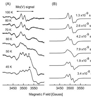
Figure 2 shows two spectra taken in the 40–120 K temperature range. The spectrum at 100 K shows an EPR signal which is observed between 100 and 120 K without changes. The lineshape of this signal resembles the so-called slow-type signal, which is associated with Mo(V) ions in several Mo-containing hydroxylases of the xanthine oxidase family [1, 3]. Slow-type EPR signals present almost-axial symmetry with resonances split by a
Fig. 1 Variable-temperature EPR spectra (a,b, andc) taken of the dithionate-reduced state ofD.alaskensisFDH. The signalsFe/S I
andIIanddashed linesare simulations. The EPR parameters used in the spectral simulation are given in Table 1
Table 1 EPR parameters obtained by simulation in D. alaskensis
FDH. Hyperfine parameters (A) and linewidths (in parentheses) are given in gauss (1 G=0.1 mT);g-values ±0.001,A-values ±1
g1 g2 g3 A1 A2 A3
Mo 1.959(8) 1.968(8) 1.971(8) 14 16 16 W 1.916(15) 1.933(13) 1.955(12)
Fe/S I 1.882(21) 1.946(12) 2.046(13) Fe/S II 1.868(80) 1.913(90) 2.066(70)
Fig. 2 EPR spectra taken of the dithionate-reduced state of D.
alaskensisFDH in the 40–120 K temperature range, together with spectral simulation. Experimental conditions: microwave power, 2 mW (a) and 0.6 mW (b); modulation amplitude, 4 Gpp;
single proton. D. alaskensis FDH presents EPR parameters (Table 1) within the typical values found for Mo(V) species giving slow-type signals, and in particular they are identical to those found in the D. gigas AOR [23]. This suggests that the structural characteristics of the molybdenum site ofD.alaskensisFDH are similar to the one found inD.gigasAOR [24].
The spectra below 70 K showed the emergent Fe/S I EPR signal described above together with another rhombic signal, with all the g-values lower than 2 (see Fig. 2b). This signal has larger deviations from the free-electron g-value and larger linewidth compared to the ones associated with Mo(V) species, pointing to tungsten in a d1 configuration. The lack of observation of the hyperfine interaction with the 183W nucleus (I=1/2, 14.4% natural abundance) may be due to the small intensity of this signal. However, this type of hyperfine splitting has not been observed in several other W pro-teins [2]. The broadening of the W(V) ion signal at
70 K is unusual for an ion in a d1configuration and
suggests that the W(V) ion is magnetically coupled to another faster relaxing paramagnet. EPR studies per-formed on model systems suggest a correlation between thegavvalue and the number of thiolate ligands in oxo-tungsten(V) species [25]. According to this correlation, the W center inD.alaskensis FDH (gav=1.934) should
be bound to four thiolates.
The EPR parameters obtained for the tungsten site suggest a bis-dithiolene coordination, as observed in other Mo- and W-containing proteins of the DMSO reductase family [3]. On the other hand, EPR parameters obtained for Mo(V) ions indicate a metal site coordi-nation similar to the one observed in D. gigas AOR, where just one pterin molecule is linked to the molyb-denum atom. The pterin cofactor of the proteins belonging to the DMSO reductase family shows a coordination geometry that is highly flexible. Three structures have been reported for DMSO reductases, two from R. capsulatus [26, 27] and one from R. spheroides [28]. These structures showed that one of the pterin molecules can be partially or totally uncoordi-nated. Our EPR data suggest that the Mo site of D. alaskensis FDH is coordinated by only one pterin mol-ecule. Therefore the Mo site inD.alaskensisFDH might have an analogous structure to the Mo AOR site from D. gigas, which would explain the striking similarity between the EPR spectra of both proteins.
Analysis of the magnetic interactions
As seen in Fig. 2, the resonance lines of the Mo(V) ion signal are split at low temperatures, which indicates that Mo(V) ions are magnetically coupled to another para-magnetic center. The W and Mo signals are typical of mononuclear Mo(W) atoms in a d1configuration, which discards the possibility of a magnetic interaction be-tween both metal ions. Therefore the splitting of the Mo(V) signal must arise from the iron-sulfur clusters.
Both dipolar and superexchange interactions have been suggested to be responsible for such couplings in me-talloproteins. Dipolar couplings predict an anisotropic splitting that depends on the angle between the spin–spin distance and the external magnetic field, although a zero splitting at the principalg-values of the Mo(V) ion signal can be obtained at the ‘‘trimagic’’ angle [29]. In contrast, superexchange interactions between two anisotropic paramagnets predict an isotropic splitting with a mag-nitude ofJ(Hex=JS1S2) when |J|<|DgbB|, where Dgis
the difference between the effective g-values of both centers, b is the Bohr magneton, and B the external magnetic field. In the case we are considering where the resonances of one of the centers are split by hyperfine interactions with a species with nuclear spin (I=1/2), it is straightforward to show that this condition is |J|<|DgbB±½A|, whereA is the hyperfine parameter.
Furthermore, magnetic interactions depend on temper-ature when one of the species of the interacting pair has a relaxation rate (T1) faster than the other one. This may
produce an enhancement of the relaxation rate of the slowly relaxing paramagnetic center and/or a tempera-ture dependence of the splitting of the resonance lines [30, 31, 32, 33].
Figure 3A shows that the Mo(V) resonances are fully split at temperatures below 50 K, partially collapsed in the 55–80 K range, and fully collapsed at 100 K (no linewidth changes were observed above 100 K and be-low 50 K). The fully split spectra, for example the one shown in Fig. 2b, were accounted for by assuming an isotropic splitting of the Mo(V) resonances, which indicates that superexchange is the magnetic interaction operating between the interacting centers. Therefore, the
temperature variation of the spectra associated with Mo(V) ions (Fig. 3) were simulated by assuming a di-meric model composed of two interactingS=1/2 centers relaxing independently [20], including in addition the hyperfine coupling with a nucleus of nuclear spinI=1/2 at one of the centers, both centers being magnetically coupled only by exchange interaction (Fig. 3). The powder-like spectra were computed after obtaining the absorption spectrum of this system with parameters representing all the interactions, allowing for these to vary according to the dimer orientation with reference to the external magnetic field. In this model the relaxation times T2 contribute only to the linewidth of the
indi-vidual resonances of each paramagnetic center. A good agreement between experimental and simulated spectra was obtained using the relaxation times evaluated for Fe/S I (see below), the g-values for the Mo(V) ion given in Table 1, and an exchange parameter J=1.2·10)3cm)1.
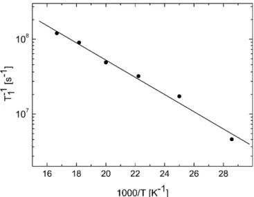
Figure 4 shows the temperature variation of the T1
values associated with the Fe/S I center. The T1values
were evaluated following a method described elsewhere [34], which is applicable when the unbroadened spec-trum shows at least one relatively well-defined peak of nearly gaussian shape, as is the case of the g1 and g3
resonances of the Fe/S I center. The difference between the relaxation times evaluated from both peaks varies by less than 6% and therefore the mean value was utilized to obtain Fig. 4. These data were least-squares fitted assuming an Orbach mechanism [1/T1exp()D/
kT) for D>>kT], wherekis BoltzmannÕs constant and D=184±12 cm)1is the energy of an excited electronic
state [35]. This value is within the magnitude order reported for iron-sulfur clusters of the [4Fe-4S] type [36].
The good agreement obtained between the experi-mental and simulated spectra indicates that the
iron-sulfur center identified as Fe/S I is the center that interacts magnetically with the molybdenum center and therefore is situated in the a-subunit of the protein. This suggests the existence of a suitably oriented chemical path connecting both centers. Although no splitting was detected at the W signal, this signal is not detectable above 70 K, which reveals an unusual relaxation behavior for the W(V) ions. This suggests that W(V) species are also likely to be magnetically coupled to the Fe/S I center. The relaxation timesT1of
the remaining Fe/S centers could not be evaluated be-cause of the superposition of the spectra. However, the fact that these centers are not detected at temperatures above 30 K indicates that the associated T1 are even
lower than that of Fe/S I, and therefore unable of producing the changes observed at the Mo(V) ion signal.
Conclusions
The main goals of the present study can be focused on two aspects: the simultaneous detection of Mo and W and the assignment of the iron-sulfur center proximal to the pterin cofactor. The former constitutes one of the few examples of an enzyme of the vast members of the molybdo- and tungstoenzymes that can incorporate both metal atoms. The latter utilizes EPR together with a semiempirical model to analyze the magnetic interac-tions between paramagnetic centers. This model was demonstrated to be very useful to gain insight into the structural aspects of metalloproteins having exchange-coupled paramagnetic centers.
AcknowledgementsThis work was supported by Fundac¸a˜o para a Cieˆncia e Tecnologia (Portugal) POCTI/BME/36152/99 and CAI+D-UNL (Argentina). MJF thanks the British Council for financial support for travel to Portugal.
References
1. Hille R (1996) Chem Rev 96:2757–2816
2. Johnson MK, Rees DC, Adams MWW (1996) Chem Rev 96:2817–2839
3. Roma˜o MJ, Kna¨blein J, Huber R, Moura JJG (1997) Prog Biophys Mol Biol 68:121–144
4. Almendra MJ, Brondino CD, Gavel O, Pereira AS, Tavares P, Bursakov S, Duarte R, Caldeira J, Moura JJG, Moura I (1999) Biochemistry 38:16366–16372
5. de Bok FAM, Hagedoorn PL, Silva PJ, Hagen WR, Schiltz E, Fritsche K, Stams E, Fritsche K, Stams AJM (2003) Eur J Biochem 270:2476–2485
6. Costa C, Teixeira M, LeGall J, Moura JJG, Moura I (1997) J Biol Inorg Chem 2:198–208
7. George GN, Costa C, Moura JJG, Moura I (1999) J Am Chem Soc 121:2625–2626
8. Raaijmakers H, Teixeira S, Dias JM, Almendra MJ, Brondino CD, Moura I, Moura JJG, Roma˜o MJ (2001) J Biol Inorg Chem 6:398–404
9. Raaijmakers H, Macieira S, Dias JM, Teixeira S, Bursakov S, Huber R, Moura JJ, Moura I, Romao MJ (2002) Structure 10:1261–1272
10. Dias JM, Than ME, Humm A, Huber R, Bourenkov GP, Bartunik HD, Bursakov S, Calvete J, Caldeira J, Carneiro C, Moura JJG, Moura I, Roma˜o MJ (1999) Structure 7:65–77 11. Boyington JC, Gladyshev VN, Khangulov SV, Stadtman TC,
Sun PD (1997) Science 275:1305–1308
12. Bursakov SA, Liu MY, Payne WJ, LeGall J, Moura I, Moura JJG (1995) Anaerobe 1:55–60
13. Khangulov SV, Gladyshev VN, Dismukes GC, Stadtman TC (1998) Biochemistry 37:3518–3528
14. Feio MJ (2000) PhD thesis. University of Portsmouth, UK 15. Postgate JR, Kent HM, Robson RL, Chesshyre JA (1984)
J Gen Microbiol 130:1597–1601
16. Lowry OH, Rosenbrough NJ, Farr AL, Randall RJ (1951) J Biol Chem 193:265–273
17. Gremer L, Meyer O (1996) Eur J Biochem 238:862–866 18. Frunzke K, Heiss B, Meyer O, Zumft WG (1993) FEMS
Microbiol Lett 113:241–246
19. Atherton MM (1973) Electron spin resonance: theory and applications. Wiley, New York
20. Salikhov KM, Galeev RT, Voronkova VK, Yablokov YV, Legendziewicz J (1997) Appl Magn Reson 14:457–472 21. Stewart LJ, Bailey S, Bennet B, Charnock JM, Garner CD,
McAlpine AS (2000) J Mol Biol 299:593–600
22. Deaton JC, Solomon EI, Watt GD, Wetherbee PJ, Durfor CN (1987) Biochem Biophys Res Commun 149:424–430
23. Turner N, Barata B, Bray RC, Deistung J, Le Gall J (1987) Biochem J 243:755–761
24. Roma˜o MJ, Archer M, Moura I, Moura JJG, LeGall J, Engh R, Schneider M, Hof P, Huber R (1995) Science 167:1170–1176 25. Barnard KR, Gable RW, Wedd AG (1997) J Biol Inorg Chem
2:623–633
26. McAlpine AS, McEwan AG, Bailey S. (1998) J Mol Biol 275:613–623
27. Schneider F, Lo¨we J, Huber R, Schindelin H, Kisker C, Kna¨blein J (1996) J Mol Biol 263:53–69
28. Schindelin H, Kisker C, Hilton J, Rajagopalan KV, Rees DC (1996) Science 272:1615–1621
29. Abragam A (1961) The principles of nuclear magnetism. Oxford Univesity Press, Oxford
30. Hirchs DJ, Beck WF, Innes JB, Brudvig GW (1992) Bio-chemistry 31:532–541
31. Hirchs DJ, Beck WF, Lynch JB, Que L Jr, Brudvig GW (1992) J Am Chem Soc 114:7475–7481
32. Andrade SLA, Brondino CD, Feio MJ, Moura I, Moura JJG (2000) Eur J Biochem 267:2054–2061
33. Caldeira J, Belle V, Asso M, Guigliarelli B, Moura I, Moura JJG, Bertrand P (2000) Biochemistry 39:2700–2707
34. Bertrand P, Roger G, Gayda JP (1980) J Magn Reson 40:539– 549
35. Pilbrow JR (1990) Transition ion electron paramagnetic resonance. Clarendon Press, Oxford