BASIC RESEARCH
Hot water extract of
Chlorella vulgaris
induced DNA
damage and apoptosis
Yasmin Anum Mohd Yusof,ISuhana Md. Saad,IISuzana Makpol,I Nor Aripin Shamaan,III Wan Zurinah Wan NgahI
IDepartment of Biochemistry, Faculty of Medicine, Universiti Kebangsaan Malaysia, Jalan Raja Muda Abdul Aziz, Kuala Lumpur, Malaysia.IIFaculty of
Health and Life Sciences, Management and Science University, Selangor Darul Ehsan, Malaysia.IIIFaculty of Medicine & Allied Health Sciences, Universiti Sains Islam Malaysia, Persiaran MPAJ, Jalan Pandan Utama, Kuala Lumpur, Malaysia.
OBJECTIVES:The aim of this study was to determine the antiproliferative and apoptotic effects of hot water extracts ofChlorella vulgarison hepatoma cell line HepG2.
INTRODUCTION: The search for food and spices that can induce apoptosis in cancer cells has been a major study interest in the last decade.Chlorella vulgaris, a unicellular green algae, has been reported to have antioxidant and anti-cancer properties. However, its chemopreventive effects in inhibiting the growth of cancer cells have not been studied in great detail.
METHODS:HepG2 liver cancer cells and WRL68 normal liver cells were treated with various concentrations (0-4 mg/ ml) of hot water extract ofC. vulgarisafter 24 hours incubation. Apoptosis rate was evaluated by TUNEL assay while DNA damage was assessed by Comet assay. Apoptosis proteins were evaluated by Western blot analysis.
RESULTS:Chlorella vulgarisdecreased the number of viable HepG2 cells in a dose dependent manner (p,0.05), with an IC50 of 1.6 mg/ml. DNA damage as measured by Comet assay was increased in HepG2 cells at all concentrations ofChlorella vulgaristested. Evaluation of apoptosis by TUNEL assay showed thatChlorella vulgaris
induced a higher apoptotic rate (70%) in HepG2 cells compared to normal liver cells, WRL68 (15%). Western blot analysis showed increased expression of pro- apoptotic proteins P53, Bax and caspase-3 in the HepG2 cells compared to normal liver cells WRL68, and decreased expression of the anti-apoptotic protein Bcl-2.
CONCLUSIONS: Chlorella vulgaris may have anti-cancer effects by inducing apoptosis signaling cascades via an increased expression of P53, Bax and caspase-3 proteins and through a reduction of Bcl-2 protein, which subsequently lead to increased DNA damage and apoptosis.
KEYWORDS: HepG2; Chlorella vulgaris; DNA damage; chemopreventive; apoptosis.
Yusof YAM, Saad SM, Makpol S, Shamaan NA, Ngah WZW. Hot water extract ofChlorella vulgarisinduced DNA damage and apoptosis. Clinics. 2010;65(12):1371-1377.
Received for publication onOctober 7, 2010;First review completed onOctober 26, 2010;Accepted for publication onOctober 26, 2010
E-mail: [email protected]
Tel.: 603-9289 7297
INTRODUCTION
Hepatocellular carcinoma (HCC) is the fifth most com-mon malignancy worldwide.1 In Malaysia, HCC is the
thirteenth most common cancer affecting the population.2
The etiology of liver cancer is multi factorial; some of the well known risk factors include hepatitis B and C viral infection, exposure to chemicals such as aromatic hydro-carbons, and the ingestion of aflatoxin B1.3-5
The process of carcinogenesis involves an initiating event which induces genetic damage, followed by survival and
progression of selected clones of the transformed cells to form tumors. Apoptosis, a form of programmed cell death, has been associated with delaying or inhibiting cancer growth.6,7 Much of cancer research over the past two decades has focused on genes that, when mutated, act in either a dominant or recessive manner to enhance pro-liferation with dysregulation of apoptosis that is respons-ible for initiating cancer and its progression.8,9 Many emerging data in recent years have shown that dietary chemopreventive agents preferentially inhibit growth of cancer cells by targeting signaling molecules, such as caspases, that subsequently lead to the induction of apoptosis.7 Some of these dietary agents include
epigallo-catechin-3-gallate (EGCG), found in green tea,10curcumin in
turmeric,11genistein in soybeans,12lycopene in tomatoes,13 anthocyanins in pomegranates14 and isothiocyanates in broccoli.15
There is a large body of evidence that links DNA damage and apoptosis.16,17 The tumor suppressor protein p53 provides an important link between DNA damage and apoptosis as it has been shown to mediate the upregulation of apoptotic proteins such as Bax, caspase-3 and -8, Noxa, PUMA and p53AIP.18
Chlorella vulgaris, a unicellular green algae, has been widely used as a food supplement and credited with high antioxidant and therapeutic abilities.19,20 The supplement
can be taken in the form of tablets, capsules, as a food additive or extracted as a liquid. Some health claims and benefits include improvement in the control of hyperten-sion, fibromyalgia and ulcerative colitis.21 In vivo studies have revealed the significant antitumor and antigenotoxic efficacy ofC. vulgaris.20A study by Hasegawaet. alshowed
that a glycoprotein, designated ARS-2, found inC. vulgaris extract has an anti-cancer effect on mice-induced fibrosar-coma.22
To the best of our knowledge, the effect ofC. vulgarison hepatoma cells has not been studied in great detail. In this study, we investigated the anti-cancer effect of C. vulgaris extract on the hepatoma cell line HepG2 by evaluating changes in proliferation, DNA damage and apoptosis.
MATERIALS AND METHODS
Culturing and Extraction ofChlorella vulgaris Stock of C. vulgaris Beijerinck (strain 072) was obtained from the University of Malaya Algae Culture Collection (UMACC, Malaysia) and grown in Bold’s basal media (BBM) with a 12 hours dark 12 hours light cycle. Cells were harvested by centrifugation at 1000 rpm. The algae were dried using a freeze dryer and then mixed in distilled water at a concentration of 10% (w/v). The algal suspension was then boiled at 100
˚
C for 20 minutes using reflux method followed by centrifugation at 10 000 rpm for 20 min. The supernatant was lyophilized using a freeze dryer to obtain the powdered form ofC. vulgarisCell Culture and Treatment
Liver cancer cell line HepG2 and normal liver cell line WRL68 were maintained in Eagle’s minimal essential medium (EMEM; Flow Labaratories, Sydney Australia) supplemented with 1mM sodium pyruvate (SIGMA, St Louis, MO, USA), 2mM glutamine, 10% fetal calf serum and 100 U/ml penicillin and streptomycin at 37
˚
C in humidified 5% CO2incubator. Cell proliferation, apoptosis andcollec-tion of protein were performed when cells reached 70% confluence density.C. vulgarisextract at various concentra-tions (0-4 mg/ml) was added to cell lines 24 hours after incubation.
Cell Proliferation Assay
A 96-well plate was seeded with HepG2 and WRL68 cells at a uniform density of 26104cells per well. Twenty-four hours after incubation, cells were treated with the hot water extract ofC. vulgarisand incubated for a further 24 hours. Cells were labeled with bromodeoxyuridine (BrdU) during the last 3 hours of C. vulgaris extract treatment. The cells were fixed with denaturing solution and the incorporation of BrdU was detected by immunoreaction. After substrate solution was added to each well, the amount of BrdU incorporated was determined by measuring the absorbance
at dual wavelengths (450/690 nm) using a scanning multi-well spectrophotometer [enzyme-linked immunosorbent assay (ELISA) reader]
Apoptosis by TUNEL Assay
Apoptotic cell death was determined by Dead EndTM Colorimetric TUNEL System (Promega, Madison, WI, USA). After 24 hours of treatment withC. vulgarisextract, floating cells and adherent cells in culture were collected in a tube, trypsinized, centrifuged and washed in phosphate buffered saline (PBS). Cells were resuspended and applied to poly-L-lysine-coated slides and air dried. Cells were fixed by immersing slides in 4% formalin in PBS for 25 minutes at room temperature. After washing with PBS, cells were permeabilized by immersing the slides in 0.2% Triton6100 solution in PBS for 5 minutes at room temperature. Cells were then equilibrated with 100ml of equilibration buffer
(2.5 mM Tris-HCl pH 6.6, 0.2 M potassium cacodylate, 2.5 mM CoCl2, 0.25 mg/mL BSA.) for 7 minutes. The
equili-brated areas were blotted with tissue paper before adding biotinylated nucleotide and TdT reaction mix (100ml) to the
slides. The slides were then covered with coverslips to ensure an even distribution of the reagent before incubating at 37
˚
C for 60 minutes in a humidified chamber. Coverslips were removed and the slides were immersed in 26 saline-sodium citrate (SSC; saline-sodium chloride 0.15M, trisaline-sodium citrate 0.015M) buffer for 15 minutes at room temperature, and washed twice with PBS. Endogenous peroxidases were blocked by immersing the slides in 0.3% hydrogen peroxide for 4 minutes at room temperature and washed again in PBS. Streptavidin horseradish peroxidase (HRP) solution (1:500 PBS) was added to each slide and incubated for 30 minutes at room temperature. After final washing with PBS, diaminobenzedine (DAB) solution was added to the slides for 20 minutes until light brown staining was observed. Finally, each slide was mounted with DPX (BDH, England) to be examined under light microscope.DNA Damage Measurement by Comet Assay
The assay was performed as described by Singh et al.23 Thirty microlitres of the HepG2 cell suspension (,400,000 cells/ml) was mixed with 80 ml of 1% low melting point
(LMP) agarose and added to fully frosted slides that had been covered with a layer of 1% LMP agarose. Subse-quently, the slides were immersed in lysis solution [2.5M NaOH, pH10; 0.1M ethylenediaminetetraacetic acid (EDTA); 0.01M Tris; and 1% Triton6100] for 1 h at 4
˚
C, followed by electrophoresis solution (300mM NaOH; and 1mM EDTA, pH13) for 20 min to allow DNA unwinding, and electrophoresed for 20 min at 25 V and 300 mA. Finally, the slides were neutralized with 0.4M Tris buffer (pH 7.5), stained with ethidium bromide (5 mg/ml) and analyzed using a fluorescence microscope (Carl Zeiss, Go¨ttingen, Germany). Images of 50 randomly selected cells per experi-mental point were visually analyzed under the microscope.Measurement of p53, Bax, Bcl-2, Caspase-3 and -8 by Western blotting
1 mM EDTA, 20 mM NaF, 100 mM Na3VO4, 1% NP-40, 1%
Triton X-100, 1 mM phenylmethylsulfonyl fluoride (PMSF), 10 mg/ml Aprotinin and 10 mg/ml Leupeptin] (SIGMA, St
Louis, MO, USA) on ice for 30 min to lyse the cells. After centrifugation, total protein was determined using a Bio-Rad protein assay kit (Bio-Rad, Hercules, CA, USA). Protein was resolved (50mg) by 10–15% SDS-PAGE and transferred to
polyvinyl difluoride (PVDF) membranes. The membrane was blocked with blocking buffer (5% skim milk in 1% Tween 20 in 20 mM Tris-buffered saline, pH7.5) by incubating for 1 h at room temperature followed by incubation with the appro-priate primary antibody (p53, Bax, Bcl-2, caspase-3 and -8; Chemicon, Billerica, MA, USA) at dilutions of 1:1000 in blocking buffer for 2 h at room temperature. The membranes were then incubated with the respective secondary anti-bodies for 1 h and antigens were detected by using the enhanced chemiluminescence blotting detection system (GE Healthcare, Piscataway, NJ, USA).
Statistical Analysis
Results are expressed as mean¡SD with the experiment
repeated at least 3 times. Statistical evaluation was done using the Student’s t-test. Apvalue of,0.05 was considered significant.
RESULTS
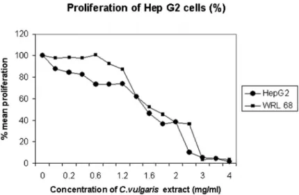
Chlorella vulgarisinhibited the proliferation of human liver cancer cells (HepG2) in a concentration-dependent manner, ranging from 0-4 mg/ml as shown by BrdU proliferation assay with a 50% reduction at 1.6 mg/ml, (IC50) (Fig. 1). The
high IC50is expected asC. vulgarisis classified as a food and
not a drug. Proliferation of normal liver cells (WRL68) was decreased when treated withC. vulgarisextract, resulting in a 50% reduction at 1.7 mg/ml. One hundred percent inhibition of proliferation of HepG2 cells and WRL68 cells was achieved at approximately 3 mg/ml and 4mg/ml, respectively.
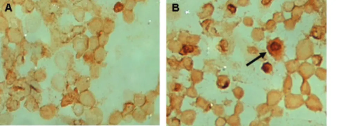
It was clearly seen that C. vulgaris induced apoptosis in HepG2 cells with distinct nuclear condensation and
blebbing of the plasma membrane, a typical characteristic of apoptotic bodies (Fig. 2).24
The percentange of apoptosis of HepG2 and WRL68 cells as induced byC. vulgarisextract at 2mg/ml is demonstrated in Figure 3; the rate of apoptosis was significantly higher (70%) in HepG2 cells compared to normal WRL68 cells (15%). WRL68 cells also underwent apoptosis but at a higher concentration ofC. vulgaris(3-4 mg/ml), perhaps as a result of the cytotoxicity effects seen at higher concentra-tions (Fig. 1)
The involvement of pro- and antiapoptotic proteins is demonstrated in Figure 4. Figure 4a shows that the level of p53 increased in a dose-dependent manner and reached maximum induction at 2 h with 2 mg/ml treatment of C. vulgaris. Interestingly, treatment withC. vulgarisresulted in increased expression of Bax (Fig. 4c), and a decreased expression of Bcl-2 (Fig. 4b) in a time-dependent manner. However, the activation of the mitochondrial apoptotic pathway in downstream caspase-8 was not significant (Fig. 4e), but a significant increase in the active form of caspase-3 was observed after 24 h of treatment with C.vulgaris in a time-dependent manner (Fig. 4d). These results demonstrate that the mitochondrial signaling path-way is involved inC. vulgaris-induced apoptosis of HepG2 cells.
DNA damage in HepG2 cells was increased byC. vulgaris treatments at all concentrations (Fig. 5). At low doses ofC. vulgarisextract, HepG2 and WRL68 cells generated smaller comets that were not significant, whilst at 2 mg/ml, C. vulgaris induced more damage in HepG2 cells (80%) compared to WRL68 (50%).
DISCUSSION
Many chemopreventive agents have been associated with antiproliferative and apoptotic effects on cancer cells because of their high antioxidant activity, targeting signaling mole-cules, and preventing or protecting cells from further damage or transformation into cancer cells.7Chlorella vulgarishas been shown to exhibit high antioxidant activity compared to other microalgae.25The antiproliferative effect ofC. vulgaris(Fig. 1)
Figure 1 -The effect ofChlorella vulgarisextract on proliferation of HepG2 and WRL68 cell lines treated with different concentrations
ofC. vulgarisextract. Data are represented as mean¡SD. * Showed significant changes (p,0.05) for n = 3 triplicate after 24 hours of
treatment with different concentrations ofC. vulgarisextract using bromodeoxyurdine (BrdU) assay with 50% reduction (IC50) seen at
could be caused by an acidic glycoprotein found to have antitumour and antioxidant properties in tumour-bearing mice.22
DNA damage is a common event in life following which, repair mechanisms and apoptosis will be activated to maintain genome integrity. The tumor-suppressor protein p53 provides an important link between DNA damage and apoptosis. DNA damage results in cell cycle arrest at checkpoints or at G1 or G2 stage to inhibit cell cycle progression and to induce apoptosis, thus protecting cells against further damage.24Antioxidants are important inhi-bitors of lipid peroxidation as a defence mechanism against oxidative damage.26Plants and microalgae have developed a protective mechanism by possessing antioxidant com-pounds mainly with phenolic moieties to eradicate the accumulation of reactive oxygen species (ROS) produced by ultraviolet (UV) radiation or heat from sunlight.26Our study showed thatC. vulgaris, with its high antioxidant activity,
protected normal WRL68 cells against severe DNA damage but induced severe DNA damage in HepG2 cells.
The induction of apoptosis is known to be an efficient strategy for cancer therapy. Many Chinese herbal remedies
Figure 2 -Comparison of the morphology of WRL68 and HepG2 cells before and after treatment with 2 mg/mlChlorella vulgaris
extract. Apoptotic cells are absent in untreated WRL68 cells (a) and in treated (2 mg/mlC. vulgaris) WRL68 cells (b). Apoptotic cells are absent in untreated HepG2 cells (c), but present in treated (2 mg/mlC. vulgaris) HepG2 cells (d). Arrow shows condensation of chromatin and cell blebbing on HepG2 cells.
Figure 3 -The effect ofChlorella vulgarisextract on apoptosis of HepG2 and WRL68 cell lines. Data are represented as mean¡SD. * showed significant changes (p,0.05) for n = 3 triplicate after 24 hours of treatment with different concentrations of C.
vulgarisextract using TUNEL assay. Per cent apoptotic cells are
scored by taking an average of 1000 cells in 10 random fields. Apoptotic cells are almost absent in untreated and at 1 mg/ml of
C. vulgarisbut present quantitatively in HepG2 cells at higher
concentrations ofC. vulgaris.
Figure 4 -Effect ofChlorella vulgarison the protein expression of P53, Bax, caspase-3 and Bcl-2. Chlorella vulgaris at 2 mg/ml (optimal dose) induced higher expressions of P53 (a), Bax (c) (proapoptotic) and caspase-3 (d) proteins in HepG2 cells compared to the control cells after 24 hours treatment. Mean-while, Bcl-2 (b) (antiapoptotic) protein expression was reduced compared to the control cells. No significant changes in the expressions of caspase-8 (e) proteins in hepatoma cells treated
with C. vulgaris (2 mg/ml) were observed after 24 hours of
such asGanoderma lucidum,Rubus coreanum,Paeoniae Radix and Phyllanthus urinaria have been shown to trigger the apoptotic pathway in MCF-7 human breast cancer cells, HT-29 human colon cancer cells, HepG2 human hepatoma cells and Lewis lung carcinoma cells, respectively.27-30 Apoptosis is modulated by antiapoptotic and proapoptotic effectors, which involve a large number of proteins. The propapoptotic and antiapoptotic members of the Bcl-2 family act as a rheostat in regulating programmed cell death and as a target of anticancer therapy.31 The ratio of
death antagonists (Bcl2, Bcl-xL) to agonists (Bax, Bcl-xs, Bad, Bid) determines whether a cell will respond to an apoptotic stimulus. Down-regulation of the death suppressor Bcl-2 protein could repress tumor growth by promoting pro-grammed cell death.32-34.
To investigate the mechanisms of C. vulgaris-induced apoptosis, we assessed the expression and activity of tumor suppressor protein P53 and a host of pro- and antiapoptotic proteins: Bcl-2, Bax, and caspases-3 and -8. Our results showed that C. vulgaris mediated apoptosis in a P53-dependent
Figure 5 -Detection of DNA damage in HepG2 and WRL68 cells as measured by Comet assay after 24 hours treatment withChlorella
vulgaris. Data are represented as mean¡SD for n = 3 triplicate after 24 hours of treatment withC. vulgarisextract. (a) HepG2 cells
manner with increased expression of Bax and decreased expression of Bcl-2 proteins in a time-dependent manner. The activation of p53 and related family members can either enforce cell cycle arrest or induce apoptosis. The p53-dependent responses are directed against the damaged cell to protect the organism. The rules that govern the choice between growth, arrest and apoptosis are likely to be enforced by other proteins that can antagonize or synergize with p53 to regulate apoptosis.35 The maximal increase of Bax expression in HepG2 cells after 18 h incubation withC. vulgaris may be the result of the increase of P53 accumulation that peaked after 2 h.
As a herbal extract, C. vulgaris contains a variety of compounds including antioxidants and a glycoprotein that may act on different pathways of tumor cell growth and survival, triggering an antagonistic effect of Bax and Bcl-2.
Apoptotic signals are generally believed to be mediated through a hierarchy of caspase activation controlled by one of two distinct pathways that are associated with either mitochondrial caspase-8 or -9. The initiating caspases then converge on the central effector caspases, caspases-3 and -7. Although we did not find significant activation of down-stream caspase-8 there was, however, a significant increase of the active form of caspase-3 after 24 h of treatment withC. vulgaris, in a time-dependent manner. These results demon-strated that the mitochondrial signaling pathway is involved in C. vulgaris-induced apoptosis of HepG2 cells. It could be postulated that C. vulgaris did not induce the initiator caspase-8 but it may have activated the other initiator caspase-9. Among the ten different members of caspases identified in mammalian cells, caspase-3 may serve as a general mediator of apoptosis. When cells are under-going apoptosis, executioner or effector caspase-3 triggers cellular proteins such as poly (ADP-ribose) polymerase and DNA fragmentation factor, resulting in the characteristic changes of apoptosis.36Caspase-3 is synthesized as a 33 kDa inactive proenzyme that requires proteolytic activation. Our results showed that a high level of proenzyme of caspase-3 was present in untreated tumor cells, and active caspase-3 gradually increased after C. vulgaristreatment, suggesting that C. vulgaris induced apoptosis through a caspase-3-dependent mechanism.
CONCLUSION
The results of the present study suggest that the antic-ancer mechanism ofC. vulgarisin hepatoma cells (HepG2) is by inhibiting DNA synthesis, triggering DNA damage and inducing apoptosis. This is shown by the reduced incor-poration of BrdU into replicating DNA of HepG2 cells and an increase in the number of DNA damage-inducing and apoptotic proteins, Bax and caspase-3 in HepG2 cells treated withC. vulgaris. The possible mechanism is speculated to be an increase in P53, Bax and caspase-3 expression that would subsequently lead to apoptosis.
ACKNOWLEDGEMENTS
This research was supported by a grant from Ministry of Science, Technology and Innovation (MOSTI)
REFERENCES
1. Bosch FX, Ribes J, Diaz M, Cleries R. Primary liver cancer: Worldwide incidence and trends. Gastroenterology. 2004;127:5-16, doi: 10.1053/j. gastro.2004.09.011.
2. Gerard LCC, Halimah Y. Second Report of the National Cancer Incidence in Malaysia, National Cancer Registry, Ministry of Health Malaysia. 2003 3. Sheu JC. Molecular mechanism of hepatocarcinogenesis. J Gastroenterol
Hepatol. 1997;12:S309–13, doi: 10.1111/j.1440-1746.1997.tb00514.x. 4. Tsai JF, Chang WY, Jeng JE, Ho MS, Lin ZY, Tsai JH. Hepatitis B and C
virus infection as risk factors for liver cirrhosis and cirrhotic hepatocel-lular carcinoma: A case-control study. Liver. 1994;14:98-102.
5. Jackson PE, Groopman JD. Aflatoxin and liver cancer. Baillieres Clin Gastroenterol. 1999;13:545-55, doi: 10.1053/bega.1999.0047.
6. Barr PJ, Tomei LD. Apoptosis and its role in human disease. Biotechnology. 1994;12:487-93, doi: 10.1038/nbt0594-487.
7. Khan N, Afaq F, Mukhtar H. Apoptosis by dietary factors: the suicide solution for delaying cancer growth. Carcinogenesis. 2007;28:233-9, doi: 10.1093/carcin/bgl243.
8. Lowe SW, Lin AW. Apoptosis in cancer. Carcinogenesis. 2000; 21:489-95, doi: 10.1093/carcin/21.3.485.
9. Zeiss CJ. The apoptosis-necrosis continuum: insights from genetically altered mice. Vet Pathol. 2003;40:481-95, doi: 10.1354/vp.40-5-481. 10. Brusselmans K, De Schrijver E, Heyns W, Verhoeven G, Swinnen JV.
Epigallocatechin-3-gallate is a potent natural inhibitor of fatty acid synthase in intact cells and selectively induces apoptosis inprostate cancer cells.Int J Cancer. 2003; 106:856-62, doi: 10.1002/ijc.11317. 11. Gao X, Deeb D, Jiang H, Liu YB, Dulchavsky SA, Gautam SC. Curcumin
differentially sensitizes malignant glioma cells to TRAIL/Apo2L-mediated apoptosis through activation of procaspases and release of cytochrome C mitochondria. J Exp Ther Oncol. 2005;5:39-48.
12. Su SJ, Chow NH, Kung ML, Hung TC, Chang KL. Effects of soy isoflavones on apoptosis induction and G2-M arrest in human hepatoma cells involvement of caspase-3 activation, Bcl-2 and Bcl-XL down-regulation, and Cdc2 kinase activity. Nutr Cancer. 2006;45:113-23, doi: 10.1207/S15327914NC4501_13.
13. Liu C, Russel RM, Wang X-D. Lycopene supplementation prevents smoke-induced changes in p53, p53 phosphorylation, cell proliferation, and apoptosis in the gastric mucosa of ferrets. J Nutr. 2006;136:106-11. 14. Larrosa M, Tomas-Barberan FA, Espin JC. The dietary hydrolysable
tanin punicalagin releases ellagic acid that induces apoptosis in human colon adenocarcinoma Caco-2-cells by using the mitochondrial pathway. J Nutr Biochem. 2006; 17:611-25, doi: 10.1016/j.jnutbio.2005.09.004. 15. Zhang Y. Cancer-preventive isothiocyanates: measurement of human
exposure and mechanism of action. Mutat Res. 2004; 173-90.
16. Wang JYJ. DNA damage and apoptosis. Cell Death Differ. 2001;8:1047-8, doi: 10.1038/sj.cdd.4400938.
17. Norbury CJ, Zhivotovsky B. DNA damage and apoptosis. Oncogene. 2004;23:2797–808, doi: 10.1038/sj.onc.1207532.
18. Benchimol S. P53-dependent pathway of apoptosis. Cell Death Differ. 2001;8:1049-51, doi: 10.1038/sj.cdd.4400918.
19. Herrero M, Alvarez PJM, Senorans FJ, Cifuentes A, Ibanez E. Optimization of accelerated solvent extraction of antioxidants from
Spirulina platensesmicroalgae. Food Chem. 2005;93: 417-23, doi: 10.1016/ j.foodchem.2004.09.037.
20. Yasukawa K, Akihisa T, Kanno H, Kaminaga T, Izumida M, Sakoh T, et al. Inhibitory effects of sterols isolated fromChlorella vulgarison 12-o-tetradecanoyl phorbol-13-acetate-induced inflammation and tumor promotion in mouse skin. Biol Pharm Bull. 1996;19:573-6.
21. Merchant RE, Andre CA. A review of recent clinical trials of the nutritional supplementChlorella pyrenoidosa in the treatment of fibro-myalgia, hypertension, and ulcerative colitis. Altern Ther Health Med. 2001;7:79-91.
22. Hasegawa T, Matsuguchi T, Noda K, Tanaka K, Kumamoto S, Shoyama Y, et al. Toll-like receptor 2 is at least partly involved in the antitumor activity of glycoprotein fromChlorella vulgaris. Int J Immunopharmacol. 2002;2: 579-89., doi: 10.1016/S1567-5769(02)00002-4
23. Singh NP, McCoy MT, Tice RR Schneider EL. A simple technique for quantitation of low levels of DNA damage in individual cells. Exp Cell Res. 1988;175:184-91, doi: 10.1016/0014-4827(88)90265-0.
24. Bortner CD, Oldenburg NBE, Cidlowski JA. The role of DNA fragmentation in apoptosis. Trends Cell Biol. 1995;5:21-6, doi: 10.1016/ S0962-8924(00)88932-1.
25. Rodriguez-Garcia I, Guil-Guerrero JL. Evaluation of the antioxidant activity of three microalgal species for use as dietary supplements and in the preservation of foods. Food Chem. 2008;108:1023-6, doi: 10.1016/j. foodchem.2007.11.059.
26. Frei B. Efficacy of dietary antioxidants to prevent oxidative damage and inhibit chronic disease. J. Nutr. 2004; 134:3196S-3198S.
27. Hu H, Ahn NS, Yang X, Lee YS. Kang KS.Ganoderma lucidumextract induces cell cycle arrest and apoptosis in MCF-7 human breast cancer cell. Int J Cancer. 2001;102:250-3, doi: 10.1002/ijc.10707.
28. Kim EJ, Lee YJ, Shin HK, Park JHY. Induction of apoptosis by the aqueous extract ofRubus coreanumin HT-29 human colon cancer cells. Nutrition. 2005;21:1141-8, doi: 10.1016/j.nut.2005.02.012.
30. Huang ST, Yang RC, Yang LJ, Lee PN, Pang JHS.Phyllanthus urinaria
triggers the apoptosis and bcl-2 down-regulation in Lewis lung carcinoma cells. Life Sci. 2003;72:1705-16, doi: 10.1016/S0024-3205(03)00016-X.
31. Goodsell DS. The molecular perspective: Bcl-2 and apoptosis. Stem Cells. 2002;20:355–6, doi: 10.1634/stemcells.20-4-355.
32. Kluck RM, Bossy-Wetzel E, Green DR, Newmeyer DD. The release of cytochrome c from mitochondria: a primary site for Bcl-2 regulation of apoptosis. Science. 1997;275:1132–6, doi: 10.1126/science.275.5303.1132.
33. Zhivotovsky B, Orrenius S, Brustugun OT, Doskeland SO. Injected cyto-chrome c induces apoptosis. Nature. 1998;391: 449–50, doi: 10.1038/35060. 34. Antonsson B, Martinou JC. The Bcl-2 protein family. Exp Cell Res.
2000;256:50–7, doi: 10.1006/excr.2000.4839.
35. Benchimol S. P53-dependent pathway of apoptosis. Cell Death Differ. 2001;8:1049-51, doi: 10.1038/sj.cdd.4400918.