in Mice Reveals Renal Colonization as a Stealth Escape
from the Blood Defenses and Antibiotics
Gwenn Ratet1,2, Fre´de´ric J. Veyrier1,2,3, Martine Fanton d’Andon1,2, Xavier Kammerscheit1,2,4, Marie-Anne Nicola5, Mathieu Picardeau6, Ivo G. Boneca1,2, Catherine Werts1,2*
1Institut Pasteur, Unite´ Biologie et Ge´ne´tique des parois bacte´riennes, Paris, France,2INSERM, e´quipe Avenir, Paris, France,3Institut Pasteur, Unite´ des infections bacte´riennes invasives, Paris, France,4ENS Cachan, de´partement de Biologie, Paris, France,5Institut Pasteur, Plate forme d’imagerie dynamique, Paris, France,6Institut Pasteur, Unite´ Biologie des Spiroche`tes, Paris, France
Abstract
Leptospira (L.) interrogansare bacteria responsible for a worldwide reemerging zoonosis. Some animals asymptomatically carryL. interrogansin their kidneys and excrete bacteria in their urine, which contaminates the environment. Humans are infected through skin contact with leptospires and develop mild to severe leptospirosis. Previous attempts to construct fluorescent or bioluminescent leptospires, which would permitin vivo visualization and investigation of host defense mechanisms during infection, have been unsuccessful. Using a firefly luciferase cassette and random transposition tools, we constructed bioluminescent chromosomal transformants in saprophytic and pathogenic leptospires. The kinetics of leptospiral dissemination in mice, after intraperitoneal inoculation with a pathogenic transformant, was tracked by bioluminescence using live imaging. For infective doses of 106to 107bacteria, we observed dissemination and exponential growth of leptospires in the blood, followed by apparent clearance of bacteria. However, with 26108 bacteria, the septicemia led to the death of mice within 3 days post-infection. In surviving mice, one week after infection, pathogenic leptospires reemerged only in the kidneys, where they multiplied and reached a steady state, leading to a sustained chronic renal infection. These experiments reveal that a fraction of the leptospiral population escapes the potent blood defense, and colonizes a defined number of niches in the kidneys, proportional to the infective dose. Antibiotic treatments failed to eradicate leptospires that colonized the kidneys, although they were effective againstL. interrogansif administered before or early after infection. To conclude, mice infected with bioluminescentL. interrogansproved to be a novel model to study both acute and chronic leptospirosis, and revealed that, in the kidneys, leptospires are protected from antibiotics. These bioluminescent leptospires represent a powerful new tool to challenge mice treated with drugs or vaccines, and test the survival, dissemination, and transmission of leptospires between environment and hosts.
Citation:Ratet G, Veyrier FJ, Fanton d’Andon M, Kammerscheit X, Nicola M-A, et al. (2014) Live Imaging of BioluminescentLeptospira interrogansin Mice Reveals Renal Colonization as a Stealth Escape from the Blood Defenses and Antibiotics. PLoS Negl Trop Dis 8(12): e3359. doi:10.1371/journal.pntd.0003359 Editor:Jenifer Coburn, Medical College of Wisconsin, United States of America
ReceivedJune 12, 2014;AcceptedOctober 18, 2014;PublishedDecember 4, 2014
Copyright:ß2014 Ratet et al. This is an open-access article distributed under the terms of the Creative Commons Attribution License, which permits unrestricted use, distribution, and reproduction in any medium, provided the original author and source are credited.
Data Availability:The authors confirm that all data underlying the findings are fully available without restriction. All relevant data are within the paper and its Supporting Information files.
Funding:GR was recipient of a Ph.D fellowship from DIM Malinf Ile de France. This study was supported by the Institut Pasteur. CW’s salary was supported in part by an ERC grant (PGNfromshapetoVIR202283, to IGB). The funders had no role in study design, data collection and analysis, decision to publish, or preparation of the manuscript.
Competing Interests:The authors have declared that no competing interests exist. * Email: cwerts@pasteur.fr
Introduction
Leptospirosis is a worldwide zoonosis transmitted by chronically infected animals, through excretion of bacteria in their urine, contaminating the soil and water. PathogenicLeptospira interrogans (L. interrogans)are spiral motile bacteria that enter a diverse range of hosts through broken skin and mucosa. Infected humans develop either a flu-like, usually mild illness, or severe disease with renal, liver and heart failure, and eventually pulmonary hemorrhages that may be fatal. Leptospirosis is a reemerging and under-diagnosed neglected zoonotic disease, that may increase worldwide because of global warming and uncontrolled urbanization [1]. In East Asia, leptospirosis is a serious health issue affecting paddy field workers, and people living in regions often flooded after typhoons. Frequent outbreaks of leptospirosis occur in developing countries, where a
L. interrogansin transparent Zebrafish embryos, demonstrating fast uptake of leptospires by phagocytic cells [11]. However to our knowledge, no dynamic study of the in vivo dissemination of leptospires has been undertaken in mammals. Two teams have succeeded in constructing genetically modified leptospires species expressing either fluorescent GFP or mRFP proteins [12] or luciferase from a luxCDABE cassette fromPhotorhabdus lumines-cens[13]. These labeled strains proved to be useful tools to easily enumerate the bacteria or for use inin vitroassays. However, these constructs did not allow the monitoring of infection with L. interrogansin cells or in animals. In contrast, a recent study showed the feasibility of using the firefly luciferase gene under the control of a strong promoter to obtain luminescentBorreliaspecies and track the bacteria in vivo in mice [14]. Therefore, we chose to construct bioluminescent leptospires, introducing a cassette expressing the firefly luciferase gene under the control of a strong leptospiral promoter, by transposon insertion in the chromosome of leptospires [10]. Since the reaction catalyzed by the firefly luciferase in the presence of its substrate, D-luciferin, requires ATP to emit photons, only metabolically active and therefore live bacteria expressing the firefly luciferase are bioluminescent in the presence of D-luciferin.
Until recently, mice have not been regarded as a good model for leptospirosis, as they are considered resistant to acute leptospirosis, typically surviving the infection [8]. However, we and another group have demonstrated thatL. interrogans-infected C57BL/6J mice develop mild fibrosis, associated with chronic renal carriage of leptospires [15,16]. Hence, in this work we used bioluminescent strains ofL. interrogansto visualize and characterize the course of leptospirosis in mice, and study the efficacy of antibiotic treatments, administered at different phases of the infection.
Materials and Methods
Bacterial strains and culture conditions
L. biflexasevorar Patoc strain Patoc (Paris) andL. interrogans
serovar Manilae strain L495 were used in this study as described earlier [17]. Bacteria were grown in
Ellinghausen-McCullough-Johnson-Harris (EMJH) medium (Bio-Rad) at 28uC without agitation. EMJH agar plates were obtained by solidification of EMJH medium with 1.2% noble agar (Difco). Leptospires were counted using a Petroff-Hauser chamber. Antibiotics were usedin vitroat the following concentrations: 100mg/mL ampicillin (Amp, MP Biomedical); 25mg/ml kanamycin (Km, Sigma-Aldrich); 50mg/ml spectinomycin (Spc, Sigma-Aldrich). Thymidine (dT, Sigma-Aldrich) and diaminopimelate (DAP, Sigma-Aldrich) were added when necessary at the final concentration of 0.3 mM.
Construction of pCj::FlgB plasmid
The firefly luciferase gene was amplified from the vector p5L3::Km [18] using the LucNcoIF and LucPstIR primers (Table 1) and ligated into pBAD28 (Life Technologies) using NcoI and PstI restriction enzymes to generate pBAD::Luc. A strong leptospiral promoter flgBp, encompassing the upstream region of the flagellar basal-body rod protein, has been amplified fromL. interrogansFiocruz L1-130 DNA (location chromosome 1 342199-341959) using specific primers (Table 1). The promoter was independently subcloned into pBAD::Luc using NcoI and NheI, resulting in the pFlgB::Luc plasmid expressing the firefly luciferase gene under the control of the leptospiral promoter. The luciferase cassette was recovered from the pFlgB::Luc plasmid using the NheI/PvuI enzymes. Plasmid pCj::FlgB was generated by cloning the blunt-ended luciferase cassette into the blunt-ended PacI restriction site of the suicide conjugative plasmid pCjTKS2 [19].
Transposon mutagenesis and conjugation betweenE. coliand leptospires
Random transposition of the luciferase cassette was conducted as previously described [20]. Briefly, pCj::pFlgB plasmid was transformed into E. coli b2163 (DAP2, [21]) by heat shock at 42uC for 45 s and transformants were selected on Luria Bertani medium plates containing kanamycin and DAP. L. interrogans
andL. biflexastrains were grown to mid exponential phase (26107
bacteria/mL) in liquid EMJH. Conjugation betweenE. colib2163 carrying pCj::pFlgB and leptospiral strains was done as previously described [22] and the transformants selected on EMJH plates containing kanamycin. The unknown insertion junction fragment of each transformant was determined by semi-random PCR and sequenced as described [22].
Infection experiments with leptospires
Female mice (7- to 10-wk old) were used in this study. Female Albino C57BL/6J mice were purchased from Charles Rivers (Chatillon-sur-Chalaronne, L’Arbresle Cedex, France). Balb/c and C57BL/6J mice were from Janvier (Le Genest, France).
Infections with leptospiral strains and bioluminescent mutants were conducted as described earlier [17]. Just before infection, bacteria in late exponential phase (around 108leptospiresperml) were centrifuged at room temperature for 25 min at 32506g, resuspended in endotoxin-free PBS, and counted using a Petroff-Hauser chamber. 200ml of leptospires in PBS were injected via the intraperitoneal route into mice.
Bioluminescence imaging
D-luciferin potassium salt (Caliper Life Sciences), the substrate of Firefly luciferase, was dissolved in PBS at a concentration of 30 mg/ml. 10 minutes before imaging, 100ml (3 mg) of D-luciferin was given to mice by intraperitoneal injection. Mice were then anesthetized using a constant flow of 2.5% isoflurane mixed with oxygen and air as recommended by the manufacturer,
Author Summary
using an XGI-8 anesthesia induction chamber (Xenogen Corp.). Mice were maintained in the anesthesia chamber for at least 5 min to allow adequate dissemination of the injected substrate. Bacterial infection images were acquired using an IVIS Spectrum system (Xenogen Corp., Alameda, CA) according to instructions from the manufacturer. Analysis and acquisition were performed using Living Image 3.1 software (Xenogen Corp.). Images were acquired using 5 min of integration time with a binning of 8 and with the emission filter in the ‘‘open’’ mode. All other parameters were held constant. Quantification was performed using a region of interest defined manually (whole mouse or kidneys) and the results were expressed as photons (P) per second (s) per cm2per steradian (SR). Non-infected control mice and infected mice were separately imaged in order to prevent hypothetical contamination. At different time points after infection, 50ml of blood was collected at the retro-mandibular sinus, added to 20ml of 100 mM EDTA and imaged immediately after addition of 10ml of 3 mg/ml D-luciferin in PBS, to detect viable bacteria, or 10ml of a solution of 3 mg/ml D-luciferin in PBS complemented with 1 mM ATP, to detect both live and dying bacteria. Same procedure was used to image bioluminescent L. interrogans in EMJH medium or in urine, collected holding the mouse above a sterile Petri dish. Forex vivoanalyses, mice were euthanized, and the kidneys and organs removed, cut and imaged for 5 min after immersion in D-luciferin solution (3 mg/ml in PBS), then stored at280uC for subsequent detection of leptospires by q-PCR.
Antibiotic treatments
Penicillin G (Sigma), ciprofloxacin (Fluka/Sigma-Aldrich), azith-romycin (Azi, RTC Sigma) and doxycycline (Sigma-Aldrich) were used at the equivalent human dose of 150 000 units/kg/day, 50 mg/kg/day, 25 mg/kg/day, and 3,3 mg/kg/week, respectively. Antibiotics were diluted in endotoxin-free PBS (Biowhittaker). To solubilize ciprofloxacin and azithromycin, the pH was dropped to 6 with 1N HCL. Antibiotic solutions were sterilized by filtration (0.45mm) and a volume of 100ml was administered daily to,20 g
albino C57BL/6J mice via the intraperitoneal (IP) route for 5 consecutive days, either beginning one day post infection (dpi), or 3 dpi, or for 7 consecutive days during the chronic phase of infection. For prophylaxis experiments, a single IP injection of azithromycin or doxycycline was carried out 2 days prior to the infection.
Leptospiral loads
The leptospiral burden in urine, blood and organs was determined by quantitative real-time PCR (q-PCR), as described [15]. The Maxwell 16 automat was used to extract total DNA from a drop of urine (5 to 100ml) or from 50ml of blood, using the cell LEV DNA, or blood DNA purification kits (Promega), respectively. Total DNA from organs was extracted with the DNAeasy tissue Qiagen kit after mechanical disruption with
metallic beads. Primers and probe designed in thelpxAgene ofL. interrogans strain Fiocruz [23] were used to specifically detect pathogenicLeptospira spp[15].L. biflexawas detected using the classical RNA 16S gene q-PCR, as described [8]. q-PCR reactions were run on a Step one Plus real-time PCR apparatus using the absolute quantification program (Applied Biosystems), with the following conditions for FAM TAMRA probes: 50uC for 2 min, 95uC for 10 min, followed by 40 cycles with denaturation at 95uC for 15 s and annealing temperature 60uC for 1 min, according to the manufacturer’s instructions. Results were expressed as the number of leptospires per 100ml of urine, 50ml of blood, orper
200 ng of total DNA extracted from organs.
Ethics statement
All protocols were reviewed by the Institut Pasteur (Paris, France), the competent authority, for compliance with the French and European regulations on Animal Welfare and with Public Health Service recommendations. This project has been reviewed and approved (# 2013-0034) by the Institut Pasteur ethic committee (CETEA#89).
Statistical analysis
Statistical analysis was performed using Graph Pad Prism software. The unpaired ttest, (two-tailed P values) was used to compare two groups at the same time point. Values are expressed as mean6standard error of the mean (SEM). Apvalue,0.05 was considered significant. p values: *p,0.05, **p
,0.01, ***p , 0.001.
Results
Production of bioluminescent transformants ofL. interrogans and L. biflexa
Bioluminescent transformants of L. interrogansand L. biflexa
were successfully obtained after chromosomal insertion of the firefly luciferase cassette by random transposition, as previously described [20]. All kanamycin resistant individual colonies of leptospires selected on agar plates after conjugation were checked for bioluminescence. The chromosomal insertion site of the transposed luciferase cassette was determined after amplification of the flanking regions by semi nested PCR and sequencing of the PCR products (Table 2), as previously described [22].
A total of 63 bioluminescent transformants of L. interrogans
serovar Manilae were obtained with theflgBp promoter upstream of the luciferase cassette and therefore named ‘‘MFlum’’. The chromosomal insertion was localized for 11 transformants, and found to be intergenic in 3 clones (MFlum1/9/28). Fourteen transformants were obtained in theL. biflexaserovar Patoc named ‘‘PFlum’’. The insertion site was localized for 13 transformants, and found to be intergenic for one clone (PFlum7) (Table 2).
In vitrocharacterization of MFlum1
Our aim was to construct bioluminescent strains of L. interrogans to track the bacteriain vivo. Therefore, we choose to characterize the intergenic transformant MFlum1 of L. interrogans serovar Manilae for which the transposon insertion should not affect the expression of surrounding genes. No difference ofin vitrogrowth in liquid EMJH was found between MFlum1 and the parental Manilae strain (Fig. 1A), with respective generation times of 18.8360.98 h and 18.0161.04 h (n = 10 independent experiments,p: 0,73), suggesting that the insertion of the cassette was not deleterious to the bacteria, grownin vitro. The light emitted by MFlum1 in the presence of the D-luciferin substrate, followed the growth curve (Fig. 1A). The emitted light Table 1.Primer list for cloning.
Primer name Sequence (59to39)
LucNcoIF GTACCATGGAAGACGCCAAAAACATAAAG
LucPstIR ATACAGCTGCAGCCTACAATTTGGACTTTC
pFlgBF ACTGCTAGCAATAGAATTCATAATTGGAAT
pFlgBR CACCCATGGTTTCTCCCCCTTCTCAAAA
was strictly proportional to the number of enumerated leptospires harvested in exponential phase, with no saturation up to 107 bacteria in 100ml of PBS (Fig. 1B). The limit of detection was 102 MFlum1 in 100ml of PBS (Fig. 1B). In comparison, bacteria from a four-month old culture, were only barely bioluminescent (Fig. 1B). Because they retained their shape under the dark field microscope, we considered the possibility that these nearly metabolically inactive bacteria could still emit light if ATP was provided in addition to the substrate. As ATP is negatively charged, it does not penetrate viable bacteria, however it could reveal the activity of luciferase released into the buffer, or still associated with senescent bacteria with altered membranes. To test this hypothesis, we repeated the dose-response experiments in the presence of D-luciferin and ATP. Indeed, the addition of ATP partially restored the bioluminescence of the bacteria harvested from the old culture, demonstrating that some luciferase protein was still active, and suggesting the recent viability of the bacteria that released the luciferase (Fig. 1B). By contrast, the addition of ATP did not change the bioluminescence of MFlum1 harvested at the exponential phase of growth, suggesting that all the bacteria were metabolically active in exponential phase (Fig. 1B). As we aimed to study septicemia, we verified that blood did not interfere with the emitted light. Bioluminescence of MFlum1, harvested from an exponential culture, was equivalent when diluted in blood or PBS (Fig. 1B). Together these experiments suggest the
feasibility of detecting in blood both live and dying MFlum1 still expressing active luciferase, by imaging in presence of D-luciferin and ATP.
Acute infection of mice with virulent bioluminescentL. interrogans
Albino C57BL/6J mice, allowing live imaging because of their white fur, were infected with MFlum1 or as a control, with the bioluminescent saprophyticL. biflexaPFlum7. Mice were infected through the intraperitoneal (IP) route with 26108 bacteria, the infective dose used in our previous studies with L. interrogans
strain Fiocruz [8,17]. Dynamic bioluminescence imaging showed that 30 min after infection, both MFlum1 and PFlum7 bacteria were present in the peritoneal cavity (Fig. 1C, lower panel). One day post infection (dpi), the bioluminescence of the PFlum7 strain decreased sharply to a weak but significantly higher level than the background of uninfected mice (Fig. 1C). The PFlum7 signal decreased further to background levels 14 dpi, without recurrence of the bacteria one month post infection (Fig. S1 in S1 Text), showing that, as expected, the saprophytic strain was not infectious. The bioluminescence of L. interrogans MFlum1 decreased until 1 dpi, corresponding to the dissemination of bacteria in the whole circulation (Fig. 1C, lower panel), then exponentially increased. However, mice infected with MFlum1 died or were sacrificed 2 or 3 dpi, because they suffered from acute Table 2.List of bioluminescent leptospires transformants obtained in this study.
Strain Mutant name Location of insertiona Predicted function of mutated gene or description
L. interrogansManilae MFlum1 LMANv1_3260005 LMANv1_3260004
Conserved protein – Putative lipoprotein
MFlum2 LMANv1_3260005 Conserved exported protein of unknown function MFlum3 LMANv1_2140001 Conserved protein of unknown function MFlum4 LMANv1_4300006 O-acetylhomoserine sulfhydrylase MFlum5 LMANv1_6000004 Protein of unknown function MFlum9 LMANv1_7240005
LMANv1_7260001
Thiamine-monophosphate kinase – 30S ribosomal protein
MFlum10 LMANv1_3330001 Cytoplasmic membrane protein MFlum16 LMANv1_130012 Histidine kinase sensor protein MFlum17 LMANv1_1720002 Microbial collagenase
MFlum18 LMANv1_8780007 Two-component sensor histidine kinase VicK MFlum28 LMANv1_1890005
LMANv1_1890004
Conserved protein – 6-pyruvoyl tetrahydrobiopterin synthase
L. biflexaPatoc PFlum1 LEPBIa2303 Thiamine biosynthesis protein ThiC
PFlum2 LEPBIa2357 Hypothetical protein
PFlum3 LEPBIa0902 Putative Mn2+and Fe2+transporter (NRAMP-family transporter NCBI = ABZ93401.1)
PFlum5 LEPBIb0087 Putative acriflavine resistance protein D; putative transmembrane protein
PFlum7 LEPBIa0480 LEPBIa0481
Mannose-1-phosphate guanylyltransferase (GMP) – Conserved hypothetical protein
PFlum8 LEPBIa2172 Hypothetical protein
PFlum10 LEPBIa2309 Conserved hypothetical protein PFlum11 LEPBIa3340 Conserved hypothetical protein
PFlum12 LEPBIa2691 Citrate synthase 1
PFlum14 LEPBIa0846 DNA-binding ATP-dependent protease
aas annotated in MicroScope (http://www.genoscope.cns.fr/agc/microscope/home/index.php). italics: insertion in an intergenic region.
Figure 1. Characterization of bioluminescentLeptospiratransformants.(A) Growth curves ofL. interrogansManilae wild-type and MFlum1 strains in EMJH medium at 28uC (Left Y axis) and corresponding bioluminescence of MFlum1 (Right Y axis). (B) Comparison of bioluminescence according to known numbers of MFlum1, grown to mid-log phase or to old stationary phase (four months). Measures were done with the IVIS Spectrum machine after addition of D-luciferin, in the presence or absence of ATP. Panels A and B are representative of 6 and 2 experiments, respectively. (C) Live imaging tracking over time of 26108MFlum1 or bioluminescentL. biflexaPatoc PFlum7 injected intra-peritoneally (IP) into albino C57BL/6J mice (Left Y axis) and corresponding weight losses (Right Y axis) of MFlum1 infected mice. All bioluminescence analyses were carried out after the IP administration of D-luciferin. Data are expressed as the mean6SEM of average radiance of light measured in photons/second/cm2in
mice and imaged in the ventral view. This panel represents 3 experiments with a total of n = 12 mice infected with MFlum1, n = 4 mice infected with PFlum7, and n = 8 naı¨ve mice.pvalues (*p,0.05, **p,0.01, ***p,0.001) between infected and uninfected group. The cross indicates that the mice died or were sacrificed because of acute leptospirosis. Below are shown images of the tracking of one mouse photographed at different time post infection. (D) Live imaging tracking over time of 50ml of blood collected from the 26108MFlum1 infected mice at different time points from the
experiment shown in panel 1C (Left Y axis) and corresponding number of leptospires measured by q-PCR (Right Y axis). Data are expressed as the mean6SEM of average radiance of light measured in photons/second/cm2in 50mL of blood.pvalues (*p,0.05), between groups. This panel represents 2 experiments with a total of n = 6 mice. Below are shown the corresponding images at different dpi in the presence or the absence of ATP. Below D0 are shown images of the MFLum1 in PBS imaged just before IP injection.
leptospirosis, as mirrored by their weight loss, between 10 to 20% of their initial weight within the first 3 dpi (Fig. 1C, left scale). To exclude the possibility that the MFlum1 transformant gained virulence due to the cassette insertion, we compared the survival of C57BL/6J mice infected with the parental Manilae and MFlum1 strain. MFlum1 was virulent and killed all mice at the dose of 26108 bacteria, although attenuated compared to the Manilae
parental strain since 3 out of 4 mice survived the infection at the dose of 26107 bacteria (Table 3). These data suggest that the
biological cost of luciferase cassette expression has only minor effects on the virulence of the L. interrogansMFlum1 transfor-mant.
To ascertain whetherL. interroganswere present in the blood circulation, a 50ml drop of blood was collected every day post infection, and imagedex vivo. Data showed that bioluminescent bacteria were indeed present in the blood (Fig. 1D). The light emitted was proportional to that measured in the whole mouse, showing exponentially growing L. interrogans in the blood between 1 and 3 dpi, which was confirmed by q-PCR data (Fig. 1D). Interestingly, although the bacteria used for the inoculation glowed equally well with or without ATP (Fig. 1D, lower panel), confirming that they were all alive at the time of inoculation, more light was emitted in the blood in the presence of ATP at 1 and 2 dpi, suggesting the presence of dying bacteria in the blood. No such difference in bioluminescence with or without ATP could be observed at 3 dpi. These findings suggest that the blood defenses were active at 1 and 2 dpi, and efficiently inactivated some bacteria, but were no longer efficient 3 dpi. Together, these results suggest that mice infected with 26108L.
interrogans succumbed because of the consequences of uncon-trolled septicemia. Interestingly, the doubling time of leptospires in the blood calculated between 1 and 3 dpi was equivalent by live imaging (8.260.8 h n = 4) and qPCR (9.561.8h n = 4) and was shorter than culture in EMJH.
Biphasic model of infection of mice with virulent bioluminescentL. interrogans
The kinetics of leptospiral infection was studied in albino C57BL6/J mice after IP injection of 107 MFlum1 bacteria per
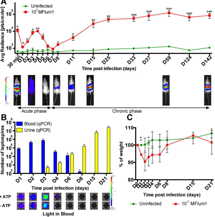
mouse, to avoid death of the mice (table 3). From 1 dpi and until 4 dpi, the bioluminescence increased, corresponding to the dissemination and rapid growth of leptospires in mice, then progressively decreased to disappear at 6 and 7 dpi, corresponding to an apparent clearance of the bacteria (Fig. 2A, lower panel). However, at 8 dpi, the bioluminescence reappeared, restricted to the kidneys and gradually increased for 1 or 2 weeks to reach a steady state (Fig. 2A). This renal chronic colonization was monitored for more than 14 months with no decrease of the bioluminescence (Fig. 2A and Fig. S2 in S1 Text). Similar observations were obtained with Balb/c mice, showing that the kinetics of leptospires dissemination and renal colonization were not unique to the C57BL6/J mice (Fig. S3 in S1 Text). Further characterization of theL. interrogans-infected albino C57BL6/J
model was performed. First, the presence of leptospires in the urine and blood was monitored throughout the first weeks post infection (Fig. 2A). q-PCR data showed that bacteria were present in the blood of mice from 1 to 8 dpi, with a peak at 3 dpi, then a gradual decrease until 8 dpi, after which no further bacteria were detected (Fig. 2B). Ex vivo light imaging of the blood samples showed that live bacteria were present in the bloodstream until 3 dpi, but were not detected afterwards. Interestingly, at 2, 3 and 4 dpi, higher levels of light were measured from blood in the presence of ATP than in the absence, suggesting that dying bacteria, or released luciferase, were present in the blood, and that blood defenses were killing some leptospires at the infectious dose of 107 MFlum1 (Fig. 2B, lower panel). The doubling time of leptospires in blood between 1 and 3 dpi was equivalent by live imaging (13.461.4 h) and q-PCR (11.861.3h), and was longer than the doubling time calculated for the lethal infection, suggesting better efficiency of the blood defense in this sub-lethal infection. The first 2 or 3 dpi were also characterized by weight loss of around 5 to 10%, followed by progressive recovery to the initial weight from 6 dpi to 8 dpi. From 15 dpi (Fig. 2C) to several months post infection (Fig. S4 in S1 Text), infected and uninfected control mice were identical in weights, suggesting an asymptom-atic chronic infection. Consistent with the increasing biolumines-cence observed in the kidneys from the second week of infection, leptospires were detected in increasing numbers in the urine from 3 dpi by q-PCR (Fig. 2B). Together, these data suggest that the course of experimental IP infection with a sublethal dose of L. interrogans is biphasic (Fig. 2A), beginning with a mild acute disease (acute phase) during the first week of infection, character-ized by septicemia associated with transient weight loss, and subsequent clearance of the bacteria from the blood, then followed from the beginning of the second week by a chronic phase corresponding to the growth of L. interrogans in the kidneys, establishment of an asymptomatic renal colonization and excre-tion of leptospires in the urine.
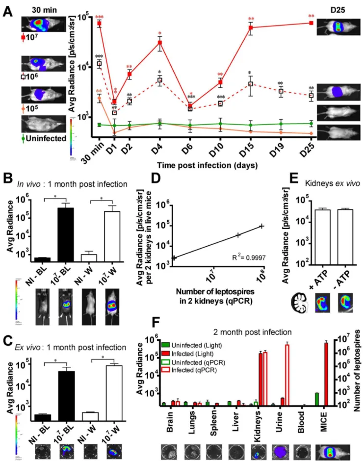
Threshold of leptospiral infective dose leading to renal colonization
We next studied the kinetics of infection in albino C57BL/6J with different doses of MFlum1 from 105 to 107 bacteria per
mouse. The kinetics of bioluminescence were similar using a dose of 106or 107leptospiresperinjection, and the kidneys of all mice were colonized (Fig. 3A). However, at a dose of 106, the extent of the bioluminescence was reduced both at the acute phase of dissemination and at the chronic phase compared to the dose of 107(Fig. 3A). At the lower dose of 105, no growth of bacteria or renal colonization occurred. These results suggest that the blood immune response is able to control a certain degree of L. interrogansinfection, with efficient clearance of the bacteria in the first dpi, following a dose of 105 or below. However, at higher infecting doses, although most of the bacteria were cleared at 6 dpi,L. interroganssucceeded in colonizing the kidneys. Hence, the threshold of the IP infection dose associated with renal
Table 3.Survival test of C57BL/6J mice to the infection withL. interrogansManilae and its mutant MFlum1.
Infected with WT strain Infected with MFlum1
26107 2
6108 2
6107 2
6108
4 days post infection 4/4 dead 4/4 dead 0/4 dead 4/4 dead
10 days post infection – – 1/4 dead –
Figure 2. Kinetics of dissemination of bioluminescent MFlum1 in mice. All the bioluminescence analyses were performed after IP administration or addition of D-luciferin. (A) Live imaging tracking of 107MFlum1 IP injected to albino C57BL/6J mice. Images below the graph show
the tracking of one infected mouse, photographed at different crucial time points. Data are expressed as the mean6SEM of average radiance of light measured in photons/second/cm2in n = 4 infected mice, imaged in the dorsal view, except for 30 min post-infection for which only imaging in the ventral view allows the visualization of the leptospiral dissemination in the peritoneal cavity.pvalues (*p,0.05, **p,0.01, ***p,0.001) between infected and uninfected groups. Images depict photographs overlaid with color representations of luminescence intensity, measured in photons/ second/cm2as indicated on the scales, where red is the most intense and purple the least intense. (B) Kinetics of leptospiral quantification by q-PCR
in blood and urine of albino C57BL/6J mice infected with 107MFlum1. Below are shown corresponding images of theex vivolive imaging of MFLum1 in blood, in the presence or absence of ATP. (C) Monitoring expressed as the percentage of weight loss of mice infected or not with 107MFlum1.
Panels B and C are representative of 2 independent experiments with a total of n = 8 mice for each group.pvalues (*p,0.05, **p,0.01) between infected and uninfected groups.
Figure 3. Threshold of infective dose of MFlum1 to obtain renal colonization.(A) Comparison of the dissemination kinetics for MFlum1 injected to albino C57BL/6J at different doses. Data are expressed as the mean6SEM of average radiance of light measured in photons/second/cm2
colonization was 106 bacteria per mouse, and interestingly, the extent of renal colonization was proportional to the infecting dose (Fig. 3A).
Infected black C57BL/6J mice can be imaged at the chronic phase
In our previous studies of innate immune recognition of leptospires, we used transgenic mice in the C57BL/6J back-ground. These mice have black fur, which prevents the measurement of emitted light. To investigate whether we could use the bioluminescentLeptospirastrains in these mice, black and albino C57BL/6J mice were infected with 107 MFlum1. One month post infection, the black mice were shaved dorsally before imaging. The levels of bioluminescence in live albino and in shaved black C57BL/6J mice were equivalent (Fig. 3B). To be sure that the infected organs were the kidneys, mice were sacrificed and kidneys removed, then transferred to a D-luciferin solution and imaged. Bioluminescence was indeed observed in the kidneys, and was equivalent in white and black mice (Fig. 3C). These results show that it is feasible to use black mice to monitor chronic renal leptospirosis using bioluminescentL. interrogans.
Bioluminescence measured during the chronic phase is proportional to bacterial load
We next wanted to investigate whether the bioluminescence measured in live mice during the chronic phase in the kidneys reflected the bacterial load. At 25 dpi, MFlum1-infected mice were imaged gating on the kidneys, then mice were sacrificed and both kidneys used for quantification of L. interrogans by q-PCR (Fig. 3D). The results showed that the number of bacteria in kidneys measured by q-PCR was strictly proportional to the emitted light in live mice. However, the emitted light was surprisingly low compared to the number of leptospires inferred by q-PCR, suggesting either that a high number of leptospires were not bioluminescent or that light emission was impaired.Ex vivoimaging of longitudinally sectioned half-kidneys, in the presence or absence of ATP, showed no difference in light (Fig. 3E), suggesting that the leptospires present in the kidneys were healthy and not in the process of being degraded (Fig. 3E, lower panel). Moreover, imaging suggested a cortical localization of bacteria in the kidney (Fig. 3E, lower panel), consistent with the known localization of leptospires in the proximal tubules of kidneys.
Kidneys are the reservoir of leptospires at the chronic phase
Because of the lower sensitivity of live imaging compared to q-PCR data in the kidneys, we wondered if, at the chronic phase,L. interrogans were restricted to kidneys as suggested by the light
images of whole mice (Fig. 3B). MFlum1 infected mice were sacrificed 2 months post infection and their organs, blood and urine collected to analyzeex vivothe presence of leptospires.Ex vivoimaging of brain, lungs, liver, spleen and blood did not reveal any bioluminescence (Fig. 3F), suggesting a low amount or absence of leptospires in these organs. The lack of bacteria in these organs and blood was further confirmed by q-PCR data (Fig. 3F). By opposition,L. interroganswere detected in infected kidneys and urine, both by light imaging and q-PCR, as expected (Fig. 3F). Altogether, these data strongly suggest that kidneys constitute the only reservoir of leptospires at the chronic phase of infection in mice.
Antibiotics administered early post-infection are efficient againstL. interrogans
We next used the bioluminescentL. interrogansto test treatments able to combat leptospirosis. First, we focused on antibiotics administered during the acute phase of infection. Penicillin G was used because it is the treatment of choice for acute leptospirosis in humans, and was administered once a day through the intraper-itoneal route for 5 consecutive days to mice already infected with 107MFlum1. Mice were treated with the antibiotic according to two protocols, either from 1 to 5 dpi, to target the dissemination phase, or from 3 until 7 dpi to let the bacteria disseminate before treatment, as performed previously [15]. Mice were imaged daily during the acute phase, and at different time points during the chronic phase. Mice were sacrificed at 25 dpi and the kidneys imaged and urine checked by q-PCR for the presence of leptospires. Administration of penicillin G from 1 dpi abrogated the biolumi-nescence in live infected mice, and no recurrence of light occurred later in the kidneys (Fig. 4A). This suggested that the bacteria had been cleared, which was confirmed as leptospires were not detected by q-PCR in the urine at 25 dpi (Fig. 4B). When administered from day 3 p.i., although the bioluminescence observed at the chronic phase was close to the background, some bioluminescence was still detected at 8 dpi. (Fig. 4A), suggesting live bacteria were present in the kidneys. Moreover, after dissection at 25 dpi, although no bioluminescence was detected in the kidneys, low levels of leptospires were detected in the urine by q-PCR (Fig. 4C). These results suggest that a few bacteria reached the kidneys before 3 dpi and escaped the D3–D5 penicillin treatment resulting in a sub detectable colonization of the kidney.
We wondered whether ciprofloxacin, a quinolone often used in urinary tract infections could be more active than penicillin to eradicate leptospires. Bioluminescence tracking showed that cipro-floxacin was active to avoid renal colonization by leptospires when administered from 1 to 5 dpi (Fig. 4D). However, the D1–D5 ciprofloxacin treatment was less active than the one with penicillin a control of infection and on the left side, images of one representative mouse at the end of the kinetics.pvalues (*p,0.05, **p,0.01, ***p,0.001) are indicated in corresponding colors for each groupversusthe uninfected group.In vivo(B) andex vivo(C) live imaging and quantification of albino (W) and black (BL) C57BL/6J mice one month post infection with (107) or without (NI) 107MFlum1. Bioluminescence imaging in dorsal view was carried out after dorsal shaving of the black mice. Below are shown corresponding images of live mice gated on the kidneys (B) andex vivo(C) tracking of half-kidneys after sacrifice and addition of D-luciferin.pvalue (*p,0.05) between infected and uninfected groups n = 4 micepergroup. (D) Correlation between renal imaging and q-PCR. Bioluminescence imaging at 25 dpi in dorsal view of live chronically infected C57BL/6J mice (from 106
or 107experiments), gated on the kidneys. Subsequently, mice were euthanized and kidneys were harvested and further processed for determination
of the leptospiral load by q-PCR. Each cross represents an individual mouse. (E)Ex vivoimaging in presence or absence of ATP of half-kidneys from 107MFlum1 infected mice one month post infection. Data are expressed as the mean
6SEM of average radiance of light measured in photons/ second/cm2for 6 half-kidneys for each group. Below are shown corresponding images of the tracking of half-kidney after sacrifice and addition of D-luciferin. On the left is shown the schematic representation of an infected kidney in longitudinal cross section. (F)Ex vivolive imaging (Left Y axis) and the corresponding number of leptospires measured by q-PCR (Right Y axis) of different organs from 107MFlum1 infected mice, one month post infection. Data are expressed as the mean6SEM of average radiance of light measured in photons/second/cm2. This panel represents 2 experiments
with a total of n = 6 mice infected with MFlum1, and n = 5 naı¨ve mice. Below are shown corresponding images of the live imaging of one representative organ or fluid and a mouse before the sacrifice. For imaging, urine was pooled from several mice, to obtain a minimum volume of 50mL.
G, since some dissemination and growing of the bacteria still occurred at the acute phase (Fig. 4D), and despite undetectable bioluminescence in the kidneys at 25 dpi, some bacteria were detected in the urines. The ciprofloxacin administered from 3 dpi was not able to block the renal colonization (Fig. 4D). Imaging of dissected kidneys and urine screening for the presence of L. interrogans confirmed these results (Figs. 4E and 4F). Together, these findings showed the feasibility of using bioluminescent L. interrogans strains to monitor the effects of drugs against
leptospirosis, and suggest that penicillin treatment, better than ciprofloxacin, is efficient to eradicate leptospires if administered in the very first days after infection.
Antibiotics administered during the chronic stage are not efficient to eradicate leptospires
To better understand whether the antibiotics could kill leptospires once they are settled in the kidneys, we next monitored the effects of both penicillin G and ciprofloxacin treatments
Figure 4. Effects of antibiotic treatments at the acute phase of leptospirosis.C57BL/6J mice were infected with 107MFlum1 (n = 4) and injected IP daily for 5 consecutive days with penicillin G (Pen) or ciprofloxacin (Cipr) from 1 until 5 dpi (D1 to D5) or from 3 until 7 dpi (D3 to D7). Thereafter, mice were sacrificed at 25 dpi. Comparison of the dissemination kinetics for 107MFlum1 injected in albino C57BL/6J mice, treated or not with penicillin G (A) or ciprofloxacin (D). Data are expressed as the mean6SEM of average radiance of light measured in photons/second/cm2in
n = 4 for each group.pvalues (+p,0.05,++p,0.01,+++p,0.001) are indicated in corresponding colors for each groupversusthe uninfected group. Panels (B) and (E) are corresponding bioluminescence images of half kidneys from mice sacrificed 25 dpi, from experiments depicted in panels (A) and (D), respectively. The arrows indicate the duration of the different treatments. Data are expressed as the mean6SEM of average radiance of light measured in photons/second/cm2gated on two half kidneys in n = 4 infected mice.pvalues (
+p,0.05,++p,0.01,+++p,0.001) are indicated in corresponding colors for each groupversusthe uninfected group. Above are shown corresponding images of theex vivotracking of corresponding half-kidneys after sacrifice and addition of D-luciferin. Images depict photographs overlaid with color representations of luminescence intensity, measured in photons/second/cm2and indicated on the scales, where red is most intense and purple is least intense. Panels (C) and (F) correspond to
leptospiral load in urine at 25 dpi, measured by q-PCR, from experiments depicted in panel (A) and (D), respectively. Data are expressed as the mean number of leptospires in 100ml of urine6SEM for n = 4 infected or uninfected mice.pvalues (*p,0.05, **p,0.01, ***p,0.001) are indicated in corresponding colors for each groupversusthe uninfected group.
administered during the chronic phase of infection. Mice infected for 25 days with 107 MFlum1, harboring the leptospires in the kidneys, were treated once a day for 7 consecutive days with the antibiotics. Interestingly, initial bioluminescence levels dropped rapidly, stabilizing on the second day of treatment at a steady state, corresponding to an approximately 10-fold reduction in biolumi-nescence compared to non-treated, infected control mice. Two days after the end of the treatment, bioluminescence began to recover in the kidneys, and was fully restored by 2 weeks post treatment (Fig. 5A). These results suggest that only a fraction of theL. interroganspopulation present in their niche, presumably the renal proximal tubule, was successfully targeted by the antibiotics, and that the remainingL. interroganswere protected and fully recolonized their niche once the treatment was terminated.
We next tested azithromycin, a macrolide antibiotic, very efficient in combating Gram-negative bacteria andL. interrogans
[24], with a very good tissue distribution [25]. Mice chronically infected with 107 MFlum1 for one month were treated for 7 consecutive days with azithromycin. The bioluminescence de-creased rapidly to background levels after 6 days of treatment, and was still not detectable 2 days after the end of the treatment, confirming the better efficacy of azithromycin compared to penicillin and ciprofloxacin to target leptospires in the kidneys (Fig. 5B). However, 6 days post treatment, the bioluminescence reappeared in the kidneys, reaching a steady state one month post treatment although at a lower level than was observed prior to treatment (Fig. 5B). To determine whether leptospires that reappeared were persistent bacteria that escaped the antibiotic, or were bacteria resistant to azithromycin, we waited for 2 months then retreated the mice for 7 days with the same concentration of azithromycin. The bioluminescence dropped rapidly to a level slightly above background, and continued to decline reaching levels close but significantly different from the background one week after the end of the treatment. However, 2 weeks post treatment, the bioluminescence again reappeared in the kidneys, at a low steady level (Fig. 5B). This second treatment was active suggesting that the bacteria did not acquire resistance to the azithromycin. These results suggest that each treatment with azithromycin succeeded to sterilize the whole leptospiral popula-tion of a relatively important number of tubules. However, in a few tubules, only a portion of the population was eliminated, and persistent bacteria were able to survive the azithromycin treatment, and refill their niche once the treatment was over.
A single injection of azithromycin protects against acute and chronic leptospirosis
Because azithromycin showed the best efficiency against L. interrogans, and has an extended half-life of more than 48 h [25], we wondered whether it could be used as a prophylactic treatment against leptospirosis. To test its efficiency against acute leptospi-rosis, naı¨ve albino C57BL/6J mice were treated with a single dose of azithromycin 2 days before infection with a lethal dose of 26108
MFlum1. Data showed that from 1 dpi, the bioluminescence increased in the infected control mice, but decreased to background levels in the azithromycin treated mice. Control infected mice suffering from acute leptospirosis had to be sacrificed at 3 dpi whereas the azithromycin pre-treated mice did not show any clinical signs of acute leptospirosis (Fig. 6). The same pre-treatment was also administered to Myd88 ko mice infected with 26108MFlum1. We previously showed that Myd88 ko mice are very sensitive to, and die from, acute leptospirosis because of a lack of TLR4 recognition [8]. The azithromycin pretreated Myd88 ko mice were protected from death induced by the lethal dose of
MFlum1, showing that sensitive hosts can also be protected by prophylactic azithromycin treatment. Importantly, 3 months after the infection, bioluminescence was not observed in the kidneys of treated mice (Fig. S5 in S1 Text). As a control, urine of all infected mice was checked by q-PCR. Urines of mice pre-treated with azithromycin were free of leptospires showing that the azithro-mycin treatment efficiently counteracted the lethal dose of L. interrogansin both resistant and susceptible hosts. These findings suggest that administration of a single dose of azithromycin can be very useful as a prophylaxis treatment against acute leptospirosis that also protects against renal colonization byL. interrogans. In comparison, an equivalent prophylactic antibiotic treatment performed with doxycycline, at the relative dose used in prophylaxis against human leptospirosis [26], was not successful to protect mice from theL. interrogans-induced death and renal colonization, since 2 out of 4 mice died from acute leptospirosis, and the 2 surviving mice harbored renal colonization (Fig. S6 in S1 Text).
Discussion
In this study, bioluminescent strains of pathogenic L. inter-rogansserovar Manilae were constructed and successfully usedin vivoto determine and characterize the kinetics of dissemination of
L. interrogansin mice. This new model proved to reproduce both acute and chronic leptospirosis and was further used to test the efficacy of antibiotics.
Pathogenic Leptospira strains are poorly transformable. Tar-geted mutagenesis in pathogenic leptospires is thus very scarce, and libraries of transposed mutants of L. interrogans are still incomplete [27]. In this study, transformation of L. interrogans
serovar Manilae, with the transposon carrying the luciferase cassette driven by the strong promoterflgBp, resulted in several random mutants, the majority of which exhibited insertions in coding sequences. For example, we obtained a mutant in the gene encoding a collagenase, which was recently shown as a virulence factor [28]. Further analysis of these mutants, or of others obtained with the strong promoter (groESp) (Table S1 in S1 Text), may increase our understanding of virulence factors inL. interrogans, includingL. interrogansserovar Copenhageni strain Fiocruz, for which we recently obtained bioluminescent transformants.
We used the intergenic transformant MFlum1 to study infection
in vivo. MFlum1 retained its virulence, suggesting that the biological cost of expression of the luciferase cassette had only a minor impact on its fitness. The bioluminescence correlated with the number of leptospires in the exponential phase of growth. The limit of detection of live imaging was around 103 bacteria/ml, corresponding to the detection of 100 MFlum1 in 100ml. This level of detection was higher than the luminescence detection limit, previously found with a luxCDABE transformant (around 104 leptospires) [13]. Compared to the luciferase expressed from the
luxCDABEoperon, the increased light emission of firefly luciferase in the presence of the exogenous D-luciferin substrate probably contributes to the ability of our bioluminescent strains to be imaged in live mice.
during the first 2 weeks post infection, although they were alive in the kidney and found by q-PCR in urine. It seems unlikely that the leptospires were dead in urine and most probably several factors
known to impact the luciferase activity may have contributed to quenching of light emission, such as isoflurane anesthesia, which inhibits luciferase activity [29], or the pH or oxygen levels [30],
Figure 5. Effect of antibiotic treatments at the chronic phase of leptospirosis.C57BL/6J mice chronically infected for 25 days (D25) with 107
MFlum1 were (A) IP injected daily for 7 days with penicillin G (Pen) (brown) or Ciprofloxacin (Cipr) (blue) from 25 until 31 dpi (T1 to T7), (B) injected daily for 7 days with azithromycin (Azi) from 25 until 31 dpi, and from 112 until 118 dpi (T1 to T7). The arrows indicate the duration of the different treatments. All the bioluminescence analyses were done after IP administration of D-luciferin. Data are expressed as the mean6SEM of average radiance of light measured in photons/second/cm2in n = 4 infected treated or untreated mice.pvalues (+p,0.05,++p,0.01,+++p,0.001) are
indicated in corresponding colors for each groupversusthe uninfected group. Below are shown images of the tracking of one untreated, infected (MFlum1) mouse and an infected treated (Azi) mouse photographed at different crucial time points. The correspondingpvalues (*p,0.05) were calculated between infected untreated group and treated group (Azi). Images depict photographs overlaid with color representations of luminescence intensity, measured in photons/second/cm2and indicated on the scales, where red is most intense and purple is least intense. PT, days post treatment.
which may not be optimal or evenly distributed throughout the tubules or in urine.
In opposition to hamsters, gerbils and Guinea pigs which are accurate models for acute human leptospirosis, mice are consid-ered relatively resistant to acute leptospirosis [4]. Indeed, in previous studies, no lethality or severe clinical symptoms were observed in C57BL/6 mice after intraperitoneal infections withL. interrogansserovar Copenhageni strain Fiocruz, even at the high infective dose of 26108bacteria [8,16]. However, we and others
have shown that mice, immunosuppressed or genetically deficient for TLR4 or B cells, proved to be more sensitive, and develop lethal acute leptospirosis, demonstrating the usefulness of these murine models to unravel key components of the innate defense against leptospirosis [5,8,31–33]. Here, C57BL/6 mice infected with high doses of the bioluminescent L. interrogans serovar Manilae, suffered from major septicemia and died within a few days. This model of septicemia induced by a highly virulent L. interrogans is novel and may be very useful to understand the pathophysiological consequences of acute leptospirosis, which in humans has a high case/fatality rate [4,34]. Moreover, we showed the feasibility of this model to test therapeutic strategies interfering with the growth of bacteria in blood, such as penicillin G treatment administered early after infection, which efficiently cleared the infection in mice. In this vein, an important result was the success of the prophylactic treatment that prevented, with only one dose of azithromycin, both acute and chronic leptospirosis in resistant and susceptible Myd88 ko mice, which like humans are susceptible to leptospirosis [8]. It is tempting to hypothesize that this prophylactic treatment could be transposed to humans to avoid potential leptospirosis in different situations, such as outbreaks or athletic events.
At sublethal infective doses, the live imaging revealed a biphasic disease with a self resolving septicemia within the first week, followed by renal colonization, and chronic shedding of leptospires
in the urines, closely matching the leptospiremia and leptospiruria phases classically described leptospirosis [2]. Of note, we showed that during the chronic phase, kidneys constitute the only reservoir of leptospires, since no DNA from leptospires could be amplified by q-PCR from blood nor from other organs known to be the target of leptospires at the acute phase such as the brain, liver, lungs and spleen [2]. Moreover, live imaging allowed us to roughly map the presence of leptospires in the renal cortex of the kidney, which is in accordance with the reported localization of leptospires in the proximal tubules of the nephrons [35–37].
The innate blood defense consists in phagocytes, antimicrobial peptides, natural IgM and complement proteins, that together participate in the clearance of microbes. L. interrogans are known to escape the human complement system by several mechanisms [38,39]. In mice, our study suggests that L. interrogans are well controlled by the blood defense, at an infectious dose of up to 105 bacteria, since at this dose, we observed a fast decrease of live bacteria and no further growth of leptospires. For higher infectious doses of 106 or 107 bacteria, leptospires were able to exponentially grow in the blood in the first 3 dpi, showing that the blood defense was partially ineffective, presumably because it was overwhelmed. In agree-ment with this hypothesis, we showed that the blood defenses inactivated some leptospires in the first days following the infection. Indeed,ex vivo live imaging of blood the presence or absence of ATP, highlighted the presence of metabolically inactive bacteria still harboring or releasing active luciferase. Moreover, the doubling time of MFlum1 in blood was higher in the sub-lethal infection than in the lethal infection, showing that the blood defense more efficiently combated the 107 leptospires than 26108. The peak of bacteremia in blood occurring 3 dpi and strikingly, the subsequent complete clearance of leptospires from the blood, is in accordance with our previous study, which showed that a specific IgM response against leptospiral LPS was
Figure 6. Azithromycin prophylactic treatment against leptospirosis.8 C57BL/6J mice were infected with 26108MFlum1 and 4 of them were
injected IP two days before infection (T-2) with azithromycin (Azi). As controls, 4 uninfected mice were treated with azithromycin. All bioluminescence analyses were performed after IP administration of D-luciferin. Data are expressed as the mean6SEM of average radiance of light measured in photons/second/cm2in n = 4 mice imaged in the dorsal view, except for 30 min post-infection (D0) for which only imaging in the ventral view allows
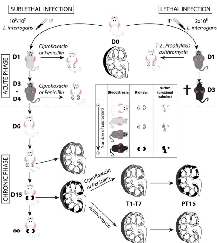
Figure 7. Model of acute and chronic leptospirosis in mice.This figure depicts the course of leptospirosis in mice following an IP infection with a lethal dose of bioluminescent MFlum1, leading to a septicemia or with a sub-lethal dose leading to a chronic leptospirosis, and the effects of different antibiotics administered at the acute (upper part of the figure) or chronic phase (lower part of the figure) of the leptospiral infection. Mice depicted without kidneys represent mice at the acute phase of infection. Mice depicted with kidneys represent mice at the chronic phase. Inside the kidneys schemed in longitudinal cross-section, the niches colonized by leptospires, presumably the proximal part of renal tubules (proximal tubules) are depicted by small circles. A grey color scale indicates the degree of leptospiral infection, where white means free of leptospires and black means a maximum of infection or colonization. The cross indicates that the mice died or were sacrificed because of acute leptospirosis. D1 to D15: days post-infection. T1-T7 duration of antibiotic treatments. PT15: 15 days post treatment. T-2 prophylaxis treatment 2 days prior to post-infection.
already mounted 3 dpi, and was partially protective against leptospires [8].
The fact that antibiotic treatments efficiently blocked renal colonization if administered 1 dpi, but partially failed if admin-istered 3 dpi, together with the fact that leptospires were alive in the blood only during the first 3 dpi, suggests that the window of time for leptospires to escape the blood defense and reach their niche, is restricted to the exponential growth phase, between the first and third dpi. Moreover, the dose response and azithromycin-treatment experiments suggest that the extent of renal colonization by leptospires depends on the initial early access of a few leptospires to a restricted number of niches (Fig. 7). Indeed, a lower dose of infection reduced the burden of stable renal colonization. At the very beginning of the chronic phase, the regular increase of bioluminescence suggested that leptospires were protected from the immune response, which was highly effective since all leptospires disappeared from the circulation. Hence, within 2 to 3 weeks, the leptospires replicated to fill up their niche, reaching a steady level where the shedding and replication rates were in equilibrium. Since no increase in renal colonization was observed over time, even in heavily colonized mice, our results also suggest that there was no cross colonization of new nephrons. Together these results indirectly suggest that the potent IgG response, known to occur in natural or experimental leptospirosis [8,40], efficiently contained the lepto-spires in their niche.
Antibiotic treatments with penicillin or ciprofloxacine were effective againstL. interrogansif administered early after infection. However, administered at the chronic phase, both penicillin and ciprofloxacin failed to eradicate all the leptospires from the tubules. Therefore once the treatment was over, the leptospires regrew in the tubules and the levels of renal colonization 15 days post treatment were identical to those before treatment (Fig. 7). These results are in line with data from the literature, showing that penicillin or other antibiotics proved efficient in patients, only when given early after the onset of leptospirosis [41]. By contrast, azithromycin efficiently cleared the infection in most of the renal tubules, and leptospires persisted in only a few tubules that were subsequently fully recolonized (Fig. 7).
Doxycycline has been used in the past as a weekly chemopro-phylaxis treatment in different situations of outbreaks [42,43], but it did not efficiently eradicate leptospires in prophylaxis treatment in our mouse model of lethal infection. This result is in line with a recent study showing that prophylaxis with a single dose of 200 mg of doxycycline in humans may not be 100% effective to protect against development of acute leptospirosis [26]. Moreover, although doxycycline was previously shown to clear leptospires more efficiently than ampicillin and ofloxacin in hamsters when administered early post infection [44], it did not efficiently clear leptospires from the hamsters kidneys when administered 4 dpi [45]. Together, these data suggest that doxycycline would have been less effective than azithromycin for clearance of leptospires at the chronic phase in mice. However, our results have to be carefully considered since we did not take into account the pharmacokinetic-pharmacodynamic parameters of antibiotics that differ between mice and humans [46]. Indeed, both ciprofloxacin and doxycycline have reduced half-lives in serum of mice compared to humans. Moreover, the daily doses of antibiotics used in our study were in the high range of what is usually administered to patients [41], but mice were injected only once a day, whilst patients usually receive antibiotics twice a day. Therefore, it is still possible that a longer course of azithromycin or adequate doses of ciprofloxacin and doxycycline would have been more efficient in eradicating the leptospires.
Leptospires have been shown to establish biofilmsin vitro[47]. InL. interrogansinfected mice, we observed at the chronic phase, strong labeling of the lumen of tubules with LipL32 antibodies [15], close to the dense colonization of renal tubules by leptospires previously observed in experimentally infected rats [48]. Biofilms are known to confer bacterial resistance to antibiotics [49] and our hypothesis is that the localized resistance of leptospires to antibiotics would be linked to biofilm formation in the proximal renal tubules of mice. Further studies aiming to test new therapeutic treatments, combining antibiotics and drugs to disperse biofilms, should be envisioned to potentiate the renal elimination of leptospires.
Chronic renal carriage of leptospires has recently been shown in mammals other than rodents, such as sea lions [50], cats [51], dogs [52] and most strikingly, in humans [53,54]. The dogma that some rodents, such as rats, are a chronic carrier of the disease, whereas other animals are accidental hosts, may be simplistic, and asymptomatic carriage in humans may have been largely overlooked. Our results should alert about the potential incom-plete elimination of leptospires after antibiotherapy, and the risk of chronic renal carriage of leptospires in both veterinary and human medicine. A recent study showed that most patients that have been hospitalized for acute leptospirosis still exhibited diverse symptoms associated with leptospirosis such as myalgia and headaches, 2 years after treatment [55]. These patients should be investigated through urine testing for potential chronic renal carriage of leptospires. In Taiwan, the incidence of chronic kidney disease is higher than in other parts of Asia, and may be linked to asymptomatic leptospirosis, since almost 10% of hospitalized patients with chronic kidney disease were found seropositive for leptospires, without reported history of the disease [56]. It would also be instructive to test these seropositive patients for urinary shedding of leptospires.
We studied here only the kinetics of infection upon intra-peritoneal injection of leptospires, which may reflect part of the infectious process, but not the initial events of leptospires penetrating the mucosa or the abraded skin. Further study is ongoing to better understand the consequences of more physio-logical routes of infection with pathogenicL. interrogans, without experimental breaches of the skin, such as by depositing bioluminescentL. interrogans on the eye, nose, mouth or skin. The bioluminescent strains will also be useful to study leptospirosis by live imaging in the young guinea pig, which constitutes a good animal model for acute leptospirosis, presenting pulmonary hemorrhages [6], or in the rat, the choice model of chronic leptospirosis [57]. Indeed, both Lewis rats and Hartley guinea pigs are albino and small enough to fit in the IVIS Spectrum machine. In conclusion, this study, together with our previous work revealing mild fibrosis in kidneys from mice chronically infected withL. interrogans[15], definitively demonstrates that the mouse is a good experimental model to study leptospirosis. This work also provides the proof of principle that bioluminescent Leptospira
strains will be useful tools to challenge vaccines, or to test therapeutic treatments. The study of survival, persistence and transmission of leptospires between the environment and rodents can also be envisioned.
Supporting Information
Text S1 Supporting information. Table S1. List of
biolu-minescent groESp-luciferase transformants of L. interrogans
serovar Manilae. Figure S1. Kinetics of bioluminescentL. biflexa
MFlum1 in Balb/c mice. Figure S4. Chronic renal colonization with L. interrogans does not result in weight loss. Figure S5. Controls 3 months post infection of absence of renal colonization in mice prophylactically treated with azithromycin. Figure S6. Doxycycline prophylaxis experiment.
(PDF)
Acknowledgments
We would like to thank Pr Albert I. Ko for stimulating discussions. We thank Maxime Fayon for help in sequencing the mutants. We thank Alain
Vandewalle and Richard Wheeler for critical reading of the manuscript and editing of English.
Author Contributions
Conceived and designed the experiments: CW GR FJV. Performed the experiments: GR FJV MFd XK CW. Analyzed the data: GR MAN IGB CW. Contributed reagents/materials/analysis tools: MP. Wrote the paper: CW GR.
References
1. Bharti AR, Nally JE, Ricaldi JN, Matthias MA, Diaz MM, et al. (2003) Leptospirosis: a zoonotic disease of global importance. Lancet Infect Dis 3: 757– 771.
2. Levett PN (2001) Leptospirosis. Clin Microbiol Rev 14: 296–326.
3. Yang CW, Wu MS, Pan MJ (2001) Leptospirosis renal disease. Nephrol Dial Transplant 16 Suppl 5: 73–77.
4. Ko AI, Goarant C, Picardeau M (2009) Leptospira: the dawn of the molecular genetics era for an emerging zoonotic pathogen. Nat Rev Microbiol 7: 736–747. 5. Pereira MM, Andrade J, Marchevsky RS, Ribeiro dos Santos R (1998) Morphological characterization of lung and kidney lesions in C3H/HeJ mice infected with Leptospira interrogans serovar icterohaemorrhagiae: defect of CD4+and CD8+T-cells are prognosticators of the disease progression. Exp Toxicol Pathol 50: 191–198.
6. Nally JE, Chantranuwat C, Wu XY, Fishbein MC, Pereira MM, et al. (2004) Alveolar septal deposition of immunoglobulin and complement parallels pulmonary hemorrhage in a guinea pig model of severe pulmonary leptospirosis. Am J Pathol 164: 1115–1127.
7. Levett PN, Morey RE, Galloway RL, Turner DE, Steigerwalt AG, et al. (2005) Detection of pathogenic leptospires by real-time quantitative PCR. J Med Microbiol 54: 45–49.
8. Chassin C, Picardeau M, Goujon JM, Bourhy P, Quellard N, et al. (2009) TLR4- and TLR2-mediated B cell responses control the clearance of the bacterial pathogen, Leptospira interrogans. J Immunol 183: 2669–2677. 9. Lourdault K, Aviat F, Picardeau M (2009) Use of quantitative real-time PCR for
studying the dissemination of Leptospira interrogans in the guinea pig infection model of leptospirosis. J Med Microbiol 58: 648–655.
10. Gahan CG (2012) The bacterial lux reporter system: applications in bacterial localisation studies. Curr Gene Ther 12: 12–19.
11. Davis JM, Haake DA, Ramakrishnan L (2009) Leptospira interrogans stably infects zebrafish embryos, altering phagocyte behavior and homing to specific tissues. PLoS Negl Trop Dis 3: e463.
12. Aviat F, Slamti L, Cerqueira GM, Lourdault K, Picardeau M (2010) Expanding the genetic toolbox for Leptospira species by generation of fluorescent bacteria. Appl Environ Microbiol 76: 8135–8142.
13. Murray GL, King AM, Srikram A, Sermswan RW, Adler B (2010) Use of luminescent Leptospira interrogans for enumeration in biological assays. J Clin Microbiol 48: 2037–2042.
14. Hyde JA, Weening EH, Chang M, Trzeciakowski JP, Hook M, et al. (2011) Bioluminescent imaging of Borrelia burgdorferi in vivo demonstrates that the fibronectin-binding protein BBK32 is required for optimal infectivity. Mol Microbiol 82: 99–113.
15. Fanton d’Andon M, Quellard N, Fernandez B, Ratet G, Lacroix-Lamande S, et al. (2014) Leptospira Interrogans Induces Fibrosis in the Mouse Kidney through Inos-Dependent, TLR- and NLR-Independent Signaling Pathways. PLoS Negl Trop Dis 8: e2664.
16. Ferrer MF, Scharrig E, Alberdi L, Cedola M, Pretre G, et al. (2014) Decay-accelerating factor 1 deficiency exacerbates leptospiral-induced murine chronic nephritis and renal fibrosis. PLoS One 9: e102860.
17. Lacroix-Lamande S, d’Andon MF, Michel E, Ratet G, Philpott DJ, et al. (2012) Downregulation of the Na/K-ATPase pump by leptospiral glycolipoprotein activates the NLRP3 inflammasome. J Immunol 188: 2805–2814.
18. Veyrier FJ, Boneca IG, Cellier MF, Taha MK (2011) A novel metal transporter mediating manganese export (MntX) regulates the Mn to Fe intracellular ratio and Neisseria meningitidis virulence. PLoS Pathog 7: e1002261.
19. Picardeau M (2008) Conjugative transfer between Escherichia coli and Leptospira spp. as a new genetic tool. Appl Environ Microbiol 74: 319–322. 20. Bourhy P, Louvel H, Saint Girons I, Picardeau M (2005) Random insertional
mutagenesis of Leptospira interrogans, the agent of leptospirosis, using a mariner transposon. J Bacteriol 187: 3255–3258.
21. Demarre G, Guerout AM, Matsumoto-Mashimo C, Rowe-Magnus DA, Marliere P, et al. (2005) A new family of mobilizable suicide plasmids based on broad host range R388 plasmid (IncW) and RP4 plasmid (IncPalpha) conjugative machineries and their cognate Escherichia coli host strains. Res Microbiol 156: 245–255.
22. Slamti L, Picardeau M (2012) Construction of a library of random mutants in the spirochete Leptospira biflexa using a mariner transposon. Methods Mol Biol 859: 169–176.
23. Que-Gewirth NL, Ribeiro AA, Kalb SR, Cotter RJ, Bulach DM, et al. (2004) A methylated phosphate group and four amide-linked acyl chains in leptospira interrogans lipid A. The membrane anchor of an unusual lipopolysaccharide that activates TLR2. J Biol Chem 279: 25420–25429.
24. Hospenthal DR, Murray CK (2003) In vitro susceptibilities of seven Leptospira species to traditional and newer antibiotics. Antimicrob Agents Chemother 47: 2646–2648.
25. Girard AE, Girard D, English AR, Gootz TD, Cimochowski CR, et al. (1987) Pharmacokinetic and in vivo studies with azithromycin (CP-62,993), a new macrolide with an extended half-life and excellent tissue distribution. Antimicrob Agents Chemother 31: 1948–1954.
26. Chusri S, McNeil EB, Hortiwakul T, Charernmak B, Sritrairatchai S, et al. (2014) Single dosage of doxycycline for prophylaxis against leptospiral infection and leptospirosis during urban flooding in southern Thailand: A non-randomized controlled trial. J Infect Chemother doi: 10.1016/j.jiac.2014. 07.016. [Epub ahead of print]
27. Murray GL, Morel V, Cerqueira GM, Croda J, Srikram A, et al. (2009) Genome-wide transposon mutagenesis in pathogenic Leptospira species. Infect Immun 77: 810–816.
28. Kassegne K, Hu W, Ojcius DM, Sun D, Ge Y, et al. (2014) Identification of collagenase as a critical virulence factor for invasiveness and transmission of pathogenic Leptospira species. J Infect Dis 209: 1105–1115.
29. Keyaerts M, Remory I, Caveliers V, Breckpot K, Bos TJ, et al. (2012) Inhibition of firefly luciferase by general anesthetics: effect on in vitro and in vivo bioluminescence imaging. PLoS One 7: e30061.
30. Khalil AA, Jameson MJ, Broaddus WC, Lin PS, Dever SM, et al. (2013) The Influence of Hypoxia and pH on Bioluminescence Imaging of Luciferase-Transfected Tumor Cells and Xenografts. Int J Mol Imaging 2013: 287697. 31. Adler B, Faine S (1977) Host immunological mechanisms in the resistance of
mice to leptospiral infections. Infect Immun 17: 67–72.
32. Viriyakosol S, Matthias MA, Swancutt MA, Kirkland TN, Vinetz JM (2006) Toll-like receptor 4 protects against lethal Leptospira interrogans serovar icterohaemorrhagiae infection and contributes to in vivo control of leptospiral burden. Infect Immun 74: 887–895.
33. Nally JE, Fishbein MC, Blanco DR, Lovett MA (2005) Lethal infection of C3H/ HeJ and C3H/SCID mice with an isolate of Leptospira interrogans serovar copenhageni. Infect Immun 73: 7014–7017.
34. Yang HY, Yen TH, Lin CY, Chen YC, Pan MJ, et al. (2012) Early identification of leptospirosis as an ignored cause of multiple organ dysfunction syndrome. Shock 38: 24–29.
35. Marshall RB (1976) The route of entry of leptospires into the kidney tubule. J Med Microbiol 9: 149–152.
36. Barnett JK, Barnett D, Bolin CA, Summers TA, Wagar EA, et al. (1999) Expression and distribution of leptospiral outer membrane components during renal infection of hamsters. Infect Immun 67: 853–861.
37. Sitprija V, Pipatanagul V, Mertowidjojo K, Boonpucknavig V, Boonpucknavig S (1980) Pathogenesis of renal disease in leptospirosis: Clinical and experimental studies. Kidney Int 17: 827–836.
38. Castiblanco-Valencia MM, Fraga TR, Silva LB, Monaris D, Abreu PA, et al. (2012) Leptospiral immunoglobulin-like proteins interact with human comple-ment regulators factor H, FHL-1, FHR-1, and C4BP. J Infect Dis 205: 995– 1004.
39. Wolff DG, Castiblanco-Valencia MM, Abe CM, Monaris D, Morais ZM, et al. (2013) Interaction of leptospira elongation factor tu with plasminogen and complement factor h: a metabolic leptospiral protein with moonlighting activities. PLoS One 8: e81818.
40. Adler B, de la Pena Moctezuma A (2010) Leptospira and leptospirosis. Vet Microbiol 140: 287–296.
41. Daher EF, Silva GB, Jr., de Abreu KL, Mota RM, Batista DV, et al. (2012) Leptospirosis-associated acute kidney injury: penicillin at the late stage is still controversial. J Clin Pharm Ther 37: 420–425.
42. Dechet AM, Parsons M, Rambaran M, Mohamed-Rambaran P, Florendo-Cumbermack A, et al. (2012) Leptospirosis outbreak following severe flooding: a rapid assessment and mass prophylaxis campaign; Guyana, January–February 2005. PLoS One 7: e39672.