online | memorias.ioc.fiocruz.br
Molecular characterisation and disease severity
of leptospirosis in Sri Lanka
Kanchana Kumari Bandara1,2, Manjula Weerasekera1/+, Chinthika P Gunasekara1, Nilantha Ranasinghe3, Chamil Marasinghe4, Neluka Fernando1
1Department of Microbiology 4Department of Medicine, Faculty of Medical Sciences, University of Sri Jayewardenepura, Sri Lanka 2Department of Basic Sciences, Faculty of Allied Health Sciences, General Sir John Kotelawala Defense University, Sri Lanka
3Base Hospital, Tangalle, Sri Lanka
Leptospirosis is a re-emerging zoonotic disease all over the world, important in tropical and subtropical areas. A majority of leptospirosis infected patients present as subclinical or mild disease while 5-10% may develop severe infec-tion requiring hospitalisainfec-tion and critical care. It is possible that several factors, such as the infecting serovar, level of leptospiraemia, host genetic factors and host immune response, may be important in predisposition towards severe disease. Different Leptospira strains circulate in different geographical regions contributing to variable disease sever-ity. Therefore, it is important to investigate the circulating strains at geographical locations during each outbreak for epidemiological studies and to support the clinical management of the patients. In this study immunochromatography, microscopic agglutination test and polymerase chain reaction were used to diagnose leptospirosis. Further restriction fragment length polymorphism and DNA sequencing methods were used to identify the circulating strains in two se-lected geographical regions of Sri Lanka. Leptospira interrogans, Leptospira borgpetersenii and Leptospira kirschneri
strains were identified to be circulating in western and southern provinces. L. interrogans was the predominant species circulating in western and southern provinces in 2013 and its presence was mainly associated with renal failure.
Key words: Leptospira - molecular characterisation - Sri Lanka
Leptospirosis is an endemic, zoonotic disease of public health importance in Sri Lanka (Victoriano et al. 2009). Seasonal outbreaks of leptospirosis occur annually and in 2013, 4,276 cases were reported to the Epidemiologi-cal Unit of Sri Lanka. Since Sri Lanka is predominately an agricultural country with a heavy rain fall, exposure to Leptospira is a major occupational hazard (Brenner et al. 1999). Leptospira interrogans, Leptospira santa-rosai, Leptospira kirschneri, Leptospira borgpetersenii
and Leptospira weilli have been reported from several geographical locations in Sri Lanka at different time pe-riods with varying disease severity (Brenner et al. 1999, Agampodi et al. 2012, 2014, Nwafor-Okoli et al. 2012).
Due to the highly endemic nature and associated morbidity and mortality of this disease, it is important to investigate the circulating strains at geographical loca-tions during each outbreak for epidemiological studies and to support the clinical management of the patients.
SUBJECTS, MATERIALS AND METHODS
This was a prospective hospital based study in west-ern and southwest-ern provinces in Sri Lanka between Janu-ary 2013-JanuJanu-ary 2014. All the patients more than 18 years of age, presenting with clinically suspected
lep-doi: 10.1590/0074-02760150070
Financial support: World Class University Project (PhD/01/2012), University of Sri Jayewardenepura
+ Corresponding author: [email protected] Received 18 February 2015
Accepted 19 May 2015
tospirosis according to the World Health Organization (WHO) guideline admitted to the medical wards were included in the study.
Informed consent was obtained from all suspected patients and sociodemographic data and risk factors were gathered using a pre-tested interviewer adminis-tered questionnaire. A venous blood sample of 5 mL was collected following standard procedures and aliquoted into a plain tube for serum separation and the rest added to an ethylenediamine tetraacetic acid (EDTA) tube for DNA extraction. All samples were transported at 4ºC to the Department of Microbiology, University of Sri Jayewardenepura, Sri Lanka.
IgM immunochromatographic assay and microscop-ic agglutination test (MAT) - Leptospira infection was presumptively diagnosed by detecting Leptospira spe-cific IgM using a rapid immunochromatographic assay kit (Leptocheck WB; Zephyr Biomedicals, India) fol-lowing the manufacturer’s instructions. MAT was done in order to obtain single MAT antibody titres using the genus specific Leptospira biflexa serovar Patoc 1 strain
(Medical Research Institute, Sri Lanka) and ≥ 400 titre
was considered as positive for MAT (WHO 2010).
DNA extraction - EDTA blood samples (200 µL) were used for Leptospira DNA extraction using QIAamp DNA blood mini kit (Qiagen GmbH, Germany) according to the manufacturer’s instructions. Eluted DNA was quantified and purity was checked using Nanodrop 2000/200C spec-trophotometer (Thermo Fisher Scientific, USA).
Natara-jaseenivasan et al. 2012). Amplification of isolated DNA was carried out in 50 µL volume with 0.5 µL template DNA, 5 µL 5X green GoTaq® Flexi buffer (pH 8.5)
(Pro-mega, USA), 2 mM MgCl2 (Promega),0.1 µM of each primer (F1-TCTCACCGTTCTCTAAAGTTCAAC, R1- CTGAATTCGGTTTCATATTTGCC), 0.4 mM deoxy nucleotide triphosphate (dNTP) mix (Promega) and 0.25 units of Taq DNA polymerase (Promega). L. interrogans
DNA was used as a positive control and a negative control without the template DNA were included in each PCR as-say. PCR amplification was initiated at 94ºC for 5 min followed by 45 cycles of 94ºC for 1 min, 56ºC for 1 min, 72ºC for 90 s and a final elongation step at 72ºC for 10 min with final hold at 4ºC. The resulting amplicon was 793 bp and these were stored at 4ºC until further analysis.
Restriction fragment length polymorphism (RFLP) - PCR products of flaB PCR positive patient samples were used for RFLP digestion using Hae III and Hind III re-striction enzymes (Kawabata et al. 2001). The rere-striction digestion was carried out in 20 µL of volume in a sterile microcentrifuge tube. The reaction mixture contained 10 µL of PCR product, 2 µL of 10 X RE buffer (MulticoreTM
buffer, Promega), 0.5 µL restriction enzyme (10 U/µL), 0.2 µL of acetylated bovine serum albumin (10 µg/µL) and distilled water to a final volume of 20 µL. The reac-tion mixture was incubated in an incubator at 37ºC for 5 h. The final product was subjected to electrophoresis us-ing 2% agarose gel in tris-acetate-EDTA buffer contain-ing 5 µg/mL ethidium bromide (Sigma Aldrich). Each digested PCR product was mixed with 1/5 volume of the gel loading buffer (Promega) and loaded into the agarose gel. Electrophoresis was carried out at room temperature for one and half hours. At the end of the electrophoresis the gel was visualised under ultraviolet transillumina-tor (Biometra GmbH, Germany). RFLP was done with three reference serovars: L. interrogans serovar Canicola, Icterohaemorrhagiae and Pyrogenes. An undigested PCR product, where the reaction mix was prepared without Hind 111, Hae 111 restriction enzymes, was used as a con-trol (Figs 1, 2, Lane 2).
Nested PCR - A single tube nested PCR was used to amplify 16S rDNA gene specific for pathogenic and intermediate Leptospira species. Amplification was carried out using PCR primers: rrs-outer F (51
-CTCA-GAACTAACGCTGGCGGCGCG-31), rrs-outer-R (51
-GGTTCGTTACTGAGGGTTAAAACCCCC-31),
rrs-inner-F (51-CTGGCGGCGCG T CTTA-31), rrs-inner-R
(51-GTTTTCACACCTGACTTACA-31) (Boonsilp et al.
2011). PCR master mix consisting of 0.5 µL template DNA, 5 µL 5X green GoTaq® Flexi buffer (pH 8.5)
(Pro-mega), 4 mM MgCl2 (Promega),0.2 pmol of each outer primer, 1.2 pmol of inner F, 5 pmol of inner R, 0.2 mM dNTP mix (Promega) and 0.25 units of Taq DNA poly-merase (Promega) were used in a total volume of 25 µL. PCR reaction was carried out using a thermal cycler (Techne Flexigene, UK) with an initial denaturation at 95ºC for 2 min followed by 40 cycles of 95ºC for 10 s, 67ºC for 15 s, 72ºC for 30 s, another 40 cycles of 95ºC for 10 s, 55ºC for 15 s, 72ºC for 30 s and a final elongation
Fig. 1: hae 111 digestion of Leptospira. Lane 1: 100 bp DNA marker; 2: undigested polymerase chain reaction (PCR) product; 3: Leptospira interrogans serovar Canicola (100 bp, 300 bp, 400 bp); 4: L. inter-rogans serovar Icterohaemorrhagiae (100 bp, 200 bp, 300 bp); 5: L. interrogans serovar Pyrogenes (100 bp, 300 bp, 400 bp); 6; Leptospira biflexa Patoc 1 strain; 7-13: flaB PCR positive patient samples.
step at 72ºC for 10 min. The resulting amplicon size was a 547 bp. Amplicons were visualised by gel electropho-resis using an 1.5% agarose gel. L. interrogans Serovar Canicola and Leptostpira fainei BUT 6 strain were taken as positive controls and L. biflexa Patoc 1 strain and no template control were used as the negative controls.
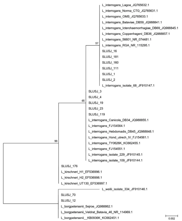
PCR products were purified using a PCR product purification kit (Promega) according to manufacturer’s protocol and sequenced bidirectionally at Macrogen Inc (South Korea). DNA sequences were obtained using 3.1 Big Dye chemistry. Individual gene sequences were aligned using Bio Edit v.7.0.9.0. Consensus sequenc-es were generated using Chromas v.5.0 and specisequenc-es were identified using National Center for Biotechnol-ogy Information (NCBI) BLAST. The gene sequences were deposited in the NCBI GenBank and accessions were obtained. Phylogenetic tree was developed using MEGA 6.0 (Fig. 3).
Ethics - Ethical approval was granted from the Ethi-cal Review Committee of University of Sri Jayewarde-nepura (application 702/12).
RESULTS
positive in 84 (50%) while 13 (7.7%) were positive by flaB PCR. Of the 168 suspected patients, 61 (36%) had
MAT titre of ≥ 1:400 (Table I) among them, 90% had a MAT titre of ≥ 800.
When the flaB PCR products were subjected to re-striction enzyme digestion by Hae III, the DNA of refer-ence strains, L. interrogans serovar Canicola and Pyro-genes (Fig. 1, Lanes 3, 5) resulted in three bands (100 bp, 300 bp and 400 bp). When the patient samples were test-ed by digestion with Hae III, three patients (Fig. 1, Lanes 8-10) had a restriction digestion pattern corresponding
to L. interrogans serovar Canicola or Pyrogenes. Hae III restriction digestion was not able to differentiate between serovars Canicola and Pyrogenes. The reference DNA from L. interrogans serovar Icterohaemorrhagiae (Fig. 1, Lane 4) resulted in 3 bands (100 bp, 200 bp and 300 bp). Two patients in our study had a similar RFLP pat-tern corresponding to serovar Icterohaemorrhagiae (Fig. 1, Lanes 7, 11). A single band of 700 bp was observed in two patients (Fig. 1, Lanes 12, 13) and they were identi-fied as L. borgpetersenii by DNA sequencing.
Hind 111 digestion resulted in three DNA fragments 100 bp, 300 bp and 350 bp in all reference strains; L. in-terrogans serovar Canicola, Icterrohaemorrgiae and Pyro-genes. All patient samples tested gave the same banding pattern (Fig. 2). Therefore Hind III was found to be less discriminative in the identification of Leptospira serovars.
Of the 84 Leptospira IgM positive patients, 12 were confirmed as leptospirosis using the nested PCR target-ing the 16S rDNA gene. Interesttarget-ingly, two IgM negative patients also gave positive results by rrs PCR. Therefore, 14 patients had confirmed leptospirosis by rrs PCR.
When risk factors were considered among the 14 lept-ospirosis confirmed patients, being a farmer (p = 0.017), outdoor laborer (p = 0.046) and contact with contaminat-ed water (p = 0.007) showcontaminat-ed a significant association with having leptospirosis. All the confirmed leptospirosis pa-tients had an exposure history prior to the onset of the dis-ease. Of these, nine patients reported exposure to contam-inated water sources (paddy/agricultural land and flood), five reported animal exposure (cattle, rats and dogs) and three had either cracked heels or wounds on their feet.
Based on sequence analysis, L. interrogans was the most common cause of disease in this study (n = 11, 78.57%) followed by L. borgpetersenii (n = 2, 14.28%) and L. kirschneri (n = 1, 7.14%). The consensus sequenc-es were submitted to GenBank and accsequenc-essions were ob-tained as shown in Table II. A BLAST search revealed 99-100% identity of our isolates to L. interrogans, L. borgpetersenii and L. kirschneri (Table II).
Phylogenetic analysis shows that L. interrogans
strains in our study were similar to the Leptospira iden-tified in the 2008 outbreak in the central province of Sri Lanka (Fig. 3). Specimens SLUSJ_1, 2, 16, 111, 160 and 181 in our study were identified as L. interrogans which were closely related to isolate 68-JF910147 identified in the 2008 outbreak while specimen SLUSJ_3, 4, 19, 23 and 119 were closely related to L. interrogans isolate 229-JF910145 and isolate 109-JF910144 which were also identified during this outbreak (Agampodi et al. 2011). Specimen SLUSJ_12 and 70 were identified as L. borg-petersenii and specimen SLUSJ_176 was identified as L. kirschneri strains (Table II).
When clinical symptoms were analysed almost all patients were febrile on admission and had prostration. Headache (57%), myalgia (57%) and muscle tenderness (43%) were the common symptoms found in all confirmed cases. Conjunctival haemorrhage was seen in 35.7% of the confirmed leptospirosis patients. Elevated blood urea was seen in 14.2% whilst serum glutamic oxaloacetic transa-minase and serum glutamic pyruvic transatransa-minase were
Fig. 2: hind 111 digestion of Leptospira. Lane 1: 100 bp DNA marker; 2: undigested polymerase chain reaction (PCR) product; 3: Leptospira interrogans serovar Canicola; 4: L. interrogans serovar Icterohae-morrhagiae; 5: L. interrogans serovar Pyrogenes; 6: Leptospira biflexa
Patoc 1 strain; 7-13: flaB PCR positive patient samples.
TABLE I
Results of the laboratory diagnosis of leptospirosis based on microscopic agglutination test (MAT)a, polymerase chain
reaction (PCR) and immunochromatographic assay (Leptocheck) identification methods
Category
(Leptospira case definition) Method Result
Patients n (%)
Definitive cases MAT + 61 (36)
PCR + 14 (8.3)
MAT and PCR + 7 (4.2) MATor PCR + 66 (39.2) MAT, PCR and
Leptocheck + 6 (3.6) Presumptive cases Leptocheck + 84 (50) Unconfirmed cases MAT, PCR and
Leptocheck - 73 (43.4)
Total - - 168 (100)
raised in 28.5% patients. Of these patients, 35.7% had leucocytosis and 57.5% had neutrophilia whilst haematu-ria (> 5 red blood cells per high power field) was seen in 35.7%. Serum creatinine levels were elevated in 7.14%. Electrocardiography changes were seen in 14.2%. Among the leptospirosis confirmed patients 28.5% required ICU treatment. Of these patients, 75%had infection due to L. interrogans and 25%had L. borgpetersenii infection. Re-nal failure was seen in 35.7% of the confirmed cases out of them, 80% were due to L. interrogans.
DISCUSSION
Leptospirosis is a widespread zoonotic infection gaining rapid importance in Sri Lanka due to the fact that the disease is associated with high morbidity and mortality (Agampodi et al. 2011, 2014, Nwafor-Okoli et al. 2012). In this study population, 50% were presump-tively identified as leptospirosis, whilst 36% were
con-firmed by MAT (titre ≥ 400) (WHO 2010) (Table I). Of
the total suspected patients, 13 were confirmed as lept-ospirosis by flaB PCR and 14 by rrs PCR, respectively,
according to the LERG guideline (WHO 2010). The rap-id immunochromatographic assay (Leptocheck) used in this study had a sensitivity of 93% (Bandara et al. 2014) while the PCR was less sensitive. The high sensitivity of rapid immunochromatographic assay may have been as-sociated with false positives. Similar observations were seen in a study done in India (Panwala et al. 2011). In this study the low PCR positivity may be explained by limited survival of the organism in the collected blood sample, immune system responses, prior use of antibiot-ics, DNA degradation during transportation and varied level of bacteraemia (Smythe et al. 2002).
RFLP has been used by several researchers to differ-entiate genotypes of Leptospira (Kawabata et al. 2001, Zakeri et al. 2010). The two restriction enzymes, Hae
TABLE II
Leptospira sequence identity related to disease complications
Specimen number
(SLUSJ_) Identity
Sequence similarity
(%) GenBank accession Disease complication
1 L. interrogans 100 KP732501 Myocarditis
2 L. interrogans 100 KP732502 Acute renal failure 3 L. interrogans strain
Canicola 100 KP732503 Acute renal failure
4 L. interrogans strain
Canicola 100 KP732504 No complications
12 L. borgpetersenni strain
sejroe 100 KP732506 Liver insufficiency
16 L. interrogans 100 KP732508 No complications 19 L. interrogans strain
Canicola 100 KP732507 Liver insufficiency
23 L. interrogans strain 100 KP732509 Liver failure 70 L. borgpetersenii strain 99 KP732510 Liver failure
111 L. interrogans 99 KP732511 Myocarditis
119 L. interrogans strain
Canicola 100 KP732512 Acute renal failure
160 L. interrogans 100 KP732513 Acute renal failure 176 L. kirschneri H2 100 KP732514 Acute renal failure 181 L. interrogans 99 KP732515 No complications
TABLE III
Comparison of selected features of leptospirosis outbreaks in Sri Lanka reported in 2008 and 2011 with the current study
Feature 2008a 2011b 2013c
Outbreak Central province North central province Western and southern provinces
Period Throughout the year Following heavy rains and
floods in first quarter of the year
Throughout the year
Predominant species Leptospira interrogans
(20/26)
Leptospira kirschneri
(26/32)
L. interrogans
(11/14)
Median duration of fever (IQR) 6 (4-8) 6 (2-8) 6 (4-8)
Renal failure (%) 13.8 21.9 35.7
Myocarditis (%) 10.3 15.6 14.3
a: Agampodi et al. (2011); b: Agampodi et al. (2014); c: current study; IQR:interquartile range.
III and Hind III, used in our study were unable to dif-ferentiate between L. interrogans serovar Canicola and Pyrogenes. However, Hae 111 digestion was more dis-criminative than Hind 111 digestion for differentiating
L. interrogans from L. borgpetersenii. Thus, its use in
et al. 2012). In the current study, SLUSJ_111 gave a posi-tive PCR with rrs, but was negaposi-tive with the flaB PCR. This can occur as a result of an intermediate strain or due to varying degree of sensitivity of the two assays. In the blast search of the amplified rrs sequence of SLUSJ_ 111 revealed an identity of 99% with L. interrogans. How-ever, there is still a possibility of this being an interme-diate strain because in the current study only a segment of rrs gene was subjected to sequencing. Intermediate species of Leptospira such as Leptospira broomii, Lep-tospira inadai, LepLep-tospira licerasiae, LepLep-tospira wollfi
and L. fainei has been reported to cause acute febrile illness (Levett 2001). However there is no documented report of intermediate strains causing leptospirosis in Sri Lanka thus far.
In this study L. interrogans strains were the most common cause of disease followed by L. borgpeterse-nii and L.kirschneri strains. Circulating L. interrogans
strains showed a 100% similarity to the 2008 strain which was isolated from central province in Sri Lanka (Agam-podi et al. 2011). The strains isolated in this study showed 100% similarity to L. interrogans which was found to be the predominant strain in the current study and had been reported in Sri Lanka in 2008 outbreak.This strain was identified as a highly virulent strain (Agampodi et al. 2013). Moreover it has been reported from China and the Andaman Islands and seems to be associated with both severe and nonsevere disease (Agampodi et al. 2013).
Among 14 confirmed leptospirosis patients, only 11 developed complications whilst four were managed in intensive care units. Renal failure was the most common (45%) complication seen in the current study as seen in 2008 study(Agampodi et al. 2011)(Table III). Further in the current study, L. interrogans was the main cause of renal failure followed by hepatic insufficiency and myo-carditis. L. borgpetersenii and L. kirschneri were not de-tected in the 2008 outbreak, but they have been reported previously during the 1960s and in the recent past from human and animal sources in Sri Lanka (Brenner et al. 1999, Koizumi et al. 2009, Agampodi et al. 2011, 2014). However, circulation of L. borgpetersenii among humans has not been well documented previously although it has been found among dairy cattle (Gamage et al. 2014). Cat-tle may be the source of infection in these two patients.
This study was conducted in the western and south-ern provinces of Sri Lanka having a different climatic, geographical and socioeconomical conditions when compared to the previous studies done in central and mid central provinces. This study highlights the evolu-tionary pattern of circulating strains in different time frames in Sri Lanka. In conclusion, L. interrogans was the predominant circulating strain in western and south-ern provinces in 2013 in Sri Lanka. The current data will contribute to determining molecular epidemiological di-versity both in Sri Lanka and globally.
ACKNOWLEDGEMENTS
To the staff members at the Department of Microbiology, University of Sri Jayewardenepura, to the consultant physi-cians, to hospital staff members in the respective hospitals in Sri Lanka, to Dr Lilani Karunanayake, Head of
Bacteri-ology division of Medical Research Institute, to Ms Rathna-mali Perera, for providing diagnostic facility for MAT, and to Dr Menaka Hapugoda, Faculty of Medicine, University of Kelaniya, for providing serological diagnostic kits.
REFERENCES
Agampodi SB, Dahanayaka NJ, Bandaranayaka AK, Perera M, Priyan-kara S, Weerawansa P, Matthias MA, Vinetz JM 2014. Regional differences of leptospirosis in Sri Lanka: observations from a flood-associated outbreak in 2011. PLoS Negl Trop Dis8: e2626.
Agampodi SB, Matthias MA, Moreno AC, Vinetz JM 2012. Utility of quantitative polymerase chain reaction in leptospirosis diagnosis: association of level of leptospiremia and clinical manifestations in Sri Lanka. Clin Infect Dis54: 1249-1255.
Agampodi SB, Moreno AC, Vinetz JM, Matthias MA 2013. Utility and limitations of direct multi-locus sequence typing on qPCR-positive blood to determine infecting Leptospira strain. Am J Trop Med Hyg88: 184-185.
Agampodi SB, Peacock SJ, Thevanesam V, Nugegoda DB, Smythe L, Thaipadungpanit J, Craig SB, Burns MA, Dohnt M, Boonsilp S, Senaratne T, Kumara A, Palihawadana P, Perera S, Vinetz JM 2011. Leptospirosis outbreak in Sri Lanka in 2008: lessons for assessing the global burden of disease. Am J Trop Med Hyg85: 471-478.
Bandara K, Gunasekara C, Weerasekara M, Ranasinghe N, Hapu-goda M, Marasinghe C, Perera N, Gunapala A, DickmaduHapu-goda N, Jayalath P, Siwagnanam FG, Fernando N 2014. Evaluation of three commercial rapid immunochromatographic kits for the pre-sumptive identification of leptospirosis in Sri Lanka. Proceed-ings of the 1st International Conference on Multidisciplinary Approaches, University of Sri Jayewardenepura, August 13-14 2014 Sri Lanka, Faculty of Graduate Studies, University of Sri Jayewardenepura, Sri Lanka, p. 196.
Boonsilp S, Thaipadungpanit J, Amornchai P, Wuthiekanun V, Chi-erakul W, Limmathurotsakul D, Day NP, Peacock SJ 2011. Mo-lecular detection and speciation of pathogenic Leptospira spp in blood from patients with culture-negative leptospirosis. BMC Infect Dis11: 338.
Brenner DJ, Kaufmann AF, Sulzer KR, Steigerwalt AG, Rogers FC, Weyant RS 1999. Further determination of DNA relatedness be-tween serogroups and serovars in the family Leptospiraceae with a proposal for Leptospira alexanderi sp. nov. and four new Lep-tospira genomospecies. Int J Syst Bacteriol49: 839-858.
Gamage CD, Koizumi N, Perera AK, Muto M, Nwafor-Okoli C, Ra-nasinghe S, Kularatne SA, Rajapakse RP, Kanda K, Lee RB, Obayashi Y, Ohnishi M, Tamashiro H 2014. Carrier status of lep-tospirosis among cattle in Sri Lanka: a zoonotic threat to public health. Transbound Emerg Dis61: 91-96.
Kawabata H, Dancel LA, Villanueva SY, Yanagihara Y, Koizumi N, Watanabe H 2001. flaB-polymerase chain reaction (flaB-PCR) and its restriction fragment length polymorphism (RFLP) analy-sis are an efficient tool for detection and identification of Lep-tospira spp. Microbiol Immunol45: 491-496.
Koizumi N, Muto M, Tanikawa T, Mizutani H, Sohmura Y, Hayashi E, Akao N, Hoshino M, Kawabata H, Watanabe H 2009. Human leptospirosis cases and the prevalence of rats harbouring Lep-tospira interrogans in urban areas of Tokyo, Japan. J Med Micro-biol58: 1227-1230.
Levett PN 2001. Leptospirosis. Clin Microbiol Rev14: 296-326.
Natarajaseenivasan K, Raja V, Narayanan R 2012. Rapid diagnosis of leptospirosis in patients with different clinical manifestations by 16S rRNA gene based nested PCR. Saudi J Biol Sci19: 151-155.
2012. Leptospira infection at the University of Peradeniya Teach-ing Hospital, Sri Lanka: clinical and laboratory investigations.
Southeast Asian J Trop Med Public Health43: 943-950.
Panwala T, Mulla S, Patel P 2011. Seroprevalece of leptospirosis in South Gujarat region by evaluating the two rapid commercial di-agnostic kits against the MAT test for detection of antibodies to
Leptospira interrogans. National Journal of Community Medi-cine2: 64-70.
Smythe LD, Smith IL, Smith GA, Dohnt MF, Symonds ML, Barnett LJ, McKay DB 2002. A quantitative PCR (TaqMan) assay for pathogenic Leptospira spp. BMC Infect Dis2: 13.
Victoriano AF, Smythe LD, Gloriani-Barzaga N, Cavinta LL, Kasai T, Limpakarnjanarat K, Ong BL, Gongal G, Hall J, Coulombe CA, Yanagihara Y, Yoshida S, Adler B 2009. Leptospirosis in the Asia Pacific Region. BMC Infect Dis9: 147.
WHO - World Health Organization 2010. Report of the first meeting of the leptospirosis burden epidemiology reference group, WHO, Geneva, 40 pp.