Evidence of leptospiral exposure in neotropical primates
rescued from illegal trade and a Zoo in Bahia, Brazil
1Daniela S. Almeida
2,3, Andréia C. dos Santos
,3, Caroline Luane R. da Silva
3,4, Arianne
P. Oriá
2, Alberto Vinicius D. Oliveira
5, Fernanda A. Libório
6, Daniel A. Athanazio
3,7*
and Melissa H. Pinna
2ABstrAct.-
Almeida D.S, Santos A.C., Silva C.L.R., Oriá A.P., Oliveira A.V.D., Libório F.A.,
Athanazio D.A. & Pinna M.H. 2016.
Evidence of leptospiral exposure in neotropical
pri-mates rescued from illegal trade and a Zoo in Bahia, Brazil.
Pesquisa Veterinária
Brasi-leira 36(9):864-868
. Departamento de Patologia e Medicina Legal, Praça XV de Novembro
s/n, Largo do Terreiro de Jesus, Salvador, BA 40025-010, Brazil. E-mail:
daa@ufba.br
Few studies have compared the seroprevalence of antileptospiral agglutinins with the
demonstration of urinary shedding of leptospires or evidence of active infection in the
bloodstreams of non-human primates.
The study population consists of 58 animals,
in-cluding d 42 monkeys from the Zoological Park of Salvador (
Parque Zoobotânico Getúlio
Vargas
), Bahia, Brazil. The study also evaluated 16 primates (
Cebus
sp.) rescued from illegal
trade that were housed in the Wildlife Rehabilitation Center of Salvador (CETAS), Bahia,
Brazil. The seroprevalence of antileptospiral antibodies was low (2%) in the animals from
the Zoo. A higher rate (31%) was observed among the animals that were rescued from
illegal trade in the state of Bahia. Even if all the blood and urine samples were negative for
leptospiral DNA fragments, the high frequency of serological evidence of exposure suggests
a potential risk of leptospirosis transmission when keeping these animals as pets.
INDEX TERMS:Leptospirosis, Leptospira, Primates.
rEsUMO.-
[
Indícios de exposição a leptospiras em
pri-matas neotropicais resgatados do comércio ilegal e de
um Zoológico da Bahia.
] Poucos estudos compararam a
soroprevalência de aglutininas antileptospira com a
de-monstração de excreção urinária de leptospiras ou
evidên-cia de infecção ativa em primatas não humanos.
A população
estudada consistiu em 58 animais, sendo 42 primatas do
Parque Zoobotânico Getúlio Vargas, Bahia, Brasil. O estudo
avaliou ainda 16 primatas (
Cebus
sp.) resgatados do tráfico
ilegal e abrigados no Centro de Triagem de Animais
Silves-tres Chico Mendes, Salvador, Bahia, Brasil. A
soroprevalên-cia de anticorpos antileptospira foi baixa (2%) nos animais
do Zoológico. Uma taxa mais elevada (31%) foi observada
nos animais resgatados do tráfico ilegal. Mesmo que todas
as amostras de sangue e urina tenham sido negativas para
DNA de leptospiras, a alta frequência de evidência de
exsição nos animais de origem selvagem indicam o risco
po-tencial da adoção de primatas como animais de estimação.
TERMOS DE INDEXAçãO:Leptospirose, Leptospira, Primatas.IntrOdUctIOn
Leptospirosis is a widespread zoonosis caused by
pathoge-nic leptospires. Humans acquire infection by direct
expo-sure to contaminated urine from mammalian reservoirs or,
more commonly, by exposure to contaminated soil or
wa-ter. The disease occurs in different settings associated with
a wide range of reservoirs, such as field mice in paddy fiel
-ds, farm animals in occupational exposures, urban rodents
1 Received on September 29, 2015.
Accepted for publication on May 31, 2016.
2 Escola de Medicina Veterinária e Zootecnia, Universidade Federal da
Bahia (UFBA), Av. Adhemar de Barros 500, Ondina, Salvador, BA
40170-110, Brazil. E-mails: dalmeidastar@ig.com.br, arianneoria@ufba.br, melis -sahp@ig.com.br
3 Centro de Pesquisa Gonçalo Moniz, Fundação Oswaldo Cruz, Rua
Wal-demar Falcão 121, Candeal, Salvador, BA 40296-710, Brazil. E-mails: ca-roline_luane@hotmail.com, deyasantos@hotmail.com, daa@ufba.br
4 Escola Baiana de Medicina e Saúde Pública, Av. Dom João VI 274,
Bro-tas, Salvador, BA 40285-001, Brazil. E-mail: caroline_luane@hotmail.com
5 Parque Zoobotânico Getúlio Vargas, Alto de Ondina s/n, Ondina,
Salva-dor, BA 40170-110, Brazil. E-mail: vinicius.dantas@inema.ba.gov.br
6 Centro de Triagem de Animais Silvestres Chico Mendes (CETAS), Rua
Fernando Pedreira s/n, Estrada das Barreiras, Cabula, Salvador, BA 41195-220, Brazil. E-mail: fvetibama@yahoo.com.br
7 Departamento de Patologia e Medicina Legal, Faculdade de Medicina,
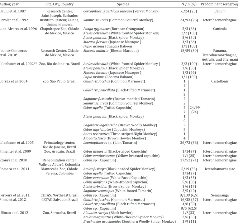
table 1. studies in Latin America on the serologic evidence of exposure to leptospires in non-human primates in captivity
Author, year Site, City, Country Species N / n (%) Predominant serogroup
Baulu et al. 1987 Research Center, Cercopithecus aethiops sabeaus (Vervet Monkey) 6/24 (25) Ballum
Saint Joseph, Barbados
Perolat et al. 1992 Instituto Pasteur, Caiena, Saimiri sciureus (Common Squirrel Monkey) 24/93 (26) Icterohaemorrhagiae
Guiana Francesa
Luna-Alvarez et al. 1996 Chapultepec Zoo, Cidade Pongo pygmaeus (Bornean Orangutan) 2/3 (66) Canicola do México, México Ateles belzebuth (White-fronted Spider Monkey) 2/2 (100)
Ateles paniscus (Black Spider Monkey) 3/6 (50) Macaca fuscata (Japanese Macaque ) 1/3 (66) Papio ursinus (Chacma Baboon) 1/1 (100)
Ibanez-Contreras Research Center, Cidade Macaca mulatta (Rhesus Macaque) 18/59 (30) Panama,
et al. 2010* do México, México Icterohaemorrhagiae,
Autralis, and Shermani
Lilenbaum et al. 2002** Zoo, Rio de Janeiro, Brazil Ateles belzebuth (White-fronted Spider Monkey ) 2/2 (100) Icterohaemorrhagiae Ateles paniscus (Black Spider Monkey) 3/6 (50)
Macaca fuscata (Japanese Macaque ) 1/3 (66) Papio ursinus (Chacma Baboon) 1/1 (100)
Corrêa et al. 2004 Zoo, São Paulo, Brazil Callithrix jacchus (Common Marmoset) 1 Castellonis
4
Callithrix penicillata (Black-tufted Marmoset) 1
2
Saguinus fuscicolis (Brown-mantled Tamarin) 2 Saimiri sciureus (Common Squirrel Monkey) 2 Cebus apella (Tufted Capuchin) 4 24/99
7 (24)
Ateles paniscus (Black Spider Monkey) 1
0
Lagothrix lagothricha (Brown Woolly Monkey) 2 Cebus nigrivitatus (Capuchin Monkeys) 5 Aotus trvirgatus (Three-striped Night Monkey) 1 Alouatta fusca (Brown Howler Monkey) 4
Lilenbaum et al. 2005 Primatology center, Leontopithecus sp. (Lion Tamarin) 26/73 (36) Icterohaemorrhagiae Rio de Janeiro, Brazil
Pimentel et al. 2009 Zoo, Aracaju, Brazil Cebus libinosus (Black-striped Capuchin) 1/14 (7) Icterohaemorrhagiae Cebus xanthosternus (Yellow-breasted capuchin) 1/4(25) Icterohaemorrhagiae Szonyi et al. 2010 Rehabilitation center, Cebus sp. (Capuchin) 37/52 (71) Icterohaemorrhagiae Valle de Aburrá, Colombia
Romero et al. 2011 Mantecaña Zoo, Cidade Ateles fusiceps (Black-headed Spider Monkey) 5/19 (33) Icterohamorhagiae
Pereira, Colombia Cebus apella (Tufted Capuchin) 1/14 (7)
Cebus capucinus (White Faced Capuchin) 1/3 (33) Cebus albifrons (White-fronted capuchin) 5/6 (83) Ateles hybridus (Brown Spider Monkey) 1/6 (17) Saguinus leoucopus (White-footed Tamarin) 2/5 (40)
Ferreira et al. 2011 CETAS, Northeast Brazil Cebus sp. (Capuchin) 9/139 (6,5) Semaranga Pinna et al. 2012 CETAS, Salvador, Brazil Callithrix jacchus (Common Marmoset) 16/28 (57) Icterohaemorrhagiae Callithrix penicillata (Black-tufted Marmoset) 4/8 (50)
Cebus sp. (Capuchin) 8/5 (62,5)
Ullman et al. 2012 Zoo, Sorocaba, Brazil Alouatta caraya (Black howler) 1/3(33) Icterohaemorrhagiae Ateles marginatus (White-cheeked Spider Monkey) 2/6 (33)
Brachyteles arachnoides (Southern Woolly Spider Monkey) 1/9 (11) Note: All studies used the cutoff of 1:100 MAT titers except for * and **, which used 1:20 and 1:200 cutoffs, respectively.
in large urban areas with poor sanitation, and wild animals
in water sports and ecotourism related exposures (Bharti
et al. 2003). Most human infections are asymptomatic or
cause mild febrile illness that is indistinguishable from
di-seases caused by other infectious agents. However, 5-10%
of human infections will become severe, resulting in the
Weil’s triad of acute renal failure, hemorrhages, and
jaundi-ce (with 5-30% fatality) or severe pulmonary hemorrhagic
syndrome (SPHS, with ≥ 50% fatality) (WHO 2003a, McBri
-de et al. 2005, Me-deiros et al. 2010).
Non-human primates usually develop a self resolving,
mild illness in naturally acquired or experimental infections
( Minette 1966, Minette & Shaffer 1968, Hambleton et al.
1980, Marshall et al. 1980), but some species may
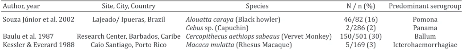
Luna-table 2. studies in Latin America on the serologic evidence of exposure to leptospires in non-human primates captured from the wild
Author, year Site, City, Country Species N / n (%) Predominant serogroup
Souza Júnior et al. 2002 Lajeado/ Ipueras, Brazil Alouatta caraya (Black howler) 46/82 (16) Pomona
Cebus sp. (Capuchin) 2/286 (2) Panama
Baulu et al. 1987 Research Center, Barbados, Caribe Cercopithecus aethiops sabeaus (Vervet Monkey) 150/501 (30) Ballum Kessler & Everard 1988 Caio Santiago, Porto Rico Macaca mulatta (Rhesus Macaque) 5/169 (3) Icterohaemorrhagiae
-Alvarez et al. 1996, Lilenbaum et al. 2002, Correa et al. 2004,
Lilenbaum et al. 2005, Souza Júnior et al. 2006, Pimentel et
al. 2009, Ibáñez-Contreras et al. 2010, Ferreira et al. 2011,
Romero et al. 2011, Szonyi et al. 2011, Pinna et al. 2012,
Ull-mann et al. 2012). Few studies have compared the
serologi-cal evidence of leptospirosis exposure with the molecular
evidence of active infection (detection of DNA fragments in
blood samples) or the renal carrier state (detection of DNA
in urine) in non-human primates (Ullmann et al. 2012). The
aim of this study was to investigate the serological evidence
of exposure and the presence of DNA fragments of
pathoge-nic leptospires in the blood and urine samples of
Neotropi-cal primates from two sources, a zoologiNeotropi-cal park and a wild
life rehabilitation center in Salvador, Bahia, Brazil.
MAtErIALs And MEthOds
In the present study, 58 of non-human primates were evaluated. The population included 42 monkeys from the Zoological Park of Salvador (Parque Zoobotânico Getúlio Vargas), Bahia, Brazil. The-se animals belonged to the following species: Cebus xanthosternos
(n=23), Cebus flavius (n=11), Alouatta caraya (n=3), Aottus sp. (n=4), and Saimiri sciureus (n=1). The study also evaluated 16 pri-mates rescued from illegal trade that were housed in the wildlife rehabilitation center of Salvador (Centro de Triagem de Animais Silvestres Chico Mendes, CETAS), Bahia, Brazil. All these animals
were identified as Cebus sp., as many of them are hybrids. Microscopic agglutination tests (MAT) were performed, ac-cording to recommended protocols (Faine et al. 1999, WHO 2003), and included twenty-three WHO reference strains and a local isolate (serovar Copenhageni strain L1130) (Ko et al. 1999,
Nascimento et al. 2004). Titers ≥1:100 were considered positive.
Polymerase chain reaction for the detection of the lipL32 gene was performed as previously described (Rojas et al. 2010, Chagas--Junior et al. 2012).
The research protocols were approved by the Research Ethics Committee of the Faculdade Franca - SP (025/2009-A). In addi-tion, they were in accordance with guidelines System Authoriza-tion and InformaAuthoriza-tion on Biodiversity the Ministry of Environment of Brazil (number 20831-1)
rEsULts And dIscUssIOn
In the present study, only one of 42 primates (2%) from the
Zoological Park of Salvador had positive serum samples,
ac-cording to the MAT. This animal was an adult female
Alouat-ta caraya (
black howler
)
that had a positive serum sample
of 1:100 with mixed reactions for the Bratislava and
Icte-rohaemorrhagiae serogroups. This animal was housed in a
cage with two other adults of the same species. The positive
animal and one negative black howler were born in the
Zo-ological Park, while the other negative adult came from
ille-gal trade (CETAS). Urine and blood samples were negative
for all 42 monkeys evaluated at the Zoological Park.
Such low seroprevalence was not expected. Salvador is
the third most populated city in Brazil, with an estimated
2.6 million inhabitants, and 60% of those inhabitants live
in slum communities (Riley et al. 2007). It is a large urban
center with poor sanitation, and incidence of severe
leptos-pirosis cases peak during rainy seasons and in association
with floods (Ko et al. 1999, Costa et al. 2001, Riley et al.
2007). Reports from the Zoological personnel indicated
that there is a high population of rodents in the park, and
rodents are frequently observed to be in contact with the
animals in their cages. In some surveys from Latin
Ameri-can zoos, the seroprevalence of antileptospiral antigens is
highly variable among different species (Table 1), and
hi-gher rates were attributed to the contact of primates with
urban rodents. Additionally, there is a high prevalence of
Icterohaemorrhagiae as the predicted infecting serogroup
in some of these studies (as implied by MAT highest titers),
and this suggests that rodents are the source of
non-prima-te infection because these serovars are known to be
selec-tively carried by urban rodents such as the Brown (
Rattus
norvegicus
) and Black rats (
R. rattus
) (Bharti et al. 2003).
In a survey of captured rats in Salvador, 80% of a total 142
animals had positive cultures for leptospires from kidney
or urine samples, and all 59 serotypes isolated by
monoclo-nal antibodies were characterized as serovar Copenhageni
(serogroup Icterohaemorrhagiae) (de Faria et al. 2008),
which is the main cause of severe leptospirosis in Salvador
and in other large urban Brazilian centers (Ko et al. 1999,
Pereira et al. 2000). It is not possible to infer that the single
Black Howler with a positive MAT acquired the infection
from exposure to rats because it had a mixed reaction with
the Bratislava serogroup, which has no known selectivity
for rodents.
In summary, the results from the Zoological Park
sug-gest that the Neotropical primates’ contact with rodents
was not associated with an increased seroprevalence of
an-tileptospiral antibodies. These species may be intrinsically
more resistant than others in terms of acquiring leptospiral
infection, or conversely, these animals may have had
pre-vious exposure in the wild that is associated with a
pro-gressive decrease in antileptospiral antibodies during the
period of captivity.
Thus, the high rate of serological evidence of Leptospira
exposure seems to be a consistent finding among animals
from the wild that are rescued from illegal trade in the
state of Bahia. It is important to note, however, that the
predicted infecting serogroup inferred by MAT highest
titers was Icterohaemorrhagiae in 84% of the cases in a
former study. In this study, the five positive samples were
distributed in the following predicted serogroups: Ballum
(1:100), Semaranga (1:200), Grippotyphosa (1:100),
Cy-nopeteri (1:100), and a mixed reaction of
Tarassovi/Autu-mnalis (1:100). Thus, while the high frequency of
Icteroha-emorrhagiae indicated that monkeys could have acquired
infection after they were caught in the wild, the wide range
of predicted serogroups in the present study indicates that
these animals may have been exposed to many potential
sources of infection in the wild. In addition to
conserva-tion concerns, these data point toward the potential risk
of keeping these animals as pets as it appears that they
may be exposed to leptospiral infection in the wild or after
entrapment.
This study also evaluated the possible association of
serological evidence of exposure with active infection and
renal carriage using PCR detection of leptospiral DNA in
blood and urine samples, respectively. None of the
evalu-ated samples yielded positive results. Further studies with
larger sample groups of animals rescued from illegal trade
are warranted to estimate the risks of infection from
inti-mate contact with exotic animals, including Neotropical
primates, when kept as pets.
cOncLUsIOns
The seroprevalence of antileptospiral antibodies was
low (2%) in the Zoological Park of Salvador, Brazil, despite
the high frequency of rodents in the area and the
endemici-ty of human leptospirosis in Salvador.
A higher rate (31%) was observed among the animals
rescued from illegal trade in the state of Bahia.
Serological evidence of exposure does not predict an
active infection or the renal carrier state in non-human
pri-mates.
Even if all the blood and urine samples were negative for
leptospiral DNA fragments, the high frequency of
serologi-cal evidence of exposure suggests a potential risk of
leptos-pirosis transmission when keeping these animals as pets.
Acknowledgements.- The authors are grateful to Victor Pereira Curvelo for technical assistance on animal capture and contention during the sur-vey performed in the Zoological Park of Salvador, Brazil.
rEfErEncEs
Baulu J., Everard C.O. & Everard J.D. 1987. Leptospires in vervet monkeys (Cercopithecus aethiops sabaeus) on Barbados. J. Wildl. Dis.23:60-66. Bharti A.R., Nally J.E., Ricaldi J.N., Matthias M.A., Diaz M.M., Lovett M.A.,
Levett P.N., Gilman R.H., Willig M.R., Gotuzzo E. & Vinetz J.M. 2003. Lep-tospirosis: a zoonotic disease of global importance. Lancet Infect. Dis. 3:757-771.
Chagas-Junior A.D., Silva C.L., Soares L.M., Santos C.S., Silva C.D., Athanazio D.A., Reis M.G., McBride F.W. & McBride A.J. 2012. Detection and
quan-tification of Leptospira interrogans in hamster and rat kidney samples:
immunofluorescent imprints versus real-time PCR. PloS one7:e32712.
Chomel B.B., Belotto A. & Meslin F.X. 2007. Wildlife, exotic pets, and emer-ging zoonoses. Emerg. Infect. Dis.13:6-11.
Correa S.H.R., Vasconcellos S.A., Morais Z., Teixeira A.A., Dias R.A., Guima-rães M.A.B.V., Ferreira F. & Neto J.S.F. 2004. Epidemiologia da Leptospi-rose em animais silvestres na Fundação Parque Zoológico de São Paulo. Braz. J. Vet. Res. Anim. Sci. 41:189-193.
Costa E., Costa Y.A., Lopes A.A., Sacramento E. & Bina J.C. 2001. Formas
gra-ves de leptospirose: aspectos clínicos, demográficos e ambientais. Revta
Soc. Bras. Med. Trop.34:261-267.
de Faria M.T., Calderwood M.S., Athanazio D.A., McBride A.J., Hartskeerl R.A., Pereira M.M., Ko A.I. & Reis M.G. 2008. Carriage of Leptospira inter-rogans among domestic rats from an urban setting highly endemic for leptospirosis in Brazil. Acta Tropica108:1-5.
Faine S.B., Adler B., Bolin C. & Perolat P. 1999. Leptospira and Leptospiro-sis. 2nd ed. MediSci, Melbourne.
Ferreira D.R.A., Laroque P.O., Wagner P.G.C., Higino S.S.S., Azevedo S.S., Rego E.W. & Mota R.A. 2011. Ocorrência de anticorpos e fatores de risco associados à infecção por Leptospira spp. em Cebus spp. mantidos em cativeiro no Nordeste do Brasil. Pesq. Vet. Bras.31:1019-1023. Hambleton P., Baskerville A., Marshall R.B., Harris-Smith P.W. & Adams
G.D. 1980. Metabolic sequelae of experimental leptospirosis in grivet monkeys. Brit. J. Exp. Pathol.61:16-21.
Ibáñez-Contreras A., Hernández-Godínez B., Torres- Barranca J.I. & Melén-dez-Vélez P. 2010. Hallazgos de anticuerpos contra Leptospira sp., se-rovariedades Panama, Lai, Australis, Shermani y Patoc, en un grupo de monos rhesus (Macaca mulatta) en condiciones de cautiverio. Archivos de Medicina Veterinaria42:101-104.
Kessler M.J. & Everard C.O.R. 1988. Leptospiral agglutinins in the Cayo Santiago macaques. Am. J. Primatol. 14:369-373.
Ko A.I., Galvao Reis M., Ribeiro Dourado C.M., Johnson Jr W.D. & Riley L.W. 1999. Urban epidemic of severe leptospirosis in Brazil. Salvador Leptos-pirosis Study Group. Lancet354:820-825.
Lilenbaum W., Monteiro R.V., Ristow P., Fraguas S., Cardoso V.S. & Fedullo L.P. 2002. Leptospirosis antibodies in mammals from Rio de Janeiro Zoo, Brazil. Res. Vet. Sci.73:319-321.
Lilenbaum W., Varges R., Moraes I.A., Ferreira A.M. & Pissinatti A. 2005. Leptospiral antibodies in captive lion tamarins (Leontopithecus sp) in Brazil. Vet. J.169: 462-464.
Luna-Alvarez M.A., Moles-Cervantes L.P., Torres-Barranca J.I. & Gual-Sill F. 1996. Serological survey of leptospirosis in captive wildlife at the Cha-pultepec Zoo in Mexico City. Veterinaria, Mexico,27:229-234.
Marshall R.B., Baskerville A., Hambleton P. & Adams G.D. 1980. Benign leptospirosis: the pathology of experimental infection of monkeys with Leptospira interrogans serovars balcanica and tarassovi. Brit. J. Exp. Pathol.61:124-131.
McBride A.J., Athanazio D.A., Reis M.G. & Ko A.I. 2005. Leptospirosis. Curr. Opin. Infect. Dis.18: 376-386.
Medeiros F.R., Spichler A. & Athanazio D.A. 2010. Leptospirosis-associa-ted disturbances of blood vessels, lungs and hemostasis. Acta Trop. 115:155-162.
Minette H.P. 1966. Leptospirosis in primates other than man. Am. J. Trop. Med. Hyg.15:190-198.
Minette H.P. & Shaffer M.F. 1968. Experimental leptospirosis in monkeys. Am. J. Trop. Med. Hyg.17:202-212.
Nascimento A.L., Ko A.I., Martins E.A., Monteiro-Vitorello C.B., Ho, P.L., Haake D.A., Verjovski-Almeida S., Hartskeerl R.A., Marques M.V., Oliveira M.C., Menck C.F., Leite L.C., Carrer H., Coutinho L.L., Degrave W.M., Dellagostin O.A., El-Dorry H., Ferro E.S., Ferro M.I., Furlan L.R., Gamberini M., Giglioti E.A., Goes-Neto A., Goldman G.H., Goldman M.H., Harakava R., Jeronimo S.M., Junqueira-de-Azevedo I.L., Kimura E.T., Kuramae E.E., Lemos E.G., Le-mos M.V., Marino C.L., Nunes L.R., Oliveira de R.C., Pereira G.G., Reis M.S., Schriefer A., Siqueira W.J., Sommer P., Tsai S.M., Simpson A.J., Ferro J.A., Camargo L.E., Kitajima J.P., Setubal J.C. & Van S.M.A. 2004. Comparative ge-nomics of two Leptospira interrogans serovars reveals novel insights into physiology and pathogenesis. J. Bacteriol.186:2164-2172.
H.L. & Marchevsky R.S. 2005. Experimental leptospirosis in marmoset monkeys (Callithrix jacchus): a new model for studies of severe pulmo-nary leptospirosis. Am. J. Trop. Med. Hyg.72:13-20.
Pereira M.M., Matsuo M.G.S., Bauab A.R., Vasconcelos S.A., Moraes Z.M., Ba-ranton G. & Saint G.I. 2000. A clonal subpopulation of Leptospira interro-gans sensu stricto is the major cause of leptospirosis outbreaks in Brazil. J. Clin. Microbiol.38:450-452.
Perolat P., Poingt J.P., Vie J.C., Jouaneau C., Baranton G. & Gysin J. 1992. Occurrence of severe leptospirosis in a breeding colony of squirrel monkeys. Am. J. Trop. Med. Hyg.46:538-545.
Pimentel J.S., Gennari S.M., Dubey J.P., Marvulo M.F.V., Vasconcellos S.A., Morais Z.M., Silva J.C.R. & Evêncio Neto J. 2009. Inquérito sorológico para toxoplasmose e leptospirose em mamíferos selvagens neotropicais do Zoológico de Aracaju, Sergipe. Pesq. Vet. Bras.29:1009-1014. Pinna M.H., Martins G., Pinheiro A.C., Almeida D.S., Oria A.P. & Lilenbaum
W. 2012. Detection of anti-Leptospira antibodies in captive nonhuman primates from Salvador, Brazil. Am. J. Primatol.74:8-11.
Riley L.W., Ko A.I., Unger A. & Reis M.G. 2007. Slum health: diseases of ne-glected populations. BMC Int. Health Hum. Rights7:2.
Rojas P., Monahan A.M., Schuller S., Miller I.S., Markey B.K. & Nally J.E.
2010. Detection and quantification of leptospires in urine of dogs: a
maintenance host for the zoonotic disease leptospirosis. Eur. J. Clin. Mi-crobiol. Infect. Dis.29:1305-1309.
Romero M.H., Astudillo M., Sanchez J.A., Gonzalez L.M. & Varela N. 2011. Anticuerpos contra Leptospira sp. en primates neotropicales y trabaja-dores de un zoológico colombiano. Revta Salud Publica13:814-823. Souza Júnior M.F., Lobato Z.I.P., Lobato F.C.F., Moreira E.C., Oiveira R.R.,
Lei-te G.G., Freitas T.D. & Assis R.A. 2006. Presença de anticorpos da classe IgM de Leptospira interrogans em animais silvestres do Estado do To-cantins, 2002. Revta Soc. Bra. Med. Trop.39:292-294.
Szonyi B., Agudelo-Florez P., Ramirez M., Moreno N. & Ko A.I. 2011. An outbreak of severe leptospirosis in capuchin (Cebus) monkeys. Vet. J. 188:237-239.
Ullmann L.S., Neto R.N.D., Teixeira R.H.F., Nunes A.V., Silva R.C., Pereira-Ri-chini V.B. & Langoni H. 2012. Epidemiology of leptospirosis at Sorocaba Zoo, São Paulo state, Southeastern Brazil. Pesq. Vet. Bras.32:1174-1178. WHO 2003. Human Leptospirosis: guidance for diagnosis, surveillance