RESEARCH ARTICLE
Identification of Non-HLA Genes Associated
with Celiac Disease and Country-Specific
Differences in a Large, International Pediatric
Cohort
Ashok Sharma1, Xiang Liu2, David Hadley2,3, William Hagopian4, Edwin Liu5, Wei-Min Chen6, Suna Onengut-Gumuscu6, Ville Simell7, Marian Rewers8, Anette-G. Ziegler9,
Åke Lernmark10, Olli Simell7, Jorma Toppari7, Jeffrey P. Krischer2, Beena Akolkar11, Stephen S. Rich6, Daniel Agardh12, Jin-Xiong She1*, TEDDY Study Group¶
1Center for Biotechnology and Genomic Medicine, Georgia Regents University, Augusta, GA, United States of America,2Pediatric Epidemiology Center, Department of Pediatrics, University of South Florida, Tampa, FL, United States of America,3Division of Population Health Sciences and Education, St George's University of London, London, United Kingdom,4Pacific Northwest Diabetes Research Institute, Seattle, WA, United States of America,5Digestive Health Institute, Children’s Hospital Colorado, University of Colorado Denver, Aurora, CO, United States of America,6Center for Public Health Genomics, University of Virginia, Charlottesville, VA, United States of America,7Department of Pediatrics, University of Turku, Turku, Finland,8Barbara Davis Center for Childhood Diabetes, University of Colorado Denver, Aurora, CO, United States of America,9Institute of Diabetes Research, Helmholtz Zentrum München, and Klinikum rechts der Isar, Technische Universität München, and Forschergruppe Diabetes e.V., Munich-Neuherberg, Germany,10Department of Clinical Sciences, Lund University/CRC, Malmö, Sweden,11 National Institutes of Diabetes and Digestive and Kidney Disorders, National Institutes of Health, Bethesda, MD, United States of America,12Diabetes and Celiac Disease Unit, Lund University, Malmo, Sweden
¶ Membership of the TEDDY Study Group is provided in the Acknowledgments.
Abstract
Objectives
There are significant geographical differences in the prevalence and incidence of celiac dis-ease that cannot be explained by HLA alone. More than 40 loci outside of the HLA region have been associated with celiac disease. We investigated the roles of these non-HLA genes in the development of tissue transglutaminase autoantibodies (tTGA) and celiac dis-ease in a large international prospective cohort study.
Methods
A total of 424,788 newborns from the US and European general populations and first-degree relatives with type 1 diabetes were screened for specific HLA genotypes. Of these, 21,589 carried 1 of the 9 HLA genotypes associated with increased risk for type 1 diabetes and celiac disease; we followed 8676 of the children in a 15 y prospective follow-up study. Genotype analyses were performed on 6010 children using the Illumina ImmunoChip. Lev-els of tTGA were measured in serum samples using radio-ligand binding assays; diagnoses
OPEN ACCESS
Citation:Sharma A, Liu X, Hadley D, Hagopian W, Liu E, Chen W-M, et al. (2016) Identification of Non-HLA Genes Associated with Celiac Disease and Country-Specific Differences in a Large, International Pediatric Cohort. PLoS ONE 11(3): e0152476. doi:10.1371/journal.pone.0152476
Editor:Yungling Leo Lee, National Taiwan University, TAIWAN
Received:November 20, 2015
Accepted:March 15, 2016
Published:March 25, 2016
Copyright:© 2016 Sharma et al. This is an open access article distributed under the terms of the
Creative Commons Attribution License, which permits unrestricted use, distribution, and reproduction in any medium, provided the original author and source are credited.
Data Availability Statement:All relevant data are within the paper and its Supporting Information files.
of celiac disease were made based on persistent detection of tTGA and biopsy analysis. Data were analyzed using Cox proportional hazards analyses.
Results
We found 54 single-nucleotide polymorphisms (SNPs) in 5 genes associated with celiac disease (TAGAP,IL18R1,RGS21,PLEK, andCCR9) in time to celiac disease analyses (10−4>P>5.8x10⁻⁶). The hazard ratios (HR) for the SNPs with the smallest P values in
each region were 1.59, 1.45, 2.23, 2.64, and 1.40, respectively. Outside of regions previ-ously associated with celiac disease, we identified 10 SNPs in 8 regions that could also be associated with the disease (P<10−4). A SNP nearPKIA(rs117128341,P= 6.5x10⁻⁸,
HR = 2.8) and a SNP nearPFKFB3(rs117139146,P<2.8x10⁻⁷, HR = 4.9) reached the
genome-wide association threshold in subjects from Sweden. Analyses of time to detec-tion of tTGA identified 29 SNPs in 2 regions previously associated with celiac disease (CTLA4,P= 1.3x10⁻⁶, HR = 0.76 andLPP,P= 2.8x10⁻⁵, HR = .80) and 6 SNPs in 5
regions not previously associated with celiac disease (P<10−4); non-HLA genes are
there-fore involved in development of tTGA.
Conclusions
In conclusion, using a genetic analysis of a large international cohort of children, we associ-ated celiac disease development with 5 non-HLA regions previously associassoci-ated with the dis-ease and 8 regions not previously associated with celiac disdis-ease. We identified 5 regions associated with development of tTGA. Two loci associated with celiac disease progression reached a genome-wide association threshold in subjects from Sweden.
Introduction
Celiac disease is strongly associated with the human leukocyte antigen (HLA) DR3–DQ2.5 (i.
e., DRB103-DQA105:01-DQB102:01) or DR4-DQ8 (DRB104-DQA103-DQB103:02)
haplotypes on chromosome 6 [1]. Moreover, there is an HLA gene-dose effect on the disease
risk as individuals carrying two copies of DR3-DQ2.5 are at a higher susceptibility for celiac
disease than those with only one copy [2,3]. Although carrying either DR3–DQ2.5 or DR4–
DQ8 is almost a necessity to develop celiac disease, these haplotypes are common in the general
population and not all carriers develop clinical disease [4]. Since the first genome-wide case/
control association study (GWAS) on celiac disease was published in 2007, a total of 40
non-HLA loci have been suggested as being associated with celiac disease[5–9]. A significant
pro-portion of the genetic predisposition comes from the HLA region (odds ratio of>5) while
non-HLA genes have modest effect sizes with an odds ratio between 1.12 and 1.36 for celiac
disease [10]. The role of these non-HLA genes have not been assessed in those with early onset
celiac disease, particularly using a prospective cohort.
Celiac disease is increasing in frequency, with significant intra- and inter-country
differ-ences in the prevalence and incidence of the disease[11]. Despite recent advances in celiac
dis-ease genetics, it remains elusive why some, but not all, individuals with the HLA risk genotypes develop celiac disease. Although the ingestion of gluten is required to trigger and maintain celiac disease, gluten exposure is nearly universal. Therefore, exposures to other environmental factors may also be important in the pathogenesis. Celiac disease is likely a multifactorial disor-der where multiple genes and multiple environmental factors interact in a complex manner. (NIEHS), Juvenile Diabetes Research Foundation
(JDRF), and Centers for Disease Control and Prevention (CDC). The complete TEDDY Study Group is listed in the Supplementary material.
Competing Interests:Dr. David Hadley is an employee of TransMed System Inc. but participated in this study as an employee of Pediatric Epidemiology Center, Department of Pediatrics, University of South Florida, Tampa, FL, USA. TransMed Systems, Inc., Cupertino, CA, USA, played no role in this study, and the specific roles of Dr. Hadley is articulated in the Author Contributions section. There are no patents, products in development, or marked products to declare. This does not alter the authors' adherence to PLOS ONE policies on sharing data or materials.
Disease risk genes may act at various stages of autoimmunity progression, with some genes playing a role early in autoantibody development, and others playing a critical role in the later stages of celiac disease development. This stage-specific contribution of different genes to the celiac disease risk is an important concept, which cannot be investigated using the cross-sec-tional case/control study design employed in all previous studies. Furthermore, genetic factors responsible for the development of tissue transglutaminase autoantibodies (tTGA) and ethnic-or country-specific differences in a genetically predisposed population have not been repethnic-orted previously.
The Environmental Determinants of Diabetes in the Young (TEDDY) is an international multicenter study that screened over 420,000 newborns from the general population in four different countries to identify children with high risk HLA genes for the development of type 1
diabetes (T1D) [12]. Recently, TEDDY demonstrated the impact of different HLA genotypes
on the risk of celiac disease as well as tTGA development, and furthermore confirmed that the HLA-DR3-DQ2/DR3-DQ2 genotype confers the single highest genetic risk for the disease
dur-ing early childhood[13]. We also found differences in risk of disease between the participating
countries that could not be explained by HLA-DR-DQ, suggesting that the risk may be influ-enced by variations in the environment and/or involvement of genes outside the HLA-DR-DQ region. One such recent finding from the TEDDY study was the protective association of
HLA-DRB10401 with celiac disease autoimmunity[14].
The present study genotyped 195,806 SNPs on ImmunoChip in 6,010 TEDDY children to identify potential genetic factors responsible for the development of early autoimmunity (tTGA development) and celiac disease as well as country-specific differences in genetic predisposition.
Results
A total of 703 subjects developed persistent tTGA and were considered“events”in the Cox
proportional hazards models for persistent tTGA. Only 317 of these 703 persistently tTGA positive children received an intestinal biopsy and 262 of the 317 subjects were confirmed to have celiac disease at the time the procedure was performed. Eleven children with positive tTGA tests at the initial time point were biopsied before they could be confirmed as having per-sistent tTGA and eight of them also had biopsy-proven celiac disease. Eighteen other children
who had persistent tTGA levels>100 units but did not have a biopsy were also considered to
have celiac disease for purposes of the study. Therefore, 288 subjects were considered as
“events”in the analysis of the time-to-celiac disease. There are known differences based upon
the family history of celiac disease, HLA-DR-DQ genotype, gender,HLA-DPB1and country of
residence. These factors were adjusted in the Cox proportional hazard models.
Analysis of reported celiac disease SNPs
A total of 69 SNPs were previously reported to be associated with celiac disease based on the
NHGRI GWAS Catalog[5,7,15–17], of which 48 were represented on the ImmunoChip (S4
Table). Risk Variants that have been reported but are not on the ImmunoChip are listed inS6 Table. In the time-to-celiac disease analysis, only one SNP (rs13015714/IL18Ron 2q12.1,
HR = 1.42, p = 1.38x10⁻⁴) attained significance after Bonferroni correction (p = 0.05/48=0.001).
Several other SNPs were close to the significance threshold of 0.001 or had p-value<0.05:
rs653178/SH2B3 (HR = 1.30; p = 0.002); rs1464510/LPP(HR = 1.28; p = 0.002); rs17035378/
PLEK(HR = 0.75; p = 0.004); rs6806528/FRMD4B(HR = 1.44; p = 0.004); rs11221332/ETS1
(HR = 1.29; p = 0.006); rs2298428/YDJC(HR = 1.27; p = 0.012); rs2327832/TNFAIP3(HR = 1.24;
p = 0.025); rs802734/PTPRK(HR = 1.21; p = 0.034); rs13098911/CCR9 (HR = 1.29; p = 0.041);
and rs10876993/CDK4 (HR = 0.84; p = 0.042).
For time-to-persistent tTGA analysis, we observed 10 SNPs with p<0.05: rs1464510/LPP
(HR = 1.16; p = 0.004); rs2298428/YDJC(HR = 1.17; p = 0.011); rs864537/CD247(HR = 0.87;
p = 0.013); rs13015714/IL18R1(HR = 1.15; p = 0.02); rs10936599/MYNN(HR = 1.15; p = 0.022);
rs11203203/UBASH3A(HR = 1.13; p = 0.027); rs11712165/CD80(HR = 1.12; p = 0.035); rs7574
865/STAT4(HR = 1.14; p = 0.036); rs2816316/RGS1(HR = 0.859; p = 0.038); and rs802734/PTP
RK(HR = 1.12; p = 0.046).
Analysis of previously reported celiac disease regions
Next, we extended our analysis to all SNPs within 400 kb up- and downstream of the 48
reported SNPs. The–log10 p-values for all SNPs in these regions are plotted inFig 1Afor
tTGA andFig 1Bfor celiac disease. Since these are analyses for candidate regions, we
consid-ered p<10−4as suggestive evidence for confirmation because multiple SNPs are tested in each
region and the SNPs are in high linkage disequilibrium.
In the tTGA plots, the two regions with strongest evidence wereCTLA4andLPP(S1 Fig). The
SNPs with smallest p-value in these two regions are: rs12990970/CTLA4(HR = 0.76; p = 1.3x10⁻⁶)
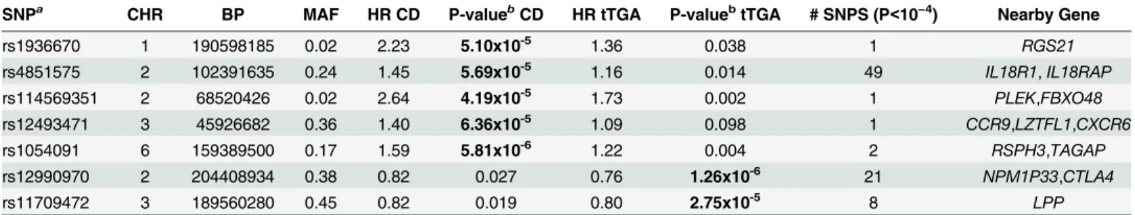
and rs11709472/LPP(HR = 0.80; P = 2.8x10⁻⁵) (Table 1). It is important to note that in our study,
the presence of the minor allele of rs12990970/CTLA4is protective (HR<1). In contrast, earlier
studies have shown thatCTLA4(rs4675374-A) is a risk factor for celiac disease (OR = 1.14)[5,18].
The Kaplan-Meier plots for the three SNPs with smallest p-values in time-to-tTGA analysis
(rs12990970/CTLA4; rs11709472/LPP;rs2925499/TNFRSF14) are shown inFig 1C.
In the celiac disease analysis, we found 5 regions (TAGAP,IL18R1,RGS21,PLEK, andCCR9)
with SNPs that had p-values<10−4: rs1054091/TAGAP(HR = 1.59; p = 5.8x10⁻⁶); rs4851575/
IL18R1(HR = 1.45; p = 5.7x10⁻⁵); rs1936670/RGS21(HR = 2.23; p = 5.1x10⁻⁵); rs114569351/
PLEK (HR = 2.64; p = 4.2x10⁻⁵); and rs12493471/CCR9 (HR = 1.40; p = 6.4x10⁻⁵) (Table 1,S2
Fig). The Kaplan-Meier plots of three SNPs with smallest p-values in the time-to-celiac disease
analysis (rs1936670/RGS21, rs12493471/CCR9, rs1054091/TAGAP) are shown inFig 1D.
SNPs associated with progression to persistent tTGA outside of the
known celiac disease regions
We then extended the analysis to include all SNPs genotyped on the ImmunoChip in search of novel SNP associations. For these analyses, 133,620 with minor allele frequencies of at least 0.01 were tested and therefore the statistical significance for any single SNP requires a
Bonferroni-corrected p<3.7x10⁻⁷. In the time-to-persistent tTGA analyses, none of the
SNPs reached this significance threshold, but 7 SNPs were identified in 5 novel celiac
disease regions with p<10−4: rs117561283/IFNG(HR = 1.81; p = 2.1x10⁻⁵); rs8013918/FOS
(HR = 0.80; p = 4.9x10⁻⁵); rs2409747/XKR6(HR = 1.37; p = 5.4x10⁻⁵); rs114157400/BANK1
(HR = 1.62; p = 8.4x10⁻⁵); and rs72717025/FCGR2A(HR = 1.84; p = 9.6x10⁻⁵) (Fig 2A;
Table 2). These SNPs are novel candidate SNPs with suggestive evidence and require further confirmation studies to rule out false positive discoveries. The Kaplan-Meier plots of three
SNPs (rs2409747/XKR6, rs117561283/IFNG, and rs8013918/FOSdiscovered in
time-to-tTGA analysis are shown inFig 2C.
SNPs associated with progression to celiac disease outside of the
known celiac disease regions
In a similar analysis using time-to-celiac disease with all SNPs, no SNP reached the
Bonfer-roni-corrected p<3.7x10⁻⁷significance threshold but 10 SNPs outside of the known celiac
Fig 1. SNPs in the previously reported celiac disease associated regions.Manhattan plot ofP-values on the−log10scale for SNPs (±400kb) previously
associated with celiac disease (A) and persistent tissue transglutaminase autoantibody (tTGA) positivity(B). HRs and p-values are calculated using three possible genotypes and adjusted for family history of celiac disease, HLA-DR-DQ genotype, gender,HLA-DPB1, population stratification (ancestral
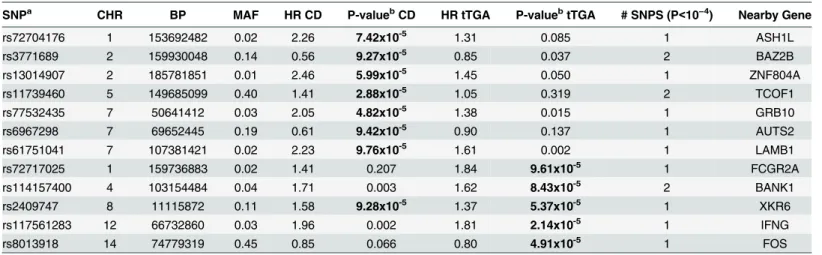
different regions: rs11739460/TCOF1(HR = 1.41; p = 2.9x10⁻⁵); rs77532435/GRB10(HR =
2.05; p = 4.8x10⁻⁵); rs13014907/ZNF804A(HR = 2.46; p = 6.0x10⁻⁵); rs72704176/ASH1L
(HR = 2.26; p = 7.4x10⁻⁵); rs3771689/
BAZ2B(HR = 0.56; p = 9.3x10⁻⁵); rs2409747/XKR6
(HR = 1.58; p = 9.3x10⁻⁵); rs6967298/AUTS2(HR = 0.61; p = 9.4x10⁻⁵); and rs61751041/
LAMB1(HR = 2.23; p = 9.8 x10⁻⁵) (Table 2). The Kaplan-Meier plots of three novel SNPs
(rs13014907/ZNF804A, rs11739460/TCOF1, and rs77532435/GRB10discovered in
time-to-celiac disease analysis are shown inFig 2D.
Country-specific genetic factors associated with progression to celiac
disease
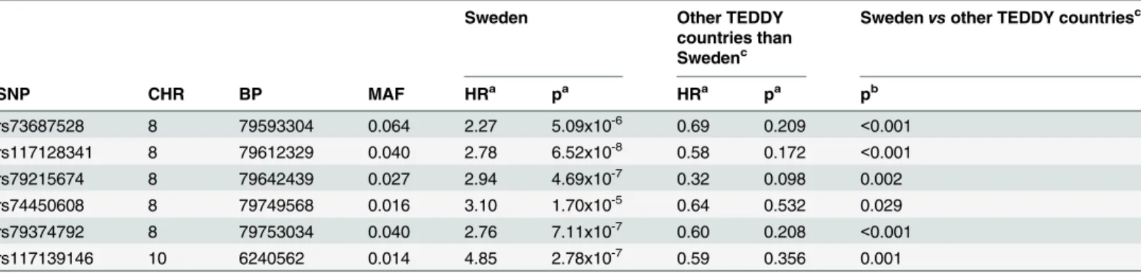
To explore country-specific genetic factors, the data with all 133,620 SNPs were analysed for each country. In the analysis of celiac disease risk among the Swedish participants, SNPs
reached the Bonferroni-corrected p<3.7x10⁻⁷significance threshold in two regions: 8q21.1 and
10p15 (Fig 3). The SNP with the smallest p-value in the 8q21.1 region was rs117128341
(p = 6.52x10⁻⁸, HR = 2.78, MAF = 0.04), a SNP in the intragenic region of the protein kinase
inhibitor alpha (PKIA) gene. The other two nearby genes in this region areZC2HC1AandIL7.
The SNP with the smallest p-value in the chromosome 10p15 region was rs117139146
(p = 2.78x10⁻⁷, HR = 4.85, MAF = 0.014). A nearby gene in this region isPFKFB3, which was
previously shown to be associated with celiac disease. Five SNPs with p<10−4map to thePKIA
region, and one SNP maps to thePFKFB3region (Table 3). In the separate analyses of two
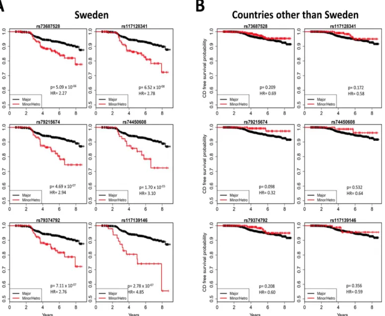
other countries, the US and Finland, none of the six SNPs reached the significance threshold. The analysis in Germany was not conducted due to small sample size. Kaplan-Meier plots of
these six SNPs for different countries (Fig 4)clearly indicated country-specific differences. The
associations of these six SNPs with celiac disease in Sweden and the other three countries are
listed inTable 3. There was no evidence of difference between US and Finland, therefore we
combined US, Finland and Germany together to compare with Sweden in the analysis of inter-action. A Cox proportional hazards model with an interaction term of the SNP with country
heterogeneity) and country of residence (as strata). The red dashed line representsp= 1x10⁻⁴. Kaplan-Meier plots of the three most significant SNPs
associated with celiac disease (C) and tTGA (D) are plotted by dividing the subjects in two groups: (i) Major homozygous (black curves) and (ii) Heterozygous combined with minor homozygous (red curves).
doi:10.1371/journal.pone.0152476.g001
Table 1. Associations with celiac disease or tissue transglutaminase autoantibody (tTGA) positivity (p<10−4), mapped to previously known regions.
SNPa
CHR BP MAF HR CD P-valueb
CD HR tTGA P-valuebtTGA # SNPS (P<10−4) Nearby Gene
rs1936670 1 190598185 0.02 2.23 5.10x10⁻⁵ 1.36 0.038 1 RGS21
rs4851575 2 102391635 0.24 1.45 5.69x10⁻⁵ 1.16 0.014 49 IL18R1,IL18RAP
rs114569351 2 68520426 0.02 2.64 4.19x10⁻⁵ 1.73 0.002 1 PLEK,FBXO48
rs12493471 3 45926682 0.36 1.40 6.36x10⁻⁵ 1.09 0.098 1 CCR9,LZTFL1,CXCR6
rs1054091 6 159389500 0.17 1.59 5.81x10⁻⁶ 1.22 0.004 2 RSPH3,TAGAP
rs12990970 2 204408934 0.38 0.82 0.027 0.76 1.26x10⁻⁶ 21 NPM1P33,CTLA4
rs11709472 3 189560280 0.45 0.82 0.019 0.80 2.75x10⁻⁵ 8 LPP
CHR: Chromosome; BP: Base Pair Position (NCBI 36.3); MAF: Minor Allele Frequency; HRCD: Hazard Ratio in celiac disease analysis; HRtTGA: Hazard Ratio in tTGA analysis. P-values<10−4are highlighted in bold
aThe data for the SNP with smallest p-value is presented from each region.
bHRs and p-value adjusted for family history of celiac disease, HLA-DR-DQ genotype, gender,HLA-DPB1, population strati
fication (ancestral heterogeneity) and country of residence (as strata).
Fig 2. Associations with risk of celiac disease and risk of persistent tissue transglutaminase autoantibody (tTGA) positivity.Manhattan plot of 133,620 SNPs with MAF>0.01, displaying theP-values on the−log10scale for SNP associations with celiac disease (A) and persistent tTGA positivity(B).
HRs and p-values are calculated using three possible genotypes and adjusted for family history of celiac disease, HLA-DR-DQ genotype, gender,
HLA-DPB1, population stratification (ancestral heterogeneity) and country of residence (as strata). The red dashed line representsp= 1x10⁻⁴, the red solid
was used to compare the effects of these six SNPs between Sweden and other countries, adjust-ing for country (Sweden vs other), gender, HLA-DPB1 genotype, HLA-DR-DQ genotype, fam-ily history of celiac disease, and population stratification. The analysis shows that the effect of
any of these SNPs in Sweden is statistically different (p<0.05) from the effect in the other
coun-tries (rs73687528: p<0.001; rs117128341: p<0.001; rs79215674: p = 0.002; rs74450608:
p = 0.029; rs79374792: p<0.001; rs117139146: p = 0.001).
Country-specific associations with progression to tTGA
In time-to-persistent tTGA analysis among subjects from Sweden, none of the SNPs reached
the Bonferroni-corrected p<3.7x10⁻⁷significance threshold, however, 9 SNPs in 9 different
genomic regions had p<10−4(S5 Table). One such SNP was rs117139146 in the region of
PFKFB3(p = 7.34x10⁻⁵, HR = 2.79, MAF = 0.014).
Discussion
HLA-DR3-DQ2.5 and DR4-DQ8 are known as the most important genetic risk factors for celiac disease; however, these two haplotypes only account for part of the genetic risk. Recently, we demonstrated that HLA can be used to assess the risk of celiac disease using the large
pro-spective TEDDY cohort [13]. This previous study clearly demonstrated an HLA gene dose
effect of HLA-DR3-DQ2.5 on the risk of celiac disease autoimmunity was doubled among het-erozygotes (HR = 2.09) but was a near 6-fold increased among homozygotes (HR = 5.70) as
compared to children carrying the lowest-risk genotype DR4-DQ8 [13]. However, another
finding of importance from this study was the difference in incidence of celiac disease between the participating countries which could not be attributed to HLA suggesting that
line represents Bonferroni correction threshold. Kaplan-Meier plots of selected SNPs associated with celiac disease (C) and persistent tTGA (D) are plotted by dividing the subjects in two groups: (i) Major homozygous (black curves) and (ii) Heterozygous combined with minor homozygous (red curves).
doi:10.1371/journal.pone.0152476.g002
Table 2. Novel associations with celiac disease or tissue transglutaminase autoantibody (tTGA) positivity (p<10−4).
SNPa CHR BP MAF HR CD P-valuebCD HR tTGA P-valuebtTGA # SNPS (P<10−4) Nearby Gene
rs72704176 1 153692482 0.02 2.26 7.42x10⁻⁵ 1.31 0.085 1 ASH1L
rs3771689 2 159930048 0.14 0.56 9.27x10⁻⁵ 0.85 0.037 2 BAZ2B
rs13014907 2 185781851 0.01 2.46 5.99x10⁻⁵ 1.45 0.050 1 ZNF804A
rs11739460 5 149685099 0.40 1.41 2.88x10⁻⁵ 1.05 0.319 2 TCOF1
rs77532435 7 50641412 0.03 2.05 4.82x10⁻⁵ 1.38 0.015 1 GRB10
rs6967298 7 69652445 0.19 0.61 9.42x10⁻⁵ 0.90 0.137 1 AUTS2
rs61751041 7 107381421 0.02 2.23 9.76x10⁻⁵ 1.61 0.002 1 LAMB1
rs72717025 1 159736883 0.02 1.41 0.207 1.84 9.61x10⁻⁵ 1 FCGR2A
rs114157400 4 103154484 0.04 1.71 0.003 1.62 8.43x10⁻⁵ 2 BANK1
rs2409747 8 11115872 0.11 1.58 9.28x10⁻⁵ 1.37 5.37x10⁻⁵ 1 XKR6
rs117561283 12 66732860 0.03 1.96 0.002 1.81 2.14x10⁻⁵ 1 IFNG
rs8013918 14 74779319 0.45 0.85 0.066 0.80 4.91x10⁻⁵ 1 FOS
CHR: Chromosome; BP: Base Pair Position (NCBI 36.3); MAF: Minor Allele Frequency; HRCD: Hazard Ratio in celiac disease analysis; HRtTGA: Hazard Ratio in tTGA analysis. P-values<10−4are highlighted in bold
aThe data for the SNP with smallest p-value is presented from each region.
bHRs and p-value adjusted for family history of celiac disease, HLA-DR-DQ genotype, gender,HLA-DPB1, population strati
fication (ancestral heterogeneity) and country of residence (as strata).
environmental factors or other genes could contribute to the disease risk. In the current study, we used the same cohort to assess the association of non-HLA genes to the progression to tTGA in addition to progression to celiac disease in early childhood. A strength of this study Fig 3. Associations with risk of celiac disease in the Swedish population. A:Manhattan plot of 133620 SNPs with MAF>0.01, displaying theP-values on the−log10scale for the SNPs associated with celiac
disease in the Swedish TEDDY population.B:Regional association plots at thePKIAlocus generated by LocusZoom, showing the significance of association and the recombination rate. Colors represent HapMap CEU linkage disequilibrium r2values with the most significantly associated SNP (rs117128341; shown in
purple).C:Pairwise LD plot for five SNPs in the region ofPKIA. The five most significant SNPs from this region are in high LD with each other.
doi:10.1371/journal.pone.0152476.g003
includes the prospective nature of the study cohort that time-to-events analyses can be
con-ducted, looking specifically in this case for genetic factors that could be related to theearly
development of celiac disease. Genetic studies have been traditionally done using cross-sec-tional case/control study design, with populations of individuals with celiac disease who have an unknown age of actual onset of autoimmunity. We know that the rate of seroconversion and subsequent development of celiac disease is high in childhood, and suspect that the yearly incidence slows down some time in adulthood. It is therefore possible that the genes involved in early onset celiac disease may be different from those involved in adult (or late) onset celiac disease. However, it may not be feasible to perform a prospective cohort study in at-risk adults due to the presumed decline in incidence.
In the first stage of analyses, we only considered the 48 SNPs previously reported to be asso-ciated with celiac disease and only one SNP was significant after Bonferroni correction.
How-ever, confirmatory evidence (p<10−4) was found for SNPs in five regions previously reported
to be associated with celiac disease (TAGAP,IL18R1,RGS21,PLEK, andCCR9). The HRs
esti-mated in this prospective cohort (HR = 1.40–2.64) are generally much higher than the odds
ratios (OR) estimated in the case control studies (OR = 1.12–1.36). TheTAGAPgene encodes a
member of the Rho GTPase-activator protein superfamily involved in T cell activation and
co-regulation withIL-2, which has been previously associated with several autoimmune diseases,
including rheumatoid arthritis [19], celiac disease [20], and multiple sclerosis[21].IL18R1is part of the cytokine receptor cluster on chromosome 2q12 which encodes for the receptors of IL18; a cytokine involved in IFN-gamma synthesis and its mRNA expression is upregulated in
active patients with celiac disease [22]. Both genes play roles in the immune response and are
therefore rational candidates for conferring risk in an autoimmune disease such as celiac disease.
The development of tTGA usually appears before the clinical onset of celiac disease and often represents the earliest stage of autoimmunity, signifying a breakdown in tolerance. The specificity of tTGA is high such that negative testing will almost certainly rule out celiac dis-ease. However the positive predictive value of the antibody especially in screened cohorts is Table 3. Six SNPs from two genomic regions significantly associated with celiac disease in Sweden.Five SNPs mapped toPKIAregion and one SNP mappedPFKFB3region.
Sweden Other TEDDY
countries than Swedenc
Swedenvsother TEDDY countriesc
SNP CHR BP MAF HRa pa HRa pa pb
rs73687528 8 79593304 0.064 2.27 5.09x10⁻⁶ 0.69 0.209 <0.001
rs117128341 8 79612329 0.040 2.78 6.52x10⁻⁸ 0.58 0.172 <0.001
rs79215674 8 79642439 0.027 2.94 4.69x10⁻⁷ 0.32 0.098 0.002
rs74450608 8 79749568 0.016 3.10 1.70x10⁻⁵ 0.64 0.532 0.029
rs79374792 8 79753034 0.040 2.76 7.11x10⁻⁷ 0.60 0.208 <0.001
rs117139146 10 6240562 0.014 4.85 2.78x10⁻⁷ 0.59 0.356 0.001
CHR: Chromosome; BP: Base Pair Position (NCBI 36.3); MAF: Minor Allele Frequency; HR: Hazard Ratio.
aHRs and p-value adjusted for family history of celiac disease, HLA-DR-DQ genotype, gender,HLA-DPB1and population stratification (ancestral
heterogeneity).
b
P-value of testing the hypothesis that the effects of the SNP are the same between Sweden and other countries from a Cox model with adjustment for family history of celiac disease, HLA-DR-DQ genotype, gender,HLA-DPB1, population stratification (ancestral heterogeneity) and country of residence (Sweden vs. other).
c
Other participating countries of TEDDY are Germany, Finland and the US.
lower, between 70–83%[23], and some may even be transient[24]. Nevertheless, individuals with only positive tTGA (even without evidence of villous atrophy) should not be disregarded.
Positive tTGA is an independent predictor of reduced bone mineral density[25], growth[26]
and mortality[27] and has been demonstrated to progress to celiac disease. In addition, many
individuals with positive celiac disease serology but normal villous morphology have been
shown to subsequently develop celiac disease in subsequent follow-up[28].
Although HLA genes are known to contribute to the development of tTGA, the contribu-tion of non-HLA genes to the development tTGA and its role in early childhood celiac autoim-munity is still not well characterized. This study suggests that there are a number of non-HLA genes potentially implicated in the development of tTGA, and that there is overlap between Fig 4. Country-specific associations with risk of celiac disease.Kaplan-Meier plots of five SNPs mapped to thePKIAregion and one SNP mapped to the
PFKFB3region, in the Swedish TEDDY population (A) and in the other TEDDY countries (B). Kaplan-Meier plots clearly indicate country-specific differences. HRs and p-values are calculated using three possible genotypes and adjusted for family history of celiac disease, HLA-DR-DQ genotype, gender,
HLA-DPB1and population stratification (ancestral heterogeneity).
doi:10.1371/journal.pone.0152476.g004
genes involved in both tTGA and celiac disease development. For example,CTLA4andLPP
are implicated in both celiac disease and tTGA development, although the association with tTGA appears to be stronger than with celiac disease. On the other hand, association evidence forRGS21,IL18R1,PLEK,CCR9,TAGAPwas only found for celiac disease (Table 1).
Our recent studies on HLA class II genes in the TEDDY cohort also demonstrated that the Swedish participants were at an increased risk for early celiac disease as compared to other
par-ticipating countries in TEDDY when adjusted for previously known risk factors[13]. We
hypothesized that this increased risk was due to variations in exposures to environmental fac-tors. However, an alternative explanation is that there could be genetic differences outside of the HLA-DR-DQ genes between Sweden and other countries which, in part, may account for differences in disease incidences. The current study tested this hypothesis and found two regions (chromosomes 8q21.1 and 10p15) with Bonferroni-corrected significance evidence in the Swedish dataset, but not in the other three countries.
The chromosome 8q21.1 region is a novel genomic interval associated with celiac disease in Sweden and contains five SNPs with strong evidence (1.7x10⁻⁵>p>6.5x10⁻⁸). It is near the
PKIAgene which encodes an extremely potent competitive inhibitor of cAMP-dependent
pro-tein kinase. It has been previously reported that intestinalPKIAgene expression was increased
among patients with untreated celiac disease [29]. Another study suggests a potential role of
cAMP-dependent protein kinase-A activation in the TNF-alpha production by gliadin-derived peptides in intestinal epithelial cells [30].
One SNP (rs117139146) located in the intragenic region of chromosome 10p15 encoding forPFKFB3(6-phosphofructo-2-kinase/fructose-2,6-biphosphatase 3) was initially identified as being associated with celiac disease through the 1000 Genomes Project using the
Immuno-Chip in 2012 [8]. The 440kB region betweenPFKFB3and Protein Kinase C Theta (PRKCQ)
has been reported in a meta-analysis to identify rheumatoid arthritis (RA) risk loci in European
populations [31], and also has been shown to be associated with T1D [32]. In a meta-analysis
of Dutch and UK data sets, shared association with thisPFKFB3/PRKCQregion was observed
in both RA and celiac disease [33]. In a study of North Americans, this region was suggestive of
an association with celiac disease, but did not reach significance[9].
Recently, two other studies have also shown region-specific associations observed in celiac disease. The prevalence of tTGA and celiac disease is lower in Russian Karelia than in Finland,
which may be associated with a lower economic status and inferior hygienic environment.[34]
Also, discrepancy of celiac disease autoimmunity between Swedish and Danish T1D cohorts suggests that regional variations in comorbidity of celiac disease in T1D is caused by difference
in exposure to environmental factors. [35]. Country-specific associations have also been
observed in other autoimmune diseases. For example,PADI4was the first non-HLA genetic
risk factor known to be associated with RA, in a Japanese population[36]. However, in Spanish,
Swedish and UK populations,PADI4polymorphisms were not associated with RA [37,38].
Gene-environment interactions probably are more important in diseases where the ingestion of a particular type of food is required to maintain or trigger the disease. Recently, it has been shown in Australia that infants of Asian-born parents are at increased risk of peanut allergy compared to infants with parents migrating from other countries, suggesting
gene-environ-ment interactions are important in food allergy [39].
and more events will likely provide more robust evidence for the newly suggested and previ-ously identified genetic factors. However, current age of our cohort allows the analysis of fac-tors involved in the earliest development of celiac autoimmunity and celiac disease, which may vary from celiac disease that develops in an older population. Our study also highlights the necessity of having another large prospective cohort like TEDDY to fully elucidate the genetic mechanism of celiac disease. It is also worth noting that the HRs presented in this analysis are based on a population of children enriched for the high-risk celiac HLAs, and the findings may not be generalizable to the general population. TEDDY is the largest and most intensive study focusing on the genetic and environmental factors as well as gene-environment interactions for
diabetes and celiac disease [12]. The current study has not explored genetic factors in the
con-text of environmental exposure data and we believe that future integrated analyses of gene-environment interactions will allow us to reveal the underlying molecular mechanism of the disease.
Material and Methods
Material
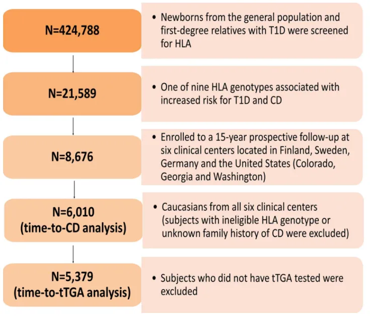
A total of 424,788 newborns from both the general population and first-degree relatives with T1D were screened for specific HLA genotypes. Of these, 21,589 had one of the nine HLA
genotypes associated with increased risk for T1D and celiac disease (S1 Table) and 8,676
eligi-ble children were enrolled to a 15-year prospective follow-up[40] (Fig 5).
The Environmental Determinants of Diabetes in the Young (TEDDY) is a prospective cohort study with the primary goal to identify environmental causes of T1D. This study was performed according to the principles of the Declaration of Helsinki. Written informed con-sent was obtained for all study participants from a parent or legal guardian. The TEDDY study was approved by local Institutional Review Boards at 6 clinical research centers (3 in the United States and 3 in Europe): University of Colorado Health Science Center, Georgia Regents Uni-versity, Pacific Northwest Diabetes Research Institute, Turku University Hospital (Finland), Institute of Diabetes Research (Germany), and Lund University (Sweden). The study is also monitored by an external evaluation committee formed by the National Institutes of Health.
Assessment of tissue transglutaminase autoantibodies (tTGA)
Sera were measured for tTGA using radioligand binding assays in two laboratories, IgA-tTGA assay at the Barbara Davis Center for Childhood Diabetes for the US samples and IgA-tTGA and IgG-tTGA assay at the University of Bristol for European samples. All positive US samples were also assayed by the Bristol lab. Levels of tTGA were expressed in arbitrary units derived
from a standard curve and were considered to be positive if the value was1.3 units. The
inter-assay coefficient of variation was 22% at both 6 units and 20 units.
Study outcomes
Persistent tTGA was defined as having two consecutive positive tTGA tests (as measured by the Bristol laboratory) taken at least three months apart. Children meeting this criterion were referred to a pediatric gastroenterologist for a clinical evaluation for celiac disease. Celiac dis-ease was defined as having an intestinal biopsy showing a Marsh score of 2 or greater by
origi-nal Marsh criteria [41]. Children who had persistent tTGA with a mean level of>100 units in
two consecutive samples but had no intestinal biopsy data, were also considered as having celiac disease for the purpose of this study [13].
Single-nucleotide polymorphism (SNP) analysis by ImmunoChip
SNPs were genotyped by the Center for Public Health Genomics at University of Virginia, using the Illumina ImmunoChip Infinium array. The ImmunoChip is a custom genotyping array of 195,806 SNPs selected from 186 regions associated with 12 autoimmune diseases. Genotype calling and quality control steps were applied to the dataset: (1) individuals with lowcall rate (<95%), or discordance with reported gender and prior genotyping were not
consid-ered in the analysis, (2) SNP markers with low call rates (<95%) were excluded, and (3)
mark-ers with allele distributions strongly deviating from Hardy-Weinberg equilibrium (HWE) in
controls (p<10−6) were discarded (except for chromosome 6 due to HLA eligibility
require-ments). This resulted in a total of 7,023 subjects with genotype data on 176,586 SNPs. Fig 5. Flow chart of study participants.The Environmental Determinants of Diabetes in the Young (TEDDY) is an international multicenter study that screened over 420,000 newborns from the general population in four different countries. The present study genotyped 195,806 SNPs on ImmunoChip in 6,010 TEDDY children to identify potential genetic factors responsible for the development of CD and country-specific differences in genetic predisposition. As shown in flow chart, a total of 6,010 subjects were included in the analysis of time-to-CD, and 5379 subjects were included in the analysis of time-to-tTGA.
Statistical analysis
The time-to-persistent tTGA and the time-to-celiac disease were the two primary outcomes analysed in this study. The time-to-persistent tTGA was defined as the age when the sample for the first tTGA positive test was collected, and the right-censored time was the age when the
participant’s last blood sample was collected for testing of tTGA. The time-to-celiac disease
was the child’s age at the time of biopsy for the diagnosis of celiac disease, or the age of the first
high-level tTGA result (defined as100 units). The right-censored time was the age of the last
TEDDY clinic visit that was confirmed to be celiac disease-free. Cox proportional hazards modelling was used to analyse the effect of each individual SNP (by genotype 0,1,2) on the out-come, after adjusting for country (as strata), gender, HLA-DPB1 genotype, HLA-DR-DQ geno-type (e.g., DR3-DQ2/DR3-DQ2, DR3-DQ2/X, DR4-DQ8/DR4-DQ8 and Other), family history (first-degree relative) of celiac disease, and principal components to account for
popu-lation stratification (ancestral heterogeneity). The Cox models were fitted using the“survival”
package in R [42]. The first four principal components were used in these analyses, calculated
from the SNP data using the SNPRelate software [43]. As the majority of the subjects were
Caucasians, and to reduce population stratification further, subjects from other races were excluded and analyses were restricted to the 6,258 Caucasians from all six clinical centers.
Subject exclusion included those who had either an ineligible HLA genotype or unknown family history (among first-degree relatives) of celiac disease. Further, subjects who did not have tTGA tested were also excluded in the analysis of time-to-persistent tTGA. As a result, 6,010 subjects were included in the analysis of time-to- celiac disease, and 5,379 subjects were
included in the analysis of time-to- persistent tTGA (Fig 5). Among the 6,010 subjects included
in the analysis of celiac disease, the median follow-up time was 5 years (interquartile range:
3.75–6.44 years). Whereas, among the 5,379 subjects included in the analysis of persistent
tTGA, the median follow-up time was 5.18 years with an interquartile range of 4.04–6.54 years.
During the follow-up, a total of 703 subjects developed persistent tTGA (US: 191 out of 1785; Finland: 167 out of 1414; Germany: 37 out of 328; Sweden: 308 out of 1852) and a total of 288 subjects were considered as having celiac disease (US: 83 out of 2028; Finland: 53 out of 1551; Germany: 9 out of 399; Sweden: 143 out of 2032). The characteristics of TEDDY participants
in the analyses of persistent tTGA (S2 Table) and celiac disease (S3 Table) are provided in the
Supplementary material.
From the 176,586 SNPs that passed quality control filters, the analysis focused on those 133,620 with minor allele frequencies of at least 0.01; thus, statistical significance for any single
SNP required a Bonferoni-corrected p<3.7x10⁻⁷. This is a highly stringent threshold as a large
number of SNPs are in linkage disequilibrium which should reduce the total number of inde-pendent tests. For the analyses of the 48 candidate SNPs that have been identified in previous studies, we considered p<10−3as suggestive evidence for confirmation (p = 0.05/48 = 10−3).
For the analyses of the 48 candidate regions, we considered p<10−4as suggestive evidence for
confirmation as multiple SNPs are tested in each region and the SNPs are in high linkage disequilibrium.
All analyses were performed using R 2.15.1. A web-based plotting toollocuszoom[44] was
used to plot HapMap CEU linkage disequilibrium r2values for additional SNPs in the
candi-date SNP regions.
Supporting Information
S1 Table. Enrolled HLA genotypes in the TEDDY study. (PDF)
S2 Table. Characteristics for Celiac Disease Autoimmunity. (PDF)
S3 Table. Characteristics for Celiac Disease. (PDF)
S4 Table. Analysis of reported celiac disease risk variants. (PDF)
S5 Table. Analysis of celiac disease risk variants only in Sweden population. (PDF)
S6 Table. Risk Variants that have been reported but are not on the IChip. (PDF)
S1 Fig. SNPs associated with tTGA risk. (PDF)
S2 Fig. SNPs associated with celiac disease risk. (PDF)
S1 Text. TEDDY Study Acknowledgments; All TEDDY sites and investigators. (PDF)
Acknowledgments
The Teddy Study Group
Lead author:Dr. Jin-Xiong She, Email:[email protected], Phone: 3410, Fax: 706-721-3688
Colorado Clinical Center:Marian Rewers, M.D., Ph.D., PI1,4,5,6,10,11, Kimberly Bautista12,
Judith Baxter9,10,12,15, Ruth Bedoy2, Daniel Felipe-Morales, Brigitte Frohnert, M.D., Patricia
Gesualdo2,6,12,14,15, Michelle Hoffman12,13,14, Rachel Karban12, Edwin Liu, M.D.13, Jill Norris,
Ph.D.2,3,12, Adela Samper-Imaz, Andrea Steck, M.D.3,14, Kathleen Waugh6,7,12,15, Hali
Wright12. University of Colorado, Anschutz Medical Campus, Barbara Davis Center for
Child-hood Diabetes.
Georgia/Florida Clinical Center:Jin-Xiong She, Ph.D., PI1,3,4,11,†, Desmond Schatz, M.
D.4,5,7,8, Diane Hopkins12, Leigh Steed12,13,14,15, Jamie Thomas6,12, Katherine Silvis2, Michael
Haller, M.D.14, Meena Shankar2, Eleni Sheehan, Melissa Gardiner, Richard McIndoe, Ph.D.,
Haitao Liu, M.D.†, John Nechtman†, Ashok Sharma, Joshua Williams, Gabriela Foghis,
Ste-phen W. Anderson, M.D.^. Medical College of Georgia, Georgia Regents University.
University of Florida,†Jinfiniti Biosciences LLC, Augusta, GA,^Pediatric Endocrine
Associ-ates, Atlanta, GA.
Germany Clinical Center:Anette G. Ziegler, M.D., PI1,3,4,11, Andreas Beyerlein Ph.D.2,
Ezio Bonifacio Ph.D.5, Michael Hummel, M.D.13, Sandra Hummel, Ph.D.2, Kristina
Foterek¥2, Mathilde Kersting, Ph.D.¥2, Annette Knopff7, Sibylle Koletzko, M.D.¶13, Claudia
Peplow12, Roswith Roth, Ph.D.9, Joanna Stock9,12, Elisabeth Strauss12, Katharina Warncke, M.
D.14, Christiane Winkler, Ph.D.2,12,15. Forschergruppe Diabetes e.V. and Institute of Diabetes
Research, Helmholtz Zentrum München, and Klinikum rechts der Isar, Technische Universität
München.Center for Regenerative Therapies, TU Dresden,¶Dr. von Hauner Children´s
Hos-pital, Department of Gastroenterology, Ludwig Maximillians University Munich,¥Research
Finland Clinical Center:Jorma Toppari, M.D., Ph.D., PI¥^1,4,11,14, Olli G. Simell, M.D., Ph.
D., PI¥^1,4,11,13, Annika Adamsson, Ph.D.^12, Heikki Hyöty, M.D., Ph.D.±6, Jorma Ilonen, M.
D., Ph.D.¥ ¶3, Miia Kähönenμ¤, Mikael Knip, M.D., Ph.D.±5, Annika Koivu¥^, Mirva
Koreasalo±§2, Kalle Kurppa, M.D., Ph.D.±13, Maria Lönnrot, M.D., Ph.D.±6, Elina
Mänty-mäki¥^, Katja Multasuo
μ¤, Juha Mykkänen, Ph.D.^¥ 3, Tiina Niininen±
12
, Mia Nyblom±,
Petra Rajala^, Jenna Rautanen±§, Anne Riikonen±, Minna Romo¥^, Satu Simell, M.D., Ph.D.^
±13, Tuula Simell, Ph.D., Ville Simell^¥13, Maija Sjöberg¥^12,14, Aino Stenius
μ¤12, Eeva
Varjo-nen¥^12, Riitta Veijola, M.D., Ph.D.μ¤14, Suvi M. Virtanen, M.D., Ph.D.±§2, Mari Åkerlund±§.
¥University of Turku,University of Tampere,μUniversity of Oulu,^Turku University
Hospi-tal, Hospital District of Southwest Finland,±Tampere University Hospital,¤Oulu University
Hospital,§National Institute for Health and Welfare, Finland,¶University of Kuopio.
Sweden Clinical Center:Åke Lernmark, Ph.D., PI1,3,4,5,6,8,10,11,15, Daniel Agardh, M.D., Ph.
D.13, Carin Andrén Aronsson2,13, Maria Ask, Jenny Bremer, Ulla-Marie Carlsson, Corrado
Cilio, Ph.D., M.D.5, Camilla Ekstrand, Emelie Ericson-Hallström2, Lina Fransson, Thomas
Gard, Joanna Gerardsson, Rasmus Håkansson, Monica Hansen, Gertie Hansson12, Susanne
Hyberg, Fredrik Johansen, Berglind Jonasdottir M.D., Linda Jonsson, Helena Elding Larsson
M.D., Ph.D.6,14, Barbro Lernmark, Ph.D., Maria Månsson-Martinez, Maria Markan,
Theodo-sia Massadakis, Jessica Melin12, Zeliha Mestan, Kobra Rahmati, Anita Ramelius, Falastin
Salami, Monica Sedig Järvirova, Sara Sibthorpe, Birgitta Sjöberg, Ulrica Swartling, Ph.D.9,12,
Erika Trulsson, Carina Törn, Ph.D.3,15, Anne Wallin, Åsa Wimar12,14, Sofie Åberg. Lund
University.
Washington Clinical Center:William A. Hagopian, M.D., Ph.D., PI1,3,4, 5, 6,7,11,13, 14, Xiang
Yan, M.D., Michael Killian6,7,12,13, Claire Cowen Crouch12,14,15, Jennifer Skidmore2, Stephen
Ayres, Kayleen Dunson, Diana Heaney, Rachel Hervey, Corbin Johnson, Rachel Lyons, Arlene Meyer, Denise Mulenga, Emma Schulte, Elizabeth Scott, Joshua Stabbert, John Willis. Pacific Northwest Diabetes Research Institute.
Pennsylvania Satellite Center:Dorothy Becker, M.D., Margaret Franciscus, MaryEllen
Dal-magro-Elias Smith2, Ashi Daftary, M.D., Mary Beth Klein, Chrystal Yates. Children’s Hospital
of Pittsburgh of UPMC.
Data Coordinating Center:Jeffrey P. Krischer, Ph.D.,PI1,4,5,10,11, Michael Abbondondolo,
Sarah Austin-Gonzalez, Rasheedah Brown12,15, Brant Burkhardt, Ph.D.5,6, Martha
Butter-worth2, David Cuthbertson, Christopher Eberhard, Steven Fiske9, Dena Garcia, Veena Gowda,
David Hadley, Ph.D.3,13, Hye-Seung Lee, Ph.D.1,2,13,15, Shu Liu, Xiang Liu, Ph.D.2,9,12, Kristian
Lynch, Ph.D.5,6,9,15, Jamie Malloy, Cristina McCarthy12,15, Wendy McLeod2,5,6,13,15, Chris
Shaffer, Laura Smith, Ph.D.9,12, Susan Smith12,15, Roy Tamura, Ph.D.1,2,13, Ulla Uusitalo, Ph.
D.2,15, Kendra Vehik, Ph.D.4,5,6,14,15, Ponni Vijayakandipan, Keith Wood, Jimin Yang, Ph.D.,
R.D.2,15. University of South Florida.
Project scientist:Beena Akolkar, Ph.D.1,3,4,5,6,7,10,11. National Institutes of Diabetes and Digestive and Kidney Diseases.
Other contributors:Kasia Bourcier, Ph.D.5, National Institutes of Allergy and Infectious
Diseases. Thomas Briese, Ph.D.6,15, Columbia University. Suzanne Bennett Johnson, Ph.D.9,12,
Florida State University. Steve Oberste, Ph.D.6, Centers for Disease Control and Prevention.
Eric Triplett, Ph.D.6, University of Florida.
Autoantibody Reference Laboratories:Liping Yu, M.D.^5, Dongmei Miao, M.D.^, Polly
Bingley, M.D., FRCP5, Alistair Williams, Kyla Chandler, Saba Rokni, Joanna Boldison,
Jacob Butterly, Gabriella Carreno, Claire Caygill, Ivey Geoghan, Anna Long, Molly
Payne, James Pearson, Sophie Ridewood, Rebecca Wyatt.^Barbara Davis Center for
Child-hood Diabetes, University of Colorado Denver,School of Clinical Sciences, University of
Bris-tol UK.
Cortisol Laboratory:Elisabeth Aardal Eriksson, M.D., Ph.D., Ing-Marie Lundgren, Ewa Lönn Karlsson, Dzeneta Nezirevic Dernroth, Ph.D. Department of Clinical Chemistry, Linkö-ping University Hospital, LinköLinkö-ping, Sweden
Dietary Biomarkers Laboratory:Iris Erlund, Ph.D.2, Irma Salminen, Jouko Sundvall, Jaana Leiviskä, Mari Lehtonen, Ph.D. National Institute for Health and Welfare, Helsinki, Finland.
HbA1c Laboratory:Randie R. Little, Ph.D., Alethea L. Tennill. Diabetes Diagnostic Labora-tory, Dept. of Pathology, University of Missouri School of Medicine.
HLA Reference Laboratory:Henry Erlich, Ph.D.3, Steven J. Mack, Ph.D., Anna Lisa Fear.
Center for Genetics, Children’s Hospital Oakland Research Institute.
Metabolomics Laboratory:Oliver Fiehn, Ph.D., Bill Wikoff, Ph.D., Brian Defelice, Dmitry Grapov, Ph.D., Tobias Kind, Ph.D., Mine Palazoglu, Luis Valdiviez, Benjamin Wancewicz, Gert Wohlgemuth, Joyce Wong. UC Davis Metabolomics Center.
Microbiome and Viral Metagenomics Laboratory:Joseph F. Petrosino, Ph.D.6. Alkek
Cen-ter for Metagenomics and Microbiome Research, Department of Molecular Virology and Microbiology, Baylor College of Medicine.
OGTT Laboratory:Santica M. Marcovina, Ph.D., Sc.D., Vinod P. Gaur, Ph.D., Northwest Lipid Metabolism and Diabetes Research Laboratories, University of Washington.
Proteomics Laboratory:Richard D. Smith, Ph.D., Thomas O. Metz, Ph.D., Charles Ansong, Ph.D., Bobbie-Jo Webb-Robertson, Ph.D., and Hugh D. Mitchell, Ph.D. Pacific Northwest National Laboratory.
Repository:Heather Higgins, Sandra Ke. NIDDK Biosample Repository at Fisher BioServices.
RNA Laboratory and Gene Expression Laboratory:Jin-Xiong She, Ph.D., PI1,3,4,11,
Rich-ard McIndoe, Ph.D., Haitao Liu, M.D., John Nechtman, Yansheng Zhao, Na Jiang, M.D. Jinfi-niti Biosciences, LLC.
SNP Laboratory:Stephen S. Rich, Ph.D.3, Wei-Min Chen, Ph.D.3, Suna OnengutGumuscu,
Ph.D.3, Emily Farber, Rebecca Roche Pickin, Ph.D., Jordan Davis, Dan Gallo, Jessica Bonnie,
Paul Campolieto. Center for Public Health Genomics, University of Virginia.
Committees:1Ancillary Studies,2Diet,3Genetics,4Human Subjects/Publicity/Publications,
5Immune Markers,6infectious Agents,7Laboratory Implementation,8Maternal Studies,9
Psy-chosocial,10Quality Assurance,11Steering,12Study Coordinators,13Celiac Disease,14Clinical
Implementation,15Quality Assurance Subcommittee on Data Quality.
Author Contributions
Conceived and designed the experiments: AS XL DH DA WH SSR JXS. Performed the experi-ments: WMC SOG SSR. Analyzed the data: AS XL DH DA WH JXS. Contributed reagents/ materials/analysis tools: VS MR AGZ ÅL OS JT JPK BA SSR. Wrote the paper: AS XL DH DA EL WH SSR JXS.
References
1. DeMarchi M, Borelli I, Olivetti E, Richiardi P, Wright P, Ansaldi N, et al. (1979) Two HLA-D and DR alleles are associated with coeliac disease. Tissue Antigens 14: 309–316. PMID:94703
2. Ploski R, Ek J, Thorsby E, Sollid LM (1993) On the HLA-DQ(alpha 1*0501, beta 1*0201)-associated susceptibility in celiac disease: a possible gene dosage effect of DQB1*0201. Tissue Antigens 41: 173–177. PMID:8362409
3. Margaritte-Jeannin P, Babron MC, Bourgey M, Louka AS, Clot F, Percopo S, et al. (2004) HLA-DQ rela-tive risks for coeliac disease in European populations: a study of the European Genetics Cluster on Coeliac Disease. Tissue Antigens 63: 562–567. PMID:15140032
5. Dubois PC, Trynka G, Franke L, Hunt KA, Romanos J, Curtotti A, et al. (2010) Multiple common vari-ants for celiac disease influencing immune gene expression. Nat Genet 42: 295–302. doi:10.1038/ng. 543PMID:20190752
6. van Heel DA, Hunt K, Greco L, Wijmenga C (2005) Genetics in coeliac disease. Best Pract Res Clin Gastroenterol 19: 323–339. PMID:15925839
7. van Heel DA, Franke L, Hunt KA, Gwilliam R, Zhernakova A, Inouye M, et al. (2007) A genome-wide association study for celiac disease identifies risk variants in the region harboring IL2 and IL21. Nat Genet 39: 827–829. PMID:17558408
8. Trynka G, Hunt KA, Bockett NA, Romanos J, Mistry V, Szperl A, et al. (2011) Dense genotyping identi-fies and localizes multiple common and rare variant association signals in celiac disease. Nat Genet 43: 1193–1201. doi:10.1038/ng.998PMID:22057235
9. Garner C, Ahn R, Ding YC, Steele L, Stoven S, Green PH, et al. (2014) Genome-Wide Association Study of Celiac Disease in North America Confirms FRMD4B as New Celiac Locus. PLoS One 9: e101428. doi:10.1371/journal.pone.0101428PMID:24999842
10. Kumar V, Wijmenga C, Withoff S (2012) From genome-wide association studies to disease mecha-nisms: celiac disease as a model for autoimmune diseases. Semin Immunopathol 34: 567–580. doi: 10.1007/s00281-012-0312-1PMID:22580835
11. Kang JY, Kang AH, Green A, Gwee KA, Ho KY (2013) Systematic review: worldwide variation in the fre-quency of coeliac disease and changes over time. Aliment Pharmacol Ther 38: 226–245. doi:10.1111/ apt.12373PMID:23782240
12. (2007) The Environmental Determinants of Diabetes in the Young (TEDDY) study: study design. Pediatr Diabetes 8: 286–298. PMID:17850472
13. Liu E, Lee HS, Aronsson CA, Hagopian WA, Koletzko S, Rewers MJ, et al. (2014) Risk of pediatric celiac disease according to HLA haplotype and country. N Engl J Med 371: 42–49. doi:10.1056/ NEJMoa1313977PMID:24988556
14. Hadley D, Hagopian W, Liu E, She JX, Simell O, Beena Akolkar A-GZ, Marian Rewers, Krischer Jeffrey P., Wei-Min Chen, Suna Onengut-Gumuscu, Teodorica L., et al. (2015) HLA-DPB1*04:01 protects genetically susceptible children from celiac disease autoimmunity in the TEDDY study. American Jour-nal of Gastroenterology.
15. Welter D, MacArthur J, Morales J, Burdett T, Hall P, Junkins H, et al. (2014) The NHGRI GWAS Cata-log, a curated resource of SNP-trait associations. Nucleic Acids Res 42: D1001–1006. doi:10.1093/ nar/gkt1229PMID:24316577
16. Ostensson M, Monten C, Bacelis J, Gudjonsdottir AH, Adamovic S, Ek J, et al. (2013) A possible mech-anism behind autoimmune disorders discovered by genome-wide linkage and association analysis in celiac disease. PLoS One 8: e70174. doi:10.1371/journal.pone.0070174PMID:23936387
17. Hunt KA, Zhernakova A, Turner G, Heap GA, Franke L, Bruinenberg M, et al. (2008) Newly identified genetic risk variants for celiac disease related to the immune response. Nat Genet 40: 395–402. doi: 10.1038/ng.102PMID:18311140
18. Holopainen P, Arvas M, Sistonen P, Mustalahti K, Collin P, Maki M, et al. (1999) CD28/CTLA4 gene region on chromosome 2q33 confers genetic susceptibility to celiac disease. A linkage and family-based association study. Tissue Antigens 53: 470–475. PMID:10372542
19. Chen R, Stahl EA, Kurreeman FA, Gregersen PK, Siminovitch KA, Worthington J, et al. (2011) Fine mapping the TAGAP risk locus in rheumatoid arthritis. Genes Immun 12: 314–318. doi:10.1038/gene. 2011.8PMID:21390051
20. Smyth DJ, Plagnol V, Walker NM, Cooper JD, Downes K, Yang JH, et al. (2008) Shared and distinct genetic variants in type 1 diabetes and celiac disease. N Engl J Med 359: 2767–2777. doi:10.1056/ NEJMoa0807917PMID:19073967
21. Patsopoulos NA, Esposito F, Reischl J, Lehr S, Bauer D, Heubach J, et al. (2011) Genome-wide meta-analysis identifies novel multiple sclerosis susceptibility loci. Ann Neurol 70: 897–912. doi:10.1002/ ana.22609PMID:22190364
22. Salvati VM, MacDonald TT, Bajaj-Elliott M, Borrelli M, Staiano A, Auricchio S, et al. (2002) Interleukin 18 and associated markers of T helper cell type 1 activity in coeliac disease. Gut 50: 186–190. PMID: 11788557
23. Hoffenberg EJ, Bao F, Eisenbarth GS, Uhlhorn C, Haas JE, Sokol RJ, et al. (2000) Transglutaminase antibodies in children with a genetic risk for celiac disease. J Pediatr 137: 356–360. PMID:10969260
24. Auricchio R, Tosco A, Piccolo E, Galatola M, Izzo V, Maglio M, et al. (2014) Potential celiac children: 9-year follow-up on a gluten-containing diet. Am J Gastroenterol 109: 913–921. doi:10.1038/ajg.2014.77 PMID:24777149
25. Simmons JH, Klingensmith GJ, McFann K, Rewers M, Ide LM, Taki I, et al. (2011) Celiac autoimmunity in children with type 1 diabetes: a two-year follow-up. J Pediatr 158: 276–281 e271. doi:10.1016/j. jpeds.2010.07.025PMID:20817171
26. Jansen MA, Kiefte-de Jong JC, Gaillard R, Escher JC, Hofman A, Jaddoe VW, et al. (2015) Growth tra-jectories and bone mineral density in anti-tissue transglutaminase antibody-positive children: the Gen-eration R Study. Clin Gastroenterol Hepatol 13: 913–920 e915. doi:10.1016/j.cgh.2014.09.032PMID: 25245626
27. Metzger MH, Heier M, Maki M, Bravi E, Schneider A, Lowel H, et al. (2006) Mortality excess in individu-als with elevated IgA anti-transglutaminase antibodies: the KORA/MONICA Augsburg cohort study 1989–1998. Eur J Epidemiol 21: 359–365. PMID:16649072
28. Kurppa K, Ashorn M, Iltanen S, Koskinen LL, Saavalainen P, Koskinen O, et al. (2010) Celiac disease without villous atrophy in children: a prospective study. J Pediatr 157: 373–380, 380 e371. doi:10. 1016/j.jpeds.2010.02.070PMID:20400102
29. Juuti-Uusitalo K, Maki M, Kainulainen H, Isola J, Kaukinen K (2007) Gluten affects epithelial differentia-tion-associated genes in small intestinal mucosa of coeliac patients. Clin Exp Immunol 150: 294–305. PMID:17888028
30. Laparra Llopis JM, Sanz Herranz Y (2010) Gliadins induce TNFalpha production through cAMP-depen-dent protein kinase A activation in intestinal cells (Caco-2). J Physiol Biochem 66: 153–159. doi:10. 1007/s13105-010-0020-zPMID:20514534
31. Raychaudhuri S, Remmers EF, Lee AT, Hackett R, Guiducci C, Burtt NP, et al. (2008) Common vari-ants at CD40 and other loci confer risk of rheumatoid arthritis. Nat Genet 40: 1216–1223. doi:10.1038/ ng.233PMID:18794853
32. Cooper JD, Smyth DJ, Smiles AM, Plagnol V, Walker NM, Allen JE, et al. (2008) Meta-analysis of genome-wide association study data identifies additional type 1 diabetes risk loci. Nat Genet 40: 1399–1401. doi:10.1038/ng.249PMID:18978792
33. Coenen MJ, Trynka G, Heskamp S, Franke B, van Diemen CC, Smolonska J, et al. (2009) Common and different genetic background for rheumatoid arthritis and coeliac disease. Hum Mol Genet 18: 4195–4203. doi:10.1093/hmg/ddp365PMID:19648290
34. Kondrashova A, Mustalahti K, Kaukinen K, Viskari H, Volodicheva V, Haapala AM, et al. (2008) Lower economic status and inferior hygienic environment may protect against celiac disease. Ann Med 40: 223–231. doi:10.1080/07853890701678689PMID:18382888
35. Adlercreutz EH, Svensson J, Hansen D, Buschard K, Lernmark A, Mortensen HB, et al. (2015) Preva-lence of celiac disease autoimmunity in children with type 1 diabetes: regional variations across the Oresund strait between Denmark and southernmost Sweden. Pediatr Diabetes 16: 504–509. doi:10. 1111/pedi.12200PMID:25131687
36. Suzuki A, Yamada R, Chang X, Tokuhiro S, Sawada T, Suzuki M, et al. (2003) Functional haplotypes of PADI4, encoding citrullinating enzyme peptidylarginine deiminase 4, are associated with rheumatoid arthritis. Nat Genet 34: 395–402. PMID:12833157
37. Caponi L, Petit-Teixeira E, Sebbag M, Bongiorni F, Moscato S, Pratesi F, et al. (2005) A family based study shows no association between rheumatoid arthritis and the PADI4 gene in a white French popula-tion. Ann Rheum Dis 64: 587–593. PMID:15485997
38. Martinez A, Valdivia A, Pascual-Salcedo D, Lamas JR, Fernandez-Arquero M, Balsa A, et al. (2005) PADI4 polymorphisms are not associated with rheumatoid arthritis in the Spanish population. Rheuma-tology (Oxford) 44: 1263–1266.
39. Koplin JJ, Peters RL, Ponsonby AL, Gurrin LC, Hill D, Tang ML, et al. (2014) Increased risk of peanut allergy in infants of Asian-born parents compared to those of Australian-born parents. Allergy.
40. Hagopian WA, Erlich H, Lernmark A, Rewers M, Ziegler AG, Simell O, et al. (2011) The Environmental Determinants of Diabetes in the Young (TEDDY): genetic criteria and international diabetes risk screen-ing of 421 000 infants. Pediatr Diabetes 12: 733–743. doi:10.1111/j.1399-5448.2011.00774.xPMID: 21564455
41. Oberhuber G (2000) Histopathology of celiac disease. Biomed Pharmacother 54: 368–372. PMID: 10989975
42. Therneau T, (2015) A Package for Survival Analysis in S.
43. Zheng X, Levine D, Shen J, Gogarten SM, Laurie C, Weir BS (2012) A high-performance computing toolset for relatedness and principal component analysis of SNP data. Bioinformatics 28: 3326–3328. doi:10.1093/bioinformatics/bts606PMID:23060615