(Nannomys) setulosus
from Coˆte d’Ivoire: Implications
for Evolution of Arenaviruses in Africa
David Coulibaly-N’Golo1,2., Bernard Allali1., Ste´phane K. Kouassi1
, Elisabeth Fichet-Calvet3, Beate Becker-Ziaja2, Toni Rieger2, Stephan O¨ lschla¨ger2, Hernri Dosso4, Christiane Denys3, Jan ter Meulen5,6, Chantal Akoua-Koffi1, Stephan Gu¨nther2*
1Laboratoire des Arbovirus/Ente´rovirus, Institut Pasteur de Coˆte d’Ivoire, Abidjan, Coˆte d’Ivoire,2Department of Virology, Bernhard-Nocht-Institute for Tropical Medicine, Hamburg, Germany,3De´partement Syste´matique et Evolution, Museum National d’Histoire Naturelle, Paris, France,4Centre de Recherche en Ecologie, Universite d’Abobo-Adjame, Abidjan, Coˆte d’Ivoire,5Institute of Virology, Philipps University, Marburg, Germany,6Merck Research Laboratories, West Point, Pennsylvania, United States of America
Abstract
This study aimed to identify new arenaviruses and gather insights in the evolution of arenaviruses in Africa. During 2003 through 2005, 1,228 small mammals representing 14 different genera were trapped in 9 villages in south, east, and middle west of Coˆte d’Ivoire. Specimens were screened by pan-Old World arenavirus RT-PCRs targeting S and L RNA segments as well as immunofluorescence assay. Sequences of two novel tentative species of the family Arenaviridae, Menekre and Gbagroube virus, were detected inHylomyscussp. andMus (Nannomys) setulosus, respectively. Arenavirus infection ofMus (Nannomys) setulosus was also demonstrated by serological testing. Lassa virus was not found, although 60% of the captured animals were Mastomys natalensis. Complete S RNA and partial L RNA sequences of the novel viruses were recovered from the rodent specimens and subjected to phylogenetic analysis. Gbagroube virus is a closely related sister taxon of Lassa virus, while Menekre virus clusters with the Ippy/Mobala/Mopeia virus complex. Reconstruction of possible virus–host co-phylogeny scenarios suggests that, within the African continent, signatures of co-evolution might have been obliterated by multiple host-switching events.
Citation:Coulibaly-N’Golo D, Allali B, Kouassi SK, Fichet-Calvet E, Becker-Ziaja B, et al. (2011) Novel Arenavirus Sequences inHylomyscussp. andMus (Nannomys) setulosusfrom Coˆte d’Ivoire: Implications for Evolution of Arenaviruses in Africa. PLoS ONE 6(6): e20893. doi:10.1371/journal.pone.0020893
Editor:Wanda Markotter, University of Pretoria, South Africa
ReceivedFebruary 9, 2011;AcceptedMay 12, 2011;PublishedJune 9, 2011
Copyright:ß2011 Coulibaly-N’Golo et al. This is an open-access article distributed under the terms of the Creative Commons Attribution License, which permits unrestricted use, distribution, and reproduction in any medium, provided the original author and source are credited.
Funding:The study was supported by grants INCO-DEV ICA4-CT2002-10050, 228292 (European Virus Archive), and Marie-Curie PIEF-GA-2009-235164 from the European Community (http://ec.europa.eu/research/fp6/index_en.cfm; http://ec.europa.eu/research/fp7/), and grant GU 883/1-1 from the German Research Foundation (DFG) (http://www.dfg.de/index.jsp). The funders had no role in study design, data collection and analysis, decision to publish, or preparation of the manuscript. The Department of Virology of the Bernhard-Nocht-Institute is a WHO Collaborating Centre for Arbovirus and Haemorrhagic Fever Reference and Research (DEU-000115).
Competing Interests:The authors have declared that no competing interests exist.
* E-mail: [email protected]
.These authors contributed equally to this work.
Introduction
Arenaviridaeare negative strand RNA viruses hosted by rodents of the superfamily Muroidea. Previous comparisons of arenavirus phylogeny with rodent host phylogeny provided several examples in which virus–host co-speciation was potentially occurring during evolution [1]. Therefore, current hypotheses assume that arena-viruses share a long-term evolutionary relationship with their rodent hosts, an evolutionary model called co-evolution. Africa is home to various Old World arenaviruses (OWA) [2,3,4,5,6,7,8], of which Lassa and Lujo virus cause hemorrhagic fever in humans [9,10,11]. Lassa virus is endemic in the countries of Nigeria, Liberia, Sierra Leone, and Guinea [1,9,12] and was recently also detected in Mali [13,14]. Whether OWAs including Lassa virus are endemic in Coˆte d’Ivoire is not known. Seroepidemiological studies and an imported case of Lassa fever indicate that arena-viruses circulate somewhere in the region comprising Ghana, Coˆte d’Ivoire, and Burkina Faso [15,16]. We conducted a systematic
search for OWAs in wildlife in Coˆte d’Ivoire. While Lassa virus was not found, two novel arenaviruses were identified and cha-racterized molecularly. The sequence data were used to gather new insights in the evolution of arenaviruses in Africa.
Results
Prefecture Divo. The six virus RNA-positive animals were genotyped by sequencing the mitochondrial cytochrome B and 16S rRNA genes. Phylogenetic analysis indicates that the rodents from Menekre presumably constitute a new Hylomyscus species (details to be reported elsewhere), while the rodents from Gbagroube areMus (Nannomys) setulosus. The virus RNA-positive
Hylomyscusspecies had been trapped in cocoa farm and forest, and the positive Mus (Nannomys) setulosus in banana plantations and forest. Both are usually forest species living in zones with high rainfall. Additional testing of allHylomyscussp. (n = 3) and allMus (Nannomys) setulosus (n = 47) that were negative in the initial screening using RT-PCRs specific for the new virus sequences did not increase the number of positive animals. Thus, virus prevalence was 100% (3/3) inHylomyscus sp. from Menekre and 12% (3/25) inMus (Nannomys) setulosusfrom Gbagroube (Table 1). Sequencing the GPC and L gene PCR fragments (,960 and 340
nucleotides, respectively) revealed that all strains from Hylomyscus
sp. constitute a new arenavirus, called Menekre virus, while all strains from Mus (Nannomys) setulosusconstitute another new are-navirus, called Gbagroube virus. Attempts to isolate the viruses in
cell culture or mice were not successful (see Materials and Methods for details). Therefore, the complete 3.5-kb S RNA segment of the viruses was sequenced directly from the rodent specimens using pan-OWA PCR primers targeting the nucleoprotein (NP) gene and a set of virus-specific primers. Full-length S RNA sequences (ex-cluding the conserved 19 nucleotides at the termini) were ob-tained for Menekre virus strain CIV1227 (3423 nucleotides) and Gbagroube virus strain CIV608 (3347 nucleotides), and partial NP sequences were obtained for Menekre virus strains CIV839 and CIV843 and Gbagroube virus strains CIV674 and CIV1290 (,1200 nucleotides). Virus and associated host sequences reported
in this article have been submitted to GenBank (Table 2). To provide additional evidence for the circulation of arena-viruses in Coˆte d’Ivoire, all PCR-negative animals captured in the country were tested for the presence of anti-OWA antibodies by immunofluorescence assay (Table 1). Lassa virus was used as an antigen due to its known cross-reactivity with immune sera from animals infected with other OWAs [3,6,18]. While the remaining threeHylomyscussp. were antibody-negative, 7 of 50Mus (Nannomys) setulosus were positive for anti-OWA IgG antibodies. All seven
Table 1.Small mammals from Coˆte d’Ivoire tested by pan-Old World arenavirus RT-PCR, Lassa virus-specific RT-PCR, and Old World arenavirus immunofluorescence assaya.
No. tested (RT-PCR-positive;antibody-positive) per sampling sitec
Genus or speciesb Ah Ba Bl Gb Ko La Me Sa To Total
Crociduraspp. 11 4 5 15 (0;1) 4 2 12 6 2 61 (0;1)
Dasymyssp. 1 – – – – – – – – 1
Gerbilliscussp. 5 – – – 1 10 – 1 – 17
Grammomyssp. 1 – – – – – – – – 1
Graphiurussp. – – 1 1 – – – – – 2
Hylomyscussp. – – 1 – – 2 3 (3;0)d – – 6 (3;0)
Lemniscomysspp. 15 2 – – 1 8 – 1 – 27
Lophuromys sikapusi 17 (0;2) 4 8 9 4 – 7 – 9 58 (0;2)
Malacomyssp. – – 1 – – 3 1 – – 5
Mastomys erythroleucus 2 – – 1 – 1 – 3 – 7
Mastomys natalensise 221 (0;1) – 97 164 12 – 104 (0;2) 128 (0;2) 3 729 (0;5)
Mus musculus – – – – – – 4 – – 4
Mus (Nannomys) baouleif – – – – – 2 – 1 – 3
Mus (Nannomys) mattheyif – – – – – – – 1 – 1
Mus (Nannomys) minutoidesf 3 4 12 (0;2) 15 1 (0;1) 2 3 (0;1) 2 7 49 (0;4)
Mus (Nannomys) musculoidesf – – – – – – – 1 – 1
Mus (Nannomys) setulosusf – 5 2 25 (3;7)g 2 3 – – 13 50 (3;7)
Praomys(formerlyMyomys)daltoni – 1 – 2 42 – – 2 27 74
Praomysspp. 5 1 4 4 9 4 – 3 2 32
Rattus rattus 14 – 6 12 – – 16 – – 48
Uranomys ruddi 46 – – – 1 4 – 1 – 52
Total 341 (0;3) 21 137 (0;2) 248 (3;8) 77 (0;1) 41 150 (3;3) 150 (0;2) 63 1228 (6;19)
aCollections with RT-PCR-positive samples as well as the respective animals are indicated in boldface.
bTaxonomic classification is based on morphological field data, if not otherwise indicated.
cThe number of positive animals is given in parentheses in the order: RT-PCR-positive; immunofluorescence assay-positive. If none of the animals was positive in either
test, only the number of tested animals is indicated. Sampling sites: Ah, Ahouakro; Ba, Bakro; Bl, Blodi; Gb, Gbagroube; Ko, Kotokossou; La, Lamto; Me, Menekre; Sa, Sanguiebo; To, Touzoukro.
dSamples CIV839, CIV843, and CIV1227 tested positive in pan-Old World arenavirus (OWA) large (L) and glycoprotein precursor (GPC) gene RT-PCRs. eThe mitochondrial cytochrome B gene of a representative subset of 73 animals, including the antibody-positive animals, from Ahouakro, Blodi, Gbagroube,
Kotokossou, Menekre, and Sanguiebo was sequenced. All animals sequenced were genetically confirmed to beMastomys natalensis.
fAnimals were classified by sequencing the cytochrome B gene. Cytogenetic, molecular, and morphometrical analysis has been published for a subset [47].
gSamples CIV608, CIV674, and CIV1290 tested positive in pan-OWA L and GPC gene RT-PCRs. Seven further animals were positive for anti-OWA IgG antibodies.
animals had been captured in Gbagroube in March, August, and November 2005, resulting in an overall seroprevalence of 28% (7 of 25) in this area. Thus, serological testing confirms arenavirus infections inMus (Nannomys) setulosusin Gbagroube. In addition, 12 animals of other species, includingCrociduraspp. (1/61),Lophuromys sikapusi (2/58), Mastomys natalensis (5/729), and Mus (Nannomys) minutoides(4/49), were seropositive (Table 1).
Genetic distance analysis revealed that Gbagroube virus is closely related to Lassa virus (Table 3). The minimum amino acid difference (uncorrectedp-distance) to Lassa virus in NP (14.1%) slightly exceeds the maximum intra-species genetic distance in OWA complex (12.1%). However, the minimum distance in GPC (9.8%) is smaller than the maximum intra-species difference within the Mopeia and lymphocytic choriomeningitis virus (LCMV) clades (11.9% and 20.9%, respectively). Menekre virus is virtually equidistant to Lassa, Gbagroube, Mopeia, Mobala, and Ippy virus (25.4–29.7% in NP and 22.2–25.5% in GPC) (Table 3). We
consider Gbagroube and Menekre virus new species of the family
Arenaviridae, since both exceed the proposed species-differentiating cut-off value of 12% amino acid difference in NP [19] and are associated with specific rodents that have so far not been described as arenavirus host species. The fact that non-identical sequences of Menekre virus and Gbagroube virus were found in three
Hylomyscussp. and threeMus (Nannomys) setulosus, respectively, and in none of the other animal species strongly suggests that the virus sequences were recovered from the natural host species. The high prevalence of anti-OWA antibodies in Mus (Nannomys) setulosus
from the Gbagroube village lend further support to this conclusion. In the phylogeny, Gbagroube virus has a sister relationship with Lassa virus in GPC, NP, and L gene trees (Fig. 2), which is consistent with the small genetic distance between both viruses. The most recent common ancestor of the two clades is supported by posterior and bootstrap values. The position of Menekre virus in the OWA trees is ambiguous, consistent with the genetic equi-distance to all viruses except Merino Walk, LCM, and Lujo virus. It shows a sister relationship with Ippy virus in GPC gene tree, with the Lassa/Gbagroube/Mopeia/Mobala virus clade in NP gene tree, and with Mobala virus in L gene tree (Fig. 2). Taken together, though the precise phylogenetic position of Menekre virus could not be determined, it appears to belong to the group of presumably apathogenic African OWAs. A sequence feature supporting this conclusion is the presence of a second stem-loop structure in the intergenic region of S RNA of Menekre virus, which otherwise occurs only in Mopeia virus [20].
The identification of arenaviruses in rodents so far not im-plicated as OWA host species led us to investigate how the new data fit with current hypotheses on arenavirus–host co-evolution [1,21]. The phylogenies of the OWAs were compared and reconciled with those of their hosts using the CoRe-PA 0.3 program [22]. As the virus phylogeny calculated with NP gene sequences was most stable and inferred by both BEAST and PhyML, it was used for the co-phylogeny analysis. The host tree was compiled from literature data [23,24,25,26,27]. All rodents known to be associated with an OWA were included [2,3,4,5,6,8,28,29]. Juxtaposition of virus and host trees showed no obvious congruence between them (Fig. 3A). Reconciliation of the virus tree with the host tree revealed that a maximum of 5 co-speciation events (joint divergence of both host and virus) might have happened during evolution. The maximum of 5 co-speciations was found by CoRe-PA irrespective of whether a pre-defined cost model was used or optimal co-phylogenies were searched for with automatically calculated cost values. In addition to 5 co-speciations, best-fit scenarios included 4 host switches (transfer of virus from one host to another host that is not itself the
Table 2.Sequences generated in this study.
Virus genes
GenBank accession no.
Host genes
GenBank accession no.
Specimen Virus GPC NP L Host Cyt-B 16S rRNA
CIV608 Gbagroube GU830848a GU830848a GU830849 Mus setulosus GU830865 GU830864
CIV674 Gbagroube GU830850 GU830851 GU830852 Mus setulosus GU830867 GU830866
CIV1290 Gbagroube GU830853 GU830854 GU830855 Mus setulosus GU830869 GU830868
CIV839 Menekre GU830856 GU830857 GU830858 Hylomyscussp. GU830845 GU830844
CIV843 Menekre GU830859 GU830860 GU830861 Hylomyscussp. GU830871 GU830870
CIV1227 Menekre GU830862a GU830862a GU830863
Hylomyscussp. GU830847 GU830846
aComplete 3.5-kb S RNA sequences. All other virus sequences are partial.
doi:10.1371/journal.pone.0020893.t002
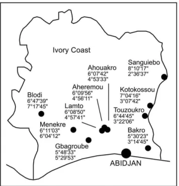
Figure 1. Map of Coˆte d’Ivoire showing the capture sites.
Latitude North and longitude West coordinates are given below the name of the villages.
immediate descendant of the source host) and 3 sorting events (apparent absence of virus in the descendants of a host that had previously been associated with virus) [30]. One of several possible co-phylogeny scenarios is shown in Fig. 3B. To test if the observed frequency of 5 co-speciations is biologically meaningful, random-ization tests implemented in CoRe-PA were performed. Reconcil-iation of randomized virus trees with the host tree showed that 5 or more speciations occur just by chance in 63% of the random co-phylogenies; the average was 4.8 events. Thus, co-speciation between OWAs and their respective hosts — the hallmark of co-evolution — was not observed at a frequency above statistical noise. In conclusion, the currently available data do not lend support to the hypothesis that arenaviruses co-evolved with their hosts in Africa. Host-switching and adaptation might have been involved in evolution.
Another piece of information on the evolutionary history of arenaviruses in Africa may be obtained by estimating the nucleotide substitution rate. The alignment of 122 partial NP sequences from OWAs, mainly collected between 1969 and 2008, contained sufficient temporal structure to estimate a global rate for the clade. The BEAST program calculated a rate of 5.161024
substitutions6site216year21 with good statistical precision: the smallest interval that contains 95% of the posterior probability density was 3.6–6.861024site216year21. Nevertheless, it may be
interpreted with caution, given that the difference in date between the samples is not large compared to the overall time scale of the reconstruction. The rate estimate for the OWA clade is in the same order of magnitude than estimates for other RNA viruses [31,32]. It implies that the ancestors of contemporary OWAs are between 3000 and 7000 years old (Fig. 4). Speciation of Menekre and Gbagroube virus was calculated to have happened within the past 2000 years. All these times are orders of magnitudes too recent to match with the speciation times of the respective rodent hosts some millions of years ago [23,25,33].
Discussion
Screening of about 1200 small mammals of various genera from Coˆte d’Ivoire provided genetic evidence for the existence two new arenavirus species, Menekre and Gbagroube virus, in Hylomyscus
sp. andMus (Nannomys) setulosus, respectively. The serological data are consistent with the circulation of Gbagroube virus in Mus (Nannomys) setulosusaround the Gbagroube village. No evidence for the spread of this virus in a larger geographical area was found. In addition, the data from the other animal species do not suggest the presence of major hot-spots of arenavirus circulation in the study area. Only few animals have been found serologically positive. Noteworthy is a shrew (Crocidurasp.) that was captured on the same trapping line like the virus-positiveMus (Nannomys) setulosus, which raises the hypothesis of a cross-species transmission of Gbagroube virus from the rodent to the shrew. The arenavirus antibodies in
Mus (Nannomys) minutoides, reaching a prevalence of nearly 10%, may point to the circulation of Kodoko virus, which has been found in this rodent species in the neighboring Guinea [5]. The lack of Lassa virus detection by PCR and the low seroprevalence of arenavirus antibodies (0.7%) in Mastomys natalensis suggest that Lassa virus is either absent (i.e. the antibodies result from infection with another arenavirus) or circulating at extremely low level in south, east, and middle west of Coˆte d’Ivoire. In 2002, the study sites were selected in areas with high rainfall in Coˆte d’Ivoire based on the assumption that rainfall is an important determinant for Lassa virus circulation (the Lassa virus endemic areas Liberia, Guinea, and Sierra Leone are characterized by high rainfall; see map at http://www.cartographie.ird.fr/images/pluvio_afrique/ pluvio1.gif). However, recent risk mapping using a larger set of environmental conditions and sophisticated mathematical model-ing predicts Lassa virus circulation rather in the west and north of Coˆte d’Ivoire [12], which is compatible with the detection of Lassa virus in the South of Mali [13,14]. Further studies in Coˆte d’Ivoire are needed to clarify whether Lassa virus is endemic in the country or not.
Related studies in Africa found two new arenaviruses (F4-8/ TZA and TZ22285/TZA) in Tanzania by genetic screening of 600 rodents [7] and one new arenavirus (Kodoko) in Guinea by genetic screening of 1500 animals [5]. Including our study, testing of about 3300 rodents from three localities in Africa resulted in the genetic identification of five new arenaviruses. Extrapolating this number in view of the great diversity and quantity of rodents on the whole African continent, it is tempting to speculate that still a considerable number of undescribed arenaviruses circulate in Africa. Some of them may have the potential to cause human disease as exemplified by Lujo virus which emerged so far one time causing several fatalities [10,11]; its host is still obscure. Whether Menekre and Gbagroube virus are able to infect humans and have pathogenic potential, is not known. In contrast to Lassa virus hosted by the commensal rodent Mastomys natalensis, they are associated with less abundant rodent species, which do not live as close to humans. However,Hylomyscussp. andMus (Nannomys) setulosusmay have contact to humans as they occur in cultivation areas. Thus, provided they can cause disease, cases might be observed rarely.
From a technical point of view, our previously described screening PCR assay targeting the L gene has proven a powerful molecular tool for discovery of new arenaviruses [17]. It led to the discovery of all the new arenaviruses in the rodent projects mentioned above. The technological base for molecular identifi-cation and characterization of unknown OWAs was further improved here by designing sets of OWA consensus primers targeting GPC and NP. These primers facilitated complete sequencing of the 3.5-kb S RNA segment of Menekre and Gbagroube virus from a few microliters of blood. The new sets of primers may prove useful in sequencing the S RNA of unknown arenaviruses in future, in particular if only limited amount of specimen is available. This also concerns F4-8/TZA, TZ22285/
Table 3.Genetic distance of Gbagroube and Menekre virus to other Old World arenaviruses.
% mean (range) uncorrectedpamino acid distance
Gbagroube virus Menekre virus
Virus NP GPC NP GPC
Lassaa 15.2 (14.1–16.1) 10.6 (9.8–11.6) 25.6 (24.7–26.8) 24.6 (23.7–25.5)
Gbagroube – – 25.4 25.5
Mopeiaa 27.4 (27.4–27.5) 22.7 (22.2–23.1) 26.6 (26.4–27.0) 22.2 (21.5–23.3)
Mobala 28.8 21.8 28.9 25.3
Menekre 25.4 25.5 – –
Ippy 29.5 29.6 29.7 24.4
Merino Walk 30.9 31.6 31.5 30.1
LCMa,b 37.2 (36.2–38.3) 42.8 (41.4–45.7) 38.1 (37.3–39.0) 43.4 (42.1–44.7)
Lujo 44.0 61.4 44.9 62.9
aThe maximum intra-species genetic distance (% uncorrectedpamino acid
distance) for Lassa, Mopeia, and lymphocytic choriomeningitis (LCM) virus clade is 11.6, 12.1, and 11.3 in NP; and 9.2, 11.9, and 20.9 in GPC, respectively. bIncluding strains hosted by
TZA, and Kodoko virus, which so far have neither been isolated nor sequenced on larger parts of the genome [5,7]. We tried different cell cultures as well as newborn mice or transgenic mice with deficiencies in the cellular and innate immune response to isolate Menekre and Gbagroube virus. The amounts of available blood and tissue were extremely small, and thus, the failure to isolate them might have been due to the low quantity of inoculum.
Technical reasons (e.g. degradation) or biological features of the viruses (e.g. receptor specificity) are not excluded as well. Never-theless, the S RNA sequences obtained directly from the speci-mens contained sufficient information for taxonomic classification of the viruses and thorough phylogenetic studies.
While the phylogenetic position of Menekre virus is somewhat ambiguous — it may stabilize with the availability of sequences of
Figure 2. Phylogenetic analysis of Old World arenaviruses including Gbagroube and Menekre virus.Phylogenies were inferred by BEAST software based on nucleotide sequences of complete GPC and NP gene, and partial L gene. Posterior values are indicated on the branches. For GPC and NP genes, the phylogeny was verified by using PhyML software (trees not shown). Branches which are not or poorly supported by PhyML (bootstrap values,0.5) are indicated with an asterisk; all other branches are supported with bootstrap values.0.5. For clarity of presentation, some distal branches were collapsed. The LCM virus main clade includes strains CH-5692, CH-5871, Armstrong, WE, Traub, MX, Pasteur, UBC-aggressive, and Marseille 12. The origin of Lassa virus strains is indicated by a prefix: SL, Sierra Leone; LIB, Liberia; GUI, Guinea; IC, Ivory Coast; NIG, Nigeria. Lassa virus lineages are indicated by roman numerals. The complete list of taxa with GenBank accession numbers is provided in Table S1.
new arenaviruses related to Menekre virus — the position of Gbagroube virus is quite clear and interesting. This virus is now the closest relative of Lassa virus. In the GPC amino acid sequence, Gbagroube virus shows nearly the same genetic distance to some Lassa virus strains (9.8% between Gbagroube virus and Lassa virus Nig08-04) than some Lassa virus strains among themselves (9.2% between Lassa virus strains CSF and Nig08-A47). However, Gbagroube virus probably is not a spillover infection of Lassa virus in Coˆte d’Ivoire or vice versa. In the phylogeny, it is basal to the Nigerian Lassa virus strains (lineages I to III) and phylogenetically far from the Lassa virus strain circulating in the area of Coˆte d’Ivoire (strain AV in lineage IV). Assuming that the dating of evolutionary events as shown in Figure 4 is roughly correct, the most recent common ancestor of
the Gbagroube and Lassa virus clades has split into the ancestors of the Nigerian Lassa virus lineages and Gbagroube virus about 1000 years ago, while Lassa virus diversified into the lineage IV strains, including strain AV, within the past 500 years. The close relationship of Gbagroube virus to the basal Lassa virus strains raises the question of whether Gbagroube virus should be con-sidered a basal strain of Lassa virus. According to the current species-differentiating cut-off value of 12% amino acid difference in NP [19], it is not Lassa virus. However, this limit may be ex-tended in future along with the availability of new virus sequences. Reconstructing the evolutionary history of viruses and timing of diversification events has become an intensively studied issue in recent years. Early hypotheses for evolution of rodent-borne viruses such as arena- and hantaviruses assumed that these viruses co-evolve with their hosts [1,34], which implies that the viruses are as old as their hosts, more than 20 Mio years [25,33]. These hypotheses were based on the association of New World and Old World viruses with New World and Old World rodents, respectively, and a certain level of congruence between virus and host phylogenetic trees. However, with the growing number of known virus species, the congruence between virus and host phylogenies decreased rather than increased. In a recently pub-lished co-phylogenetic reconciliation study, the authors could not provide evidence for co-divergence between hantaviruses and their hosts [35]. In our study, by including the novel sequences from Coˆte d’Ivoire, we show there is no congruence between OWA and host phylogeny that exceeds the level of statistical noise. While this is not direct evidence against the co-evolution hypothesis, it eliminates in part the factual basis for this hypothesis. Our analysis suggests that host-switching may have contributed to arenavirus evolution in Africa, independent of the timing of these events.
How old viruses are, is a controversial issue. Estimates of the molecular clock rate based on sequences of contemporary RNA viruses are typically in the range of 1024to 1023substitutions6
site216year21 [31,32], as we found here for the OWA clade. These rates imply that the speciation of RNA viruses took place some 1,000 or 10,000 years ago — far too recent to be compatible with a co-evolution concept. These estimates are challenged by the discovery of negative strand RNA virus sequences (filo- and bornaviruses) that had been integrated into host genomes some millions of years ago [36,37,38,39]. The long-term evolution of RNA viruses over millions of years apparently is much slower than the short-term evolution as observed with contemporary viruses, a paradox for which there is no good explanation [40,41]. A long-term evolutionary history may explain the association of New World and Old World arenaviruses with New World and Old World mice, respectively, which remained stable due to the geo-graphical constraints. However, within the African continent the signatures of co-evolution may have been obliterated by multiple host-switching and lineage extinction events.
Materials and Methods
Ethics statement
Permission to sample small mammals has been granted by the Ministe`re De´le´gue´ aupre`s du Ministre de la Solidarite´ Charge´ de la Sante´, Re´publique de Coˆte d’Ivoire (permit no. 1155 MDCS/ CAB-1/kss to the Director of the Institut Pasteur, Coˆte d’Ivoire, signed by le directeur de cabinet, Abidjan, 19 December 2002).
Wildlife trapping
Traps were placed in the fields and the houses of 9 villages in south, east, and middle west of Coˆte d’Ivoire (Table 1). Trapping sessions were performed in December 2003; March, November,
Figure 3. Historical associations between Old World arenavi-ruses and their rodent hosts. (A) Juxtaposition of host and virus tree. The rodent tree was compiled from published data [23,24,25,26,27]. The positions of Gbagroube and Menekre virus in the virus tree correspond to those in the NP gene tree shown in Fig. 2. Primary virus–host associations are indicated by dotted lines [2,3,4,5,6,8,28,29]. (B) Reconciliation of virus tree (solid lines) with host tree (dotted lines) by CoRe-PA 0.3 [22]. One of several best-fit scenarios proposed by the program is shown, which includes 5 co-speciations, 4 host-switches, and 3 sorting events [30].
and December 2004; March, August, October, and November 2005. Animals were taxonomically classified according to mor-phological criteria (weight, length of head and body, length of tail, length of hind food, length of the ear, color etc.) and dissected in the field. Blood and organ samples were stored frozen, and voucher specimens were deposited at the Museum National d’Histoire Naturelle, Paris.
RT-PCR and sequencing
Blood of 3 animals was pooled (3620ml; for Mus (Nannomys)
365ml) and RNA was prepared by using the E.Z.N.A. Blood RNA Kit (PEQLAB). Screening was performed by using two pan– OWA RT-PCRs. The first assay targeting the large (L) gene was performed as described [17]. The second assay targeting the glycoprotein precursor (GPC) gene was performed using primers 1-fwd (GCGCACCGGGGATCCTAGGC) and
OWS-1000A-rev (AGCATGTCACAAAAYTCYTCATCATG), and
the QIAGEN OneStep RT-PCR Kit (Qiagen). All pools were also tested by a Lassa virus-specific RT-PCR as described [42]. RT-PCRs specific for Menekre and Gbagroube virus sequences, respectively, were based on a diagnostic Lassa virus RT-PCR assay [43]; just the reverse primer was modified to match to Menekre virus (MenS-339-rev GTTTTTGCTGCAAGATAA-AGGCATGGTCAC) or Gbagroube virus (GbaS-339-rev GTT-TTTCGTACAAGAGAGCGGCATGGTCAT). Samples were tested individually rather than pooled in the virus-specific RT-PCRs. For sequencing the complete S RNA segment, nucleopro-tein (NP) gene fragments were amplified using the SuperScript One-Step RT-PCR Kit (Invitrogen) and the pan-OWA primer mixtures (i) OWS-2165A/B-fwd
(TCTTCAGGTCTCCCTTC-WATGTCNATCCANGT+
TCTTCAGGTCTCCCTTCWAT-GTCNATCCA) and OWS-2840A/B-rev
(AAYAAYCAGTTT-GGGACNATGCCAAG+
AAYAAYCAGTTTGGGACNATGC-C) or (ii) OWS-2380A/B-fwd
(GATGTYCTTGATGCWATG-TATGGCCANCC+
GATGTYCTTGATGCWATGTATGGC-CA) and OWS-2840A/B-rev. PCR fragments were sequenced on both stands. Remaining gaps were closed after designing <50
virus-specific primers. S RNA sequences (excluding the conserved
19 nucleotides at the termini) were assembled using SeqMan software (DNASTAR).
Phylogenetic analysis
Phylogenetic analysis included the novel sequences as well as OWA sequences available from GenBank by January 2011. Amino acid sequences of GPC, NP, and L gene were aligned by using MacVector software (MacVector). The alignment was refined manually and used for calculation of p-distance values. The cor-responding nucleotide sequences were aligned manually guided by the amino acid alignment. Recombination events within the S RNA segment were not detected by RDP3 software [44]. FindModel (http://www.hiv.lanl.gov/content/sequence/findmodel/ findmodel.html) identified the general time-reversible model of sequence evolution with a gamma distribution of among-site nu-cleotide substitution rate variation (GTR+gamma) as the substitu-tion model that best describes the data in the alignments; it was used in all phylogenetic analyses. The fraction of invariant sites was not considered because estimates for this parameter are very sensitive to the number of taxa. Phylogenies were inferred by the Bayesian Markov Chain Monte Carlo method implemented in BEAST software [45] using nucleotide sequence alignments of complete GPC gene (42 taxa, 1518 sites), complete NP gene (41 taxa, 1728 sites), and partial L gene (78 taxa, 342 sites) (see Table S1 for the list of taxa and GenBank accession numbers) with the following parameters: GTR+gamma model with strict clock (mean substitution rate fixed at 1); constant population size; 107steps with sampling every 105th step; and two independent runs combined (effective sampling size.200 for all parameters). For complete genes, the tree topology was reexamined by a maximum likelihood approach with bootstrap testing implemented in the PhyML program [46] with the parameters: GTR+gamma model and consensus of 500 bootstrap trees.
As partial NP gene sequences were available for a large number of OWA strains isolated over a period of more than 70 years, the respective alignment (122 taxa, 640 sites) contained sufficient temporal structure to estimate substitution rates and divergence times. The list of taxa with GenBank accession numbers and
Figure 4. Timing of speciation events in the Old World arenavirus clade.Phylogenies were inferred by BEAST software based on 122 partial nucleotide sequences of NP gene of viruses collected over a period of 70 years. The substitution rate was estimated from the data set. Posterior values are indicated on the branches. Blue bars indicate the smallest interval that contains 95% of the posterior probability density for a node age, i.e. the time to the most common recent ancestor of the respective clade. The year of collection is shown with the strains. The complete list of taxa with GenBank accession numbers and year of collection is provided in Table S1.
references for the collection dates is provided in Table S1. The NP gene alignment was analyzed with BEAST under the assumption of a GTR+gamma model with strict molecular clock, constant population size, and a substitution rate estimated from the data set. Four independent runs with 107 steps and sampling every 105th step were combined (effective sampling size.100 for all parameters).
Co-phylogeny scenarios were reconstructed by using CoRe-PA 0.3 [22] software with pre-defined cost values (co-speciation,22; sorting event, 1; duplication, 2; host switch, 2) or automatically calculated cost values. Randomization testing was performed with CoRe-PA using standard parameters and the above pre-defined cost values. The virus tree was randomized under the beta =21 model.
Rodent genotyping
Rodents were genotyped by amplification and sequencing the mitochondrial genes for 16S rRNA (,530 nucleotides) using
primers16Sar L (CGCCTGTTTAACAAAAACAT) and 16S-Hm (AGATCACGTAGGACTTTAAT) and cytochrome B (,1190
nucleotides) using primers L7 (ACCAATGACATGAAAAAT-CATCGTT) and H15915 (TCTCCATTTCTGGTTTACAAG-AC) [47,48]. The genes were subjected to BLAST search (http:// blast.ncbi.nlm.nih.gov/) or aligned with rodent sequences from GenBank of the superfamily Muroidea. Phylogenetic reconstruc-tion was performed by using the PhyML program [46].
Rodent serology
Lassa virus-infected cells were spread onto immunofluorescence slides, air-dried, and acetone-fixed. Rodent serum was diluted 1:20 in phosphate-buffered saline (PBS). If serum was not available in sufficient quantities, rodent organs (heart and spleen) were vigorously washed in 100ml PBS and the wash fluid was used for the assay. Organ wash fluid had previously been validated for antibody detection and shown to be equivalent to serum (E. Fichet-Calvet, unpublished). Diluted serum or organ wash fluid was incubated with the cells and bound rodent IgG was detected with anti-mouse IgG-fluorescein isothiocyanate (Dianova). Signals were visualized with a fluorescence microscope.
Virus isolation attempts
Virus isolation was attempted with blood and spleen specimens of rodents CIV839, CIV843, CIV1227, CIV608, CIV674, and
CIV1290. About 8 mm3of spleen was homogenized in 1 ml cell culture medium using a bead mill. The homogenate was cleared by centrifugation. African green monkey kidney (Vero) cells and mouse fibroblast cells (L cells) in culture vials were inoculated with blood and cleared homogenate, respectively. Cells and superna-tant were passaged 3 times. Potential virus growth was monitored in supernatant by pan-OWA RT-PCR (see above), and in cells by immunofluorescence using a mixture of OWA-specific monoclonal antibodies [49]. About 700ml spleen homogenate was also ino-culated intraperitoneally into mice lacking a functional interferon type I system as well as mature B and T cell (AR129 mice). Blood samples were taken on days 4, 7, 10, 13, and 20 post inoculation. In addition, newborn mice were inoculated intracerebrally with about 20ml of spleen homogenate. Sera from AR129 mice and brain homogenate of newborn mice were tested by pan-OWA RT-PCR and used to inoculate Vero and BHK-21 cells. Potential virus growth was monitored as described above.
Supporting Information
Table S1 Old World arenavirus strains included in the study.
The table provides GenBank accession numbers for all sequences included in the phylogenetic analyses as well as collection dates with references for the NP gene sequences.
(PDF)
Abstract S1 Translation of the abstract into French by Elisabeth
Fichet-Calvet. (PDF)
Acknowledgments
The authors thank Michel Tabi and Tape Bozoua for technical assistance in the field, the health authorities of the districts, chiefs and communities of the villages for their cooperation, Petra Emmerich for assistance with virus isolation attempts, and Armand Ehouman, Mireille Dosso, and Lamine Koivogui for their continuous support. A translation of the abstract into French is available as Abstract S1.
Author Contributions
Conceived and designed the experiments: EF-C CD JtM CA-K SG. Performed the experiments: DC-NG BA SKK BB-Z TR SO¨ HD. Analyzed the data: EF-C SO¨ CD SG. Wrote the paper: SG. Training and supervision: EF-C BB-Z HD CD CA-K SG.
References
1. Bowen MD, Peters CJ, Nichol ST (1997) Phylogenetic analysis of the Arenaviridae: patterns of virus evolution and evidence for cospeciation between arenaviruses and their rodent hosts. Mol Phylogenet Evol 8: 301–316. 2. Digoutte JP (1978) Ippy (IPPY). Am J Trop Med Hyg 27: 389–390. 3. Gonzalez JP, McCormick JB, Saluzzo JF, Herve JP, Georges AJ, et al. (1983) An
arenavirus isolated from wild-caught rodents (Praomys species) in the Central African Republic. Intervirology 19: 105–112.
4. Lecompte E, Fichet-Calvet E, Daffis S, Koulemou K, Sylla O, et al. (2006) Mastomys natalensis and Lassa fever, West Africa. Emerg Infect Dis 12: 1971–1974. 5. Lecompte E, ter Meulen J, Emonet S, Daffis S, Charrel RN (2007) Genetic identification of Kodoko virus, a novel arenavirus of the African pigmy mouse (Mus Nannomys minutoides) in West Africa. Virology 364: 178–183. 6. Wulff H, McIntosh BM, Hamner DB, Johnson KM (1977) Isolation of an
arenavirus closely related to Lassa virus from Mastomys natalensis in south-east Africa. Bull World Health Organ 55: 441–444.
7. de Bellocq JG, Borremans B, Katakweba A, Makundi R, Baird SJ, et al. (2010) Sympatric occurrence of 3 arenaviruses, Tanzania. Emerg Infect Dis 16: 692–695. 8. Palacios G, Savji N, Hui J, Travassos da Rosa A, Popov V, et al. (2010) Genomic and phylogenetic characterization of Merino Walk virus, a novel arenavirus isolated in South Africa. J Gen Virol 91: 1315–1324.
9. Gu¨nther S, Lenz O (2004) Lassa virus. Crit Rev Clin Lab Sci 41: 339–390. 10. Briese T, Paweska JT, McMullan LK, Hutchison SK, Street C, et al. (2009)
Genetic detection and characterization of Lujo virus, a new hemorrhagic fever-associated arenavirus from southern Africa. PLoS Pathog 5: e1000455.
11. Paweska JT, Sewlall NH, Ksiazek TG, Blumberg LH, Hale MJ, et al. (2009) Nosocomial outbreak of novel arenavirus infection, southern Africa. Emerg Infect Dis 15: 1598–1602.
12. Fichet-Calvet E, Rogers DJ (2009) Risk maps of Lassa fever in West Africa. PLoS Negl Trop Dis 3: e388.
13. Atkin S, Anaraki S, Gothard P, Walsh A, Brown D, et al. (2009) The first case of Lassa fever imported from Mali to the United Kingdom, February 2009. Euro Surveill 14: pii = 19145.
14. Safronetz D, Lopez JE, Sogoba N, Traore SF, Raffel SJ, et al. (2010) Detection of Lassa virus, Mali. Emerg Infect Dis 16: 1123–1126.
15. Gu¨nther S, Emmerich P, Laue T, Ku¨hle O, Asper M, et al. (2000) Imported Lassa fever in Germany: molecular characterization of a new lassa virus strain. Emerg Infect Dis 6: 466–476.
16. Akoua-Koffi C, Ter Meulen J, Legros D, Akran V, Aidara M, et al. (2006) [Detection of anti-Lassa antibodies in the Western Forest area of the Ivory Coast]. Med Trop (Mars) 66: 465–468.
17. Vieth S, Drosten C, Lenz O, Vincent M, Omilabu S, et al. (2007) RT-PCR assay for detection of Lassa virus and related Old World arenaviruses targeting the L gene. Trans R Soc Trop Med Hyg 101: 1253–1264.
18. Gu¨nther S, Hoofd G, Charrel R, Roser C, Becker-Ziaja B, et al. (2009) Mopeia virus-related arenavirus in natal multimammate mice, Morogoro, Tanzania. Emerg Infect Dis 15: 2008–2012.
20. Wilson SM, Clegg JC (1991) Sequence analysis of the S RNA of the African arenavirus Mopeia: an unusual secondary structure feature in the intergenic region. Virology 180: 543–552.
21. Hugot JP, Gonzalez JP, Denys C (2001) Evolution of the Old World Arenaviridae and their rodent hosts: generalized host-transfer or association by descent? Infect Genet Evol 1: 13–20.
22. Merkle D, Middendorf M, Wieseke N (2010) A parameter-adaptive dynamic programming approach for inferring cophylogenies. BMC Bioinformatics 11 Suppl 1: S60.
23. Lecompte E, Aplin K, Denys C, Catzeflis F, Chades M, et al. (2008) Phylogeny and biogeography of African Murinae based on mitochondrial and nuclear gene sequences, with a new tribal classification of the subfamily. BMC Evol Biol 8: 199.
24. Veyrunes F, Britton-Davidian J, Robinson TJ, Calvet E, Denys C, et al. (2005) Molecular phylogeny of the African pygmy mice, subgenus Nannomys (Rodentia, Murinae, Mus): implications for chromosomal evolution. Mol Phylogenet Evol 36: 358–369.
25. Steppan SJ, Adkins RM, Spinks PQ, Hale C (2005) Multigene phylogeny of the Old World mice, Murinae, reveals distinct geographic lineages and the declining utility of mitochondrial genes compared to nuclear genes. Mol Phylogenet Evol 37: 370–388.
26. Taylor PJ, Denys C, Mukerjee M (2004) Phylogeny of the African murid tribe Otomyini (Rodentia), based on morphological and allozyme evidence. Zoologica Scripta 33: 389–402.
27. Maree S (2002) Phylogenetic relationships and mitochondrial DNA sequence evolution in the African rodent subfamily Otomyinae (Muridae). DPhil thesis, University of Pretoria, Pretoria, viewed 2011/01/22,http://upetd.up.ac.za/ thesis/available/etd-06302005-160502/..
28. Armstrong C, Sweet LK (1939) Lymphocytic choriomeningitis: report of two cases, with recovery of the virus from gray mice (Mus musculus) trapped in the two infected households. Public Health Rep 54: 673–684.
29. Ledesma J, Fedele CG, Carro F, Lledo L, Sanchez-Seco MP, et al. (2009) Independent lineage of lymphocytic choriomeningitis virus in wood mice (Apodemus sylvaticus), Spain. Emerg Infect Dis 15: 1677–1680.
30. Page RDM, Charleston MA (1998) Trees within trees: phylogeny and historical associations. Trends in Ecology & Evolution 13: 356–359.
31. Ramsden C, Melo FL, Figueiredo LM, Holmes EC, Zanotto PM (2008) High rates of molecular evolution in hantaviruses. Mol Biol Evol 25: 1488–1492. 32. Duffy S, Shackelton LA, Holmes EC (2008) Rates of evolutionary change in
viruses: patterns and determinants. Nat Rev Genet 9: 267–276.
33. Steppan S, Adkins R, Anderson J (2004) Phylogeny and divergence-date estimates of rapid radiations in muroid rodents based on multiple nuclear genes. Syst Biol 53: 533–553.
34. Hughes AL, Friedman R (2000) Evolutionary diversification of protein-coding genes of hantaviruses. Mol Biol Evol 17: 1558–1568.
35. Ramsden C, Holmes EC, Charleston MA (2009) Hantavirus evolution in relation to its rodent and insectivore hosts: no evidence for codivergence. Mol Biol Evol 26: 143–153.
36. Taylor DJ, Leach RW, Bruenn J (2010) Filoviruses are ancient and integrated into mammalian genomes. BMC Evol Biol 10: 193.
37. Belyi VA, Levine AJ, Skalka AM (2010) Unexpected inheritance: multiple integrations of ancient bornavirus and ebolavirus/marburgvirus sequences in vertebrate genomes. PLoS Pathog 6: e1001030.
38. Horie M, Honda T, Suzuki Y, Kobayashi Y, Daito T, et al. (2010) Endogenous non-retroviral RNA virus elements in mammalian genomes. Nature 463: 84–87. 39. Katzourakis A, Gifford RJ (2010) Endogenous viral elements in animal genomes.
PLoS Genet 6: e1001191.
40. Holmes EC (2003) Molecular clocks and the puzzle of RNA virus origins. J Virol 77: 3893–3897.
41. Holmes EC (2008) Evolutionary history and phylogeography of human viruses. Annu Rev Microbiol 62: 307–328.
42. Drosten C, Gottig S, Schilling S, Asper M, Panning M, et al. (2002) Rapid detection and quantification of RNA of Ebola and Marburg viruses, Lassa virus, Crimean-Congo hemorrhagic fever virus, Rift Valley fever virus, dengue virus, and yellow fever virus by real-time reverse transcription-PCR. J Clin Microbiol 40: 2323–2330.
43. O¨ lschla¨ger S, Lelke M, Emmerich P, Panning M, Drosten C, et al. (2010) Improved detection of Lassa virus by reverse transcription-PCR targeting the 59 region of S RNA. J Clin Microbiol 48: 2009–2013.
44. Martin D, Rybicki E (2000) RDP: detection of recombination amongst aligned sequences. Bioinformatics 16: 562–563.
45. Drummond AJ, Ho SY, Phillips MJ, Rambaut A (2006) Relaxed phylogenetics and dating with confidence. PLoS Biol 4: e88.
46. Guindon S, Gascuel O (2003) A simple, fast, and accurate algorithm to estimate large phylogenies by maximum likelihood. Syst Biol 52: 696–704.
47. Kouassi SK, Nicolas V, Aniskine V, Lalis A, Cruaud C, et al. (2008) Taxonomy and biogeography of the African Pygmy mice, Subgenus Nannomys (Rodentia, Murinae, Mus) in Ivory Coast and Guinea (West Africa). Mammalia 72: 237–252.
48. Nicolas V, Querouil S, Verheyen E, Verheyen W, Mboumba JF, et al. (2006) Mitochondrial phylogeny of African wood mice, genus Hylomyscus (Rodentia, Muridae): implications for their taxonomy and biogeography. Mol Phylogenet Evol 38: 779–793.