ISJ 12: 237-245, 2015
ISSN 1824-307X
RESEARCH REPORT
Heavy metal induced biomolecule and genotoxic changes in earthworm
Eisenia fetida
G. Muthukaruppan
Department of Biotechnology, School of Bio-Engineering, SRM University, Kattankulathur 603203, Tamilnadu, India
Accepted September 16, 2015
Abstract
A vibrational [Fourier transform infrared (FTIR)] spectroscopic method was used for the structural and compositional analysis of earthworm Eisenia fetida by monitoring of metal binding and further transformations in live cells. The FTIR analyses for metals taken up by the E. fetida will be useful for analyzing the impact of the heavy metal stress on the worm metabolism. The epigeic earthworm E. fetida were exposed to 100 %, 75 %, 50 %, 30 %, 25 %, 15 % and 5 % of dried automobile service station waste mud. All the earthworms exposed in the 100 %, 75 % and 50 % concentrations didn’t survived within 10days. Further experiments were conducted with 25 %, 15 % and 5 % concentration of wastes. Each concentration level was tested with three replicates using 10 animals and the metabolic response after exposure to the heavy metal containing service station waste mud was assessed by FTIR. Furthermore we also emphasized that DNA damage was confirmed with the use of other biomarker like comet assay. The peaks at 1045, 1080, 1236 cm−1 and 1650 cm−1 represented the overall susceptibility of nucleotides, phospholipids, DNA and RNA. Nucleic acids and proteins were modified due to heavy metal accumulation. In flow-through, single cell gel electrophoresis revealed the degradation nuclear DNA. Heavy metals accumulation in the worms was measured and it was found that lead, zinc and copper accumulation increased in the treatment group. Without the use of biomarkers for identifying ecological risks of land contamination, traditional assessment would be difficult to interpret. This new FTIR based biomolecules study revealed a clear molecule shift in the exposed worms, due to heavy metal accumulation.
Key words: earthworm; Eisenia fetida; FTIR; DNA fragmentation; heavy metals and biomolecules
Introduction
Heavy metal pollution of soil is widespread across the globe and has caused biological problems, leading to potential toxicity to living organisms. Recent research found that the atmospheric input of heavy metals to agricultural systems also significantly contributed to metal loading in soil (Vidovic et al., 2005). Given the surge in passenger vehicle usage it has become difficult to avoid exposure to the metals existing in our surroundings. The determination of the toxicity of metals is difficult because of the complex nature of their interactions with biological systems.
Earthworms, among the many kinds of soil organisms are considered potential bioindicators proven their usefulness in the evaluation of metal contamination in soil. Significant positive correlations have been observed between the metal concentrations in the earthworm and the cadmium (Cd), copper (Cu), lead (Pb) and zinc (Zn)
___________________________________________________________________________
Corresponding author:
Gobi Muthukaruppan Department of Biotechnology School of Bio-Engineering SRM University
Kattankulathur 603203, Tamilnadu, India E-mail: [email protected]
concentrations in the soil (Morgan and Morgan, 1988).
Earthworm celomocytes possess the celomic fluid harboring cells, which are similar to mammalian leucocytes, are relatively easy to obtain, and may be useful to perform both bioassays on the same biological samples (Burch et al., 1999; Weeks and Svendsen, 1996).
attenuated total reflectance (ATR) mode and which, to the best of our knowledge, have not previously been reported. Furthermore, we aimed to identify and quantify the DNA fragmentation of exposed worms.
Materials and Methods
Earthworm species exposed and experimental setup
In the present study, automobile waste mud was collected from Madurai, Tamilnadu, India. The experimental beds were prepared with cow dung and automobile waste at 100 %, 75 %, 50 %, 25 %, 30 %, 15 % and 5 % levels in rectangular plastic tubs (of 12′′×17′′×51′′ size) in triplicates and control (cow dung only) was maintained. All the earthworms exposed in the 100 %, 75 % and 50 % concentrations didn’t survived within 10 days. Further experiments were conducted with 25 %, 15 % and 5 % concentration of wastes. Tests were conducted using laboratory cultured adult specimens of Eisenia fetida, each concentration level was tested with 10 worms and tests were run for a period of 35 days. After completion of 35 days, earthworms from each concentration were taken and washed with distilled water. Then, (Pokarzherskii et al., 2000) the earthworm’s gut was cleaned and then these earthworm samples were lyophilized and powdered, further these lyophilized samples were subjected for FTIR analysis. For FTIR in the absorbance mode, samples were mixed with KBr (Merck) or, for diffuse reflectance infrared Fourier transform (Shimadzu FTIR-8400S) measurements, used as dry finely ground powder in a micro sampling cup. FTIR ATR spectra were collected at 4 cm−1 of spectral resolution after 250 scans in the 400 - 4000 cm−1 range. Then spectra were baseline corrected and normalized using the absorption band at 1650 cm−1 belonging to the C–O group of protein structures. Second derivative spectra were also collected for enhancing peak resolution to identify some low resolved infrared bands.
Comet assay
Earthworm coelomocytes were obtained using the modified protocol of Reinecke and Reinecke (2004). The extrusion fluid containing cells was centrifuged and the supernatant removed. The cell pellet was suspended and washed three times in Phosphate Buffer Solution (PBS), using microcentrifugation, for 3 min at 380g. The concentration of cells in the final suspension was determined using the trypan blue exclusion method and dilutions calculated to be used for the exposures (in vitro) and for the comet assay, described below. The comet assay was conducted under yellow light, to prevent UV-induced DNA damage, and performed Nogueira et al. (2006), with a few minor modifications: normal microscope slides, not fully frosted slides, were used; the slides, were covered with the first agarose layer and left to dry to enable the adherence of the gel layer to the slides; only two layers of agarose were used (the first dried layer and the layer with the cells). Visual scoring of cellular DNA on each slide was based on
Table 1 Physico-chemical parameters of automobile
service station waste mud was analyzed and given below Parameters Values Colour Texture pH Electrical conductivity(mS/cm) Moisture content (%)
Bulk density (m/m3) Porosity (%) N (%) P (%) K (%) Cu (ppm) Pb(ppm) Cd (ppm) Zn(ppm) Fe(ppm) Brownish black Sandy clay loam 7.0-7.6 0.4-0.8 40-50 1.21- 1.32 37.8-39.2 8.4- 8.9 1.45 -1.67 12.0- 12.9 47.35-50.06 40.25-42.64 86.7-89.36 98.2-101.0 5796.7-5800.1
the categorization of 100 randomly-selected cells. Four specimens per dose were used along with the negative control. For positive control, the cells were treated with ex vivo with 100 µM H2O2 for 7 min at 0 °
C. Two slides per specimen were prepared and 100 cells per slides were scored by using autocomet software.
Residual content analysis
Earthworms from each concentration were taken and washed with distilled water. Using above said method Pokarzherskii et al. (2000) earthworm’s gut was cleaned and then these earthworms were used for further extraction. Then to assess the total metal content of samples, soil and earthworm samples from all the treatments were acid digested to determine the total amount of selected metals. Substrates used for acid digestion were dried for 48 h at 70 °C and weighed. After samples were digested in nitric acid and perchloric acid, the metal dry weight concentration was determined by using atomic absorption spectroscopy. The heavy metals concentration in medium, earthworm over 35 days and DNA damage scores at percentage of cells in each damage class in coelomocytes of E. fetida after heavy metals exposure over 35days were subjected to suitable statistical transformations and the transformed values were evaluated by one-way analysis of variance in the Microsoft Excel statistical package.
Results
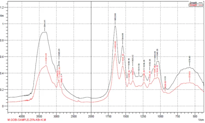
Fig. 1 FTIR-ATR over lapped spectra of E. fetida both control and 25% concentration spectra with molecular modifications are reported by a red line while control spectra are reported by black line.
Metabolic action of E. fetida against heavy metals investigated by FTIR spectroscopy
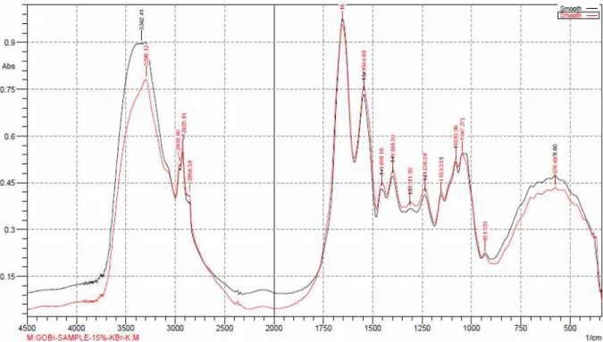
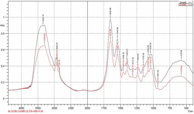
FTIR analysis offers excellent information on the nature of the bonds present on the earthworm surface and allows identification of different functional groups on the cell surface which are capable of interacting with metal ions. Changes in band frequency can also be used to estimate the relative importance of the various surface functionalities in metal sorption. The FTIR spectra of E. fetida are as shown in Figs 1 - 3 and the list of FTIR band assignment is reported in Table 2. FTIR spectroscopy shows many bands belonging to the different functional groups of worm’s biomolecules which were resulted by the structural modifications caused by the presence of pollutants. As clearly evident from Figs 1- 3, all the bands were modified by the exposure to the heavy metals containing waste.
The absorbance bands at 2925.81 cm−1 correspond respectively to the CH2 asymmetric and symmetric stretching of methylene group, which mainly monitor lipids. In the present study, the asymmetric band shift from 2925.81 to 2927.74 in 25 % levels respectively, due to heavy metal exposure. The sharp band observed at 1652.88 cm−1 and 1544.32 cm−1 corresponding to amide I and amide II vibration of structural proteins, respectively, (Casado et al., 2007). Identified Amide - I bands absorption was at 1652.88 cm−1 shifted to 1650.95 cm−1 in 25 %, 1654.81 cm−1 in 15 % and 1652.88 cm−1 in 5 % level and it was due to zinc exposure. According to peak assignment (the peaks
at 1045, 1080, 1236 cm−1 and 1650 cm−1), these peaks represent the nucleotides and phospholipids. There were shifts in these regions, which mean that heavy metals strongly affect nucleic acids. Furthermore we also emphasized that this approach should be confirmed (nucleic acid) with the use of other biomarker like comet assay.
Comet assay
Figures 4 and 5 show DNA damage scores at percentage of cells in each damage class in coelomocytes of E. fetida, after heavy metals exposure over 35 days at 25 %, 15 % and 5 % concentration. After 35 days of exposure the DNA damage, evaluated with the parameter tail DNA percentage and tail moment in the coelomocytes of E. fetida exposed to the heavy metals containing automobile service station waste. These findings clearly indicate a connection between accessible or rather available metal content and highest DNA damage in the celomocytes of E. fetida. A significant concentration-related increase in the percentage of damaged cells was observed (4.91 ± 5.3 in 5 %, 8.49 ± 10.2 in 15 %, 12.42 ± 12.72 in 25 % and 7.57 ± 10.7 in positive control). This percentage differed significantly when compared with controls, at all assayed heavy metal concentrations.
Heavy metal accumulation
Fig. 2 FTIR-ATR over lapped spectra of E. fetida both control and 15 % concentration spectra with molecular
modifications are reported by a red line while control spectra are reported by black line.
(ANOVA), the difference among treatments for contents of metals in earthworms was significant. Table 3 depicts the heavy metal contents of the automobile service station mud and the mean copper concentration of 25 %, 15 % and 5 % were 22.13 ± 0.32, 15.13 ± 0.007 and 5.34 ± 0.14 ppm, respectively. The Pb concentration was 19.4 ± 0.36, 12.48 ± 0.08, 7.06 ± 0.11, 30.96 ± 0.15, 24.6 ± 2.27 and Cd concentration was 12.7 ± 0.2 ppm, respectively. Zn and Fe content in the automobile waste was 43.03 ± 0.29, 29.59 ± 0.04, 10.4 ± 0.01, 924.23 ± 11.92, 404.57 ± 7.3 and 320.78 ± 3.65 (ppm), respectively. Differences in the mean heavy metal concentrations between the lower and higher concentrations were significant at 0.05 %. Table 2 shows that the accumulated heavy metal concentration in earthworm on 35th day, the Cu and Pb content in 25 %, 15 % and 5 % earthworm was 1.369 ± 0.042, 1.023 ± 0.04, 0.09 ± 0.001, 6.60 ± 0.095, 4.306 ± 0.02 and 2.2 ± 0.017 ppm, respectively. Similarly, the Cd, Zn and Fe content were 0.082 ± 0.004, 0.063 ± 0.006, 0.021 ± 0.001, 3.613 ± 0.006, 3.101 ± 0.003, 1.997 ± 0.013, 2.907 ± 0.004, 1.242 ± 0.3 and 0.007 ± 0.002, respectively. When compared to all heavy metals, Fe accumulated more in earthworm tissues. This could have been the result of both heavy metal accumulation with time as well as the increased service station waste mud concentration of the substrate with feed. The difference in waste concentrations in the body tissues of the earthworms between the three groups of exposure was significant (p < 0.005) at the end of the experiment.
Discussion
Metabolic action of E. fetida against heavy metals investigated by FTIR spectroscopy
The aim of this work was to evaluate the effect of heavy metals on the biomolecule changes in the earthworm E. fetida. As FTIR spectroscopy may be more sensitive to certain functional group (e.g. including polar bonds) as compared to FT-Raman spectroscopy (Kamnev et al., 2006), we attempted FTIR analyses of worms in control and in the presence of heavy metals containing medium. There are striking differences in the overall FTIR profiles between the control and heavy metal exposed worms. A most prominent feature of the metal stressed worm is the appearance of a relatively strong and well resolved CH2 asymmetric and symmetric stretching of methylene group at about 2925.81cm−1. Lipids play an important role in the membrane fluidity. By affecting the conformation of membrane proteins, they govern exposure and diffusion of membrane component (Palaniappan et al., 2010).
Fig. 3 FTIR-ATR over lapped spectra of E. fetida both control and 5 % concentration spectra with molecular modifications are reported by a red line while control spectra are reported by black line.
oxidation in the tissues with zinc exposure (Takahashi et al., 1991; Cakmak et al., 2006; Carpene et al., 2007).
The amide I and amide II bands of cellular proteins at 1656 and 1544 cm−1 are asymmetric and the test showing higher intensity and modified shapes with respect to the same band of the worms is the control. This is likely to reflect some partial changes in the secondary structure of the cellular proteins form dominating α-helix to other possible conformations (Naumann et al., 1993; Bonnina et al., 1999). This reduced amide bands in the heavy metal exposed tissues indicates important structural alteration in the existing proteins as suggested by Toyran et al. (2008). Further, the area of amide II band observed at 1544.88 cm−1 in the control tissues decreases and these changes reflect the loss of protein level of the heavy metal exposed worms. This loss of protein may be due to increased protein oxidation in the exposure worms (Takahashi et al., 1991; Cakmak et al., 2006; Akkas et al., 2007).
Nucleic acids and proteins intensity change when an external perturbation (i.e. the increased pollutant concentration) is applied to the biological system (Melin et al., 2004). This is due to accumulation of Pb, Zn and Cu in E. fetida. Some metals, such as hexavalent chromium (CrVI), manganese (Mn), and Pb, as well as Cd and arsenic (As), also reportedly inhibited the 8-oxo-Gua repair system (Bolin et al., 2006; Hodges and Chipman, 2002; Lee et al., 2005; Sava et al., 2004; Singh et al., 2009) which in turn increased the
mutation frequency. FT-IR spectroscopic technique is used to determine the biomolecular profile of micro-samples of coelomic fluid. Presence of arginine and lysine and absence of glutamic acid under toxicological condition could be considered as markers of pollutants in the environment (Joe et al., 2014).
Comet assay
The comet assay is a genotoxicity assay for the detection of DNA single strand breaks (Singh et al., 1988) in single cells. The exposure to the heavy metals containing soil seem to have caused significant DNA damages after 35 days of exposure compared with the control. The increase in DNA damages was probably caused by the production and intracellular accumulation of ROS, induced by the exposure of earthworms to heavy metals.
Table 2 List of functional groups present in FTIR spectra of E. fetida assigned according to the identified bands of absorption
Peaks
Control 25% 15% 5%
Identified bands
1045.35 1045.35 1047.27 1043.42 Phospolipids, DNA and RNA
1080.06 1081.99 1080.06 1080.06 C–O carbohydrates
1153.35 1153.35 11.53.35 1153.35 CO–O–C asymmetric stretching: mainly glycogen and nucleic acids
1236.29 1236.29 1236.29 1238.21 C–N peptide group of nucleic acids
1456.16 1452.3 1456.16 1456.16 CHfrom proteins 2 bending: mainly lipids with little contribution
1544.88 1541.02 1544.88 1544.88 Amide II: N–H bending and C–N stretching of the polypeptide and protein backbone
1652.88 1650.95 1654.81 1652.88 Amide I: C=O stretching of proteins
2925.81 2927.74 2925.81 2925.81
CH2 asymmetric stretching: mainly lipids, with little contribution from proteins, carbohydrates, nucleic acids
3342.41 3301.91 3296.12 3299.98
Amide A: mainly N–H stretching of proteins with negligible contribution from O–H stretching of intermolecular hydrogen bonding
together with increased cell proliferation and blocked apoptosis could result in tumor formation (Waalkes 2003; Waisberg et al., 2003; Hei and Filipic, 2004). Zn has an optimal intracellular range above or below which internucleosomal DNA cleavage, chromatin condensation, and nuclear fragmentation are induced (Krug, 2002). Hwang et al. (2004) and Seve et al. (2002) reported that metals such as Zn and Cd may have apoptotic and/or necrotic effects over cells of different organs.
Heavy metal accumulation
Earthworms are known to accumulate metals from the soil efficiently as observed by various authors (Ireland, 1975a, b; Labort et al., 1998; Morgan and Morgan, 1988; Wright and Stringer, 1980). The toxicity of heavy metal for earthworms increases with increasing the soil metal concentration (Marinussen et al., 1997). Earthworms predominantly take up heavy metals from soluble metal fractions (Saxe et al., 2001; Vijver et al., 2003).
As a result, the possibility for Fe metal to be bound to ions and carbonates (i.e. more soluble fractions) increases in ingested material. As a result, the metal content reduces in digested organic material due to bioaccumulations of more soluble fractions of metals in an earthworm’s gut or cutaneous tissues. In general, earthworms consume a great amount of organic waste to achieve appropriate nutrition, and during this process metals are liberated in free forms due to the enzymatic actions in their gut (Suthar, 2007). Furthermore, such available forms of metals are then absorbed by the epithelial layer of gut during the transiting of wastes through it. Bioaccumulation of high
concentration of metals is well documented (Hsu et al., 2006).
Heavy metal accumulation depends on the exposure duration whereas the accumulation of Fe, Zn, Cu, Cd and Pb is dependent upon the metabolic turnover. Thereafter metal concentrations remain constant throughout the entire life span. The present study clearly demonstrates that statistically
Fig. 5 DNA damage scores at percentage of cells in each damage class in celomocytes of E. fetida, after heavy metals exposure over 35 days at 25 %, 15 % and 5 % concentration. Significant at p < 0.05 % level
significant accumulation of Fe, Zn, Cu, Cd and Pb in the earthworm does take place as reported earlier (Honda et al., 1984). It is known that earthworm chloragosomes function as the cation exchange system capable of taking up and retaining heavy metals (Ireland, 1978; Morgan and Morgan, 1998), which are subsequently excreted by fractionation of the chloragocytes (Fischer, 1976).
Conclusion
Without the use of biomarkers for identifying ecological risks of land contamination, traditional assessment would be difficult to interpret. Therefore, the earthworm based biomarkers used in this study to measure ecological exposure to hazardous substance have identified risks to scientifically relevant organisms and indicated bioavailability of pollutants and their effects. This new FTIR based biomolecules study revealed a
clear molecular shift in the exposed worms, due to heavy metal accumulation and this is new to earthworm toxicology. The bio-molecule modification responses of heavy metals are best assessed exploiting one of the many budding in vivo techniques. Besides, the short-term biomolecule assay (FTIR study) on earthworms is an early indicator of long-term toxicity, which may result in DNA damage. Further, the diverse structural changes in the DNA, (DNA fragmentation) was visualized and quantified, which provide a promising basis for the development of sensitive biomarkers for ecotoxicological study.
Acknowledgement
This research was supported by the Department of Science and Technology (DST), New Delhi, Fast Track Young Scientist grant SR/FT/LS-007/2007. The authors would like to thank UGC-NRCBS and MKU-USIC for laboratory assistance.
Table 3 Various concentrations of accumulated heavy metals in the medium and as well as earthworm over 35
days exposure in the three different treatments setup
Heavy metals
Heavy metals concentration in the medium at the initial stage
Heavy metals concentration in the medium after 35 days exposure
Heavy metals concentration in the earthworm after 35 days exposure
25% 15% 5% 25% 15% 5% 25% 15% 5% Cu 22.13±0.32 15.03±0.07 5.34±0.14 16.24±1.39 11.93±0.11 4.723±0.52 1.369±0.04 1.023±0.04 0.09±0.00
Pb 19.4±0.36 12.48±0.08 7.06±0.11 9.21±1.00 6.94±0.15 4.176±0.16 6.606±0.09 4.306±0.02 2.2±0.01
Cd 30.96±0.15 24.6±2.27 12.7±0.2 27.14±1.69 18.68±1.03 10.31±1.11 0.82±0.00 0.063±0.0 0.021±0.00
Zn 43.03±0.29 29.59±0.04 10.4±0.01 35.55±1.08 21.0±1.91 7.207±0.41 3.613±0.00 3.101±0.0 1.997±0.01
Fe 924.23±11.92 404.57±7.3 320.78±3.65 546.05±20.1 224.7±12.6 134.4±8.78 2.907±0.00 1.242±0.3 0.007±0.0
F value 16880.8* 7567.2* 21693.7* 1993.9* 805.19* 622.09* 7072.9* 450.0* 38399*
References
Akkas SB, Severcan M, Yilmaz O, Severcan F. Effect of lipoic acid supplementation on rat brain tissue: an FT-IR spectroscopic and neutral network study. Food Chem. 105: 1281-1288, 2007.
Argov S, Sahu RK, Bernshtain E, Salman A, Shohast G, Zelig U, et al. Inflammatory bowel diseases as an intermediate stage between normal and cancer: A FTIR-microspectroscopy approach. Biopolymers 75: 384-392, 2004. Beleits C, Steiner G, Sowa MG, Baumgartner R,
Sobottka S, Schacker G, et al. Classification of human gliomas by infrared imaging spectroscopy and chemometric image processing. Vibr. Spectrosc. 38: 143-149, 2005. Bolin CM, Basha R, Cox D. Exposure to lead (Pb)
and the developmental origin of oxidative DNA damage in the aging brain. FASEB J. 20: 788-790, 2006.
Bonnina S, Bessona F, Gelhausena M, Chiericib S, Rouxa B. A FTIR spectroscopy evidence of the interactions between wheat germ agglutinin and N-acetylglucosamine residues. FEBS Lett. 456: 361-364, 1999.
Brzóska MM, Moniuszko-Jakoniuk J. Interactions between cadmium and zinc in the organism. Food Chem. Toxic. 39: 967-980, 2001.
Burch SW, Fitzpatrick LC, Goven AJ, Venables BJ, Giggleman MA. In vitro earthworm Lumbricus terrestris coelomocyte assay for use in terrestrial toxicity identification evaluation. Bull. Environ. Contam. Toxicol. 62: 547-554, 1999. Cakmak G, Dogan I, Severcan F. 17b-Estradiol
induced compositional, structural and functional changes in rainbow trout liver, revealed by FT-IR spectroscopy: a comparative study with nonylphenol. Aquat. Toxicol. 77: 53-63, 2006. Carpene E, Andreani G, Monari M, Kindt M, Isani G.
Biochemical changes during post-larval growth in white muscle of Gilthead Sea Bream (Sparus aurata) fed Zincfortified Diets. Vet. Res. Commun. 27, 215-218, 2003.
Casabé N, Piola L, Fuchs JS, Oneto ML, Pamparato L, Basack S. Ecotoxicological assessment of the effects of glyphosate and chlorpyrifos in an Argentine soya field. J. Soils Sedi. 7: 232-239, 2007.
Casado AR, Carmona P, Moreno P, Gonzalez IS. Structural changes in sardine muscle during storage: investigation by DRIFT spectroscopy. Food Chem. 103: 1024-1030, 2007.
Cohen JJ. Programmed cell death in the immune system. Adv. Immunol. 50: 55, 1991.
Cope FO, Wille JJ. In Apoptosis: The Molecular Basis of Cell Death. In: Tomei LD., Cope FO (eds), Cold Spring Harbor Laboratory Press: Cold Spring Harbor, NY, pp 61, 1991.
Darzynkiewicz Z, Bruno SG, DelBine H, Gorczyca MA, Hotz P, Lassota F, et al. Features of apoptotic cells measured by flow cytometry. Cytometry 13: 795-780, 1992.
Ellis RE, Yuan J, Horvitz HR. Mechanism and functions of cell death. Annu. Rev. Cell Biol. 7: 663-698, 1991.
Fischer E. Chloragogenzelle-Eleocyte-Transformation, induziert mit Benomyl-und
Carbofuran-Vergiftung der Lumbriciden (Oligochaeta). Zool. Anz. 197: 225-233, 1976. Hei T, Filipic M. Role of oxidative damage in the
genotoxicity of arsenic. Free Radic. Biol. Med. 37: 574-581, 2004.
Hodges NJ, Chipman JK. Down-regulation of the DNA-repair endonuclease 8-oxo-guanine DNA glycosylase 1 (hOGG1) by sodium dichromate in cultured human A549 lung carcinoma cells. Carcinogenesis 23: 55-60, 2002.
Honda K, Nasu T, Tattsukawa R. Metal distribution in the earthworm, Pheretima hilgendorfi and their variations with growth. Arch. Environ. Contam. Toxicol. 13: 427-432, 1984.
Hsu MJ, Selvaraj K, Agoramoorthy G. Taiwan’s industrial heavy metal pollution threatens terrestrial biota. Environ. Pollut. 143: 327-334, 2006.
Hwang JS, Kobayashi C, Agata K. Ikeo K, Gojobori T. Detection of apoptosis during planarian regeneration by the expression of apoptosis-related genes and TUNEL assay. Gene 333: 15-25, 2004.
Ireland MP. The effect of earthworm Dendrobaena rubida on the solubility of lead, zinc and calcium in heavy metal contaminated soil in Wales. J. Soil Sci. 26: 313-318, 1975a.
Ireland MP. Distribution of lead, zinc and calcium in Dendrobaena rubida (Oligochaeta) living in soil contaminated by base metal mining in Wales. Com. Biochem. Physiol. 52: 551-555, 1975b. Ireland MP. Heavy metal binding properties of
earthworm chloragosomes. Acta Biol. Acad. Sci. Hung. 29: 385-394, 1978.
Jimenez LA, Zenela C. Fung H. Janseen YMW, Vacek P, Charland C, et al. Role of extracellular signal-regulated protein kinases in apoptosis by asbestos and H2O2. Am. J. Physiol. 273: 1029-1035, 1997.
Aja M, Jaya M, Vijayakumaran NK, Hubert Joe I. FT-IR spectroscopy as a sentinel technology in earthworm toxicology. Spectrochimica Acta Part A Molec.. Biomolecular. Spec. 120: 534- 541, 2014.
Kamnev AA, Tugarova AV, Antonyuk LP, Tarantilis PA, Kulikov LA, Perfiliev YD, et al. Instrumental analysis of bacterial cells using vibrational and emission Mossbauer spectroscopic techniques. Anal. Chim. Acta 573-574: 445-452, 2006. Kerr JF, Wyllie AH, Currie AR. Apoptosis: a basic
biological phenomenon with wide range applications in tissue kinetics. Br. J. Cancer 26: 239-257, 1972.
Krug HF. Metals in clinical medicine: the induction of apoptosis by metal compounds. Werkstofftech 33: 770-774, 2002.
Labort F, Narbonne, JF, Ville P, Saint-Denis M, Ribera D. Acute toxicity, toxicokinetics and tissue target of lead and uranium in the clam Corbicula fluminea and the worm Eisenia fetida comparison with the fish Brachydanio rerio. Arch. Environ. Contam. Toxicol. 36: 167-178, 1998.
protein of 8-oxo-7,8-dihydro-2′-deoxyguanosine DNA glycosylase 1. Cancer Epidem. Biomarkers Prev. 14: 497-505, 2005.
Marinussen MPJC, Van Der Zee SEATM, De Haan FAM. Cu accumulation in the earthworm Dendrobaena veneta in a heavy metal (Cu, Pb, Zn) contaminated site compared to Cu accumulation in laboratory experiments. Environ. Pollut. 96: 227-233, 1997.
Melin A, Perromat A, Lorin C, Deleris G. Sensitivity of Deinococcus radiodurans to chemical aggression: investigation by one- and two-dimensional infrared spectroscopy. Vib. Spectrosc. 36: 15-22, 2004.
Miller SA, Dykes DD, Polesky HFA. Simple salting out procedure for extracting DNA from human nucleated cells. Nucleic Acids Res. 16: 1215, 1998.
Mirsal IA. Soil pollution: origin, monitoring and remediation; Springer: New York, 2004.
Morgan JE, Morgan AJ. Earthworms as biological monitors of cadmium, copper, lead and zinc in metalliferous soils. Environ. Pollut. 54: 123-138, 1988.
Morgan JE, Morgan AJ. The distribution and intracellular compartmentation of metals in the endogeic earthworm Aporrectodea caliginosa sampled from an unpolluted and a metal-contaminated site. Environ. Pollut. 99: 167-175, 1998.
Naumann D, Ch U, Schultz Goerne-Tschelnokow, Hucho F. Secondary structure and temperature behavior of the acetylcholine receptor by Fourier transform infrared spectroscopy. Biochemistry 32: 3162-3168, 1993.
Nogueira PR, Lourenço J, Mendo S, Rotchell JM. Mutation analysis of ras gene in the liver of European eel (Anguilla anguilla L.) exposed to benzo[a]pyrene. Mar. Pollut. Bull. 52: 1611-1616, 2006.
Palaniappan PLRM, Nishanth T, Renju VB. Bioconcentration of Zinc and its effect on the biochemical constituents of the gill tissues of Labeo rohita: An FT-IR study. Infrared Phys. Technol. 53: 103-111, 2010.
Pokarzhevskii AD, Nico M, Straalen V, Semenov AM. Agar as a medium for removing soil from earthworm guts. Soil Biol. Biochem. 32: 1315-1317, 2000.
Posthuma L, Hogervorst RF, Van Straalen NM. Adaptation to soil pollution by cadmium excretion in natural populations of Orchesella cincta (L.) (Collembola). Arch. Environ. Contam. Toxicol. 22: 146-156, 1992.
Raff MD. Social controls on cell survival and cell death. Nature 356: 397, 1992.
Reinecke SA, Reinecke AJ. The Comet assay as biomarker of heavy metal genotoxicity in earthworms. Arch. Environ. Contam. Toxicol. 46: 208-215, 2004.
Salman A, Erukhimovitch V, Talyshinky M, Huleihil M, Huleihel M. FTIR spectroscopic method for detection of cells infected with herpes viruses. Biopolymers (Biospectroscopy) 67: 406-412, 2002.
Sambrook J, Fritsch EF, Maniatis T. Molecular Cloning: A Laboratory Manual Cold Spring
Harbor Laboratory Press, Cold Spring Harbor, New York, pp 10.59-10.66,1989.
Sava V, Mosquera D, Song S, Cardozo-Pelaez F, Sánchez-Ramos JR. Effects of melanin and manganese on DNA damage and repair in PC12-derived neurons. Free Radic. Biol. Med. 36: 1144-1154, 2004.
Saxe JK, Impellitteri CA, Peijnenburg WJGM, Allen HE. Novel model describing trace metal concentrations in the earthworm, Eisenia andrei. Environ. Sci. Technol.35: 4522-4529, 2001. Saxena PN, Chauhan LKS, Gupta G. Cytogenetic
effects of commercial formulation of cypermethrin in root meristem cells of Allium sativum: Spectroscopic basis of chromosome damage. Toxicology 216: 244-252, 2005. Seve M, Chimienti F, Favier A. Role du zinc
intracellulaire dans la mort cellulaire programme´ e. Pathol. Biol. 50: 212-221, 2002. Singh KP, Kumari R, Pevey C, Jackson D, Du Mond
JW. Long duration exposure to cadmium leads to increased cell survival, decreased DNA repair capacity, and genomic instability in mouse testicular Leydig cells. Cancer Lett. 279: 84-92, 2009.
Singh NP, McCoy MT, Tice RR, Schneider EL. A simple technique for quantitation of low levels of DNA damage in individual cells. Exp. Cell Res. 175: 184-191, 1988.
Suthar S. Nutrient changes and biodynamics of epigeic earthworm Perionyx excavatus (Perrier) during recycling of some agriculture wastes. Bioresour. Technol. 98: 1608-1614, 2007. Takahashi H, French SM, Wong PTT. Alterations in
hepatic lipids and proteins by chronic ethanol intake: a high-pressure Fourier transforms infrared spectroscopic study on alcoholic liver disease in the rat. Clin. Exp. Res. 15: 219-223, 1991.
Toyran N, Severcan F, Severcan M, Turan B. Effect of selenium supplementation on rat heart apex and right ventricle myocardia by using FT-IR spectroscopy: a cluster analysis and neural network approach. Food Chem. 110: 590-597, 2008.
Vidovic M, Sadibasic A, Cupic S, Lausevic M. Cd and Zn in atmospheric deposit, soil, wheat, and milk. Environ. Res. 97: 26-31, 2005.
Vijver MG, Vink JPM, Miermans CJH, Van Gestel CAM Oral sealing using glue: a new method to distinguish between intestinal and dermal uptake of metals in earthworms. Soil Biol. Biochem. 35: 125-132, 2003.
Waalkes MP. Cadmium carcinogenesis. Mutat. Res. - Rev. Mutat. Res. 533: 107-120, 2003.
Waisberg M, Joseph P, Hale B, Beyersmann D. Molecular and cellular mechanisms of cadmium carcinogenesis. Toxicology 192: 95-117, 2003. Weeks JM, Svendsen C. Neutral red retention by
lysosomes from earthworm (Lumbricus rubellus) coelomocytes: A simple biomarker of exposure to soil copper. Environ. Toxic. Chem. 15: 1801-1805, 1996.