ISSN : 2248-9622, Vol. 5, Issue 2, ( Part -4) February 2015, pp.54-59
Structural Analysis Of Alfa Fibers After Chemical Treatment
Zakaria Mouallif*, Bouchaib Radi*
*(Laboratory of Mechanical Engineering, Industrial Management and Innovation, Hassan 1st University, Settat, Morocco)
ABSTRACT
Nowadays, natural fibers are used as reinforcement in composite materials. The Alfa fibers have undergone an alkaline treatment with sodium hydroxide NaOH at a concentration of 10%, during an immersion period of two days. After drying, the Fourier transform infrared spectroscopy by attenuated total reflection (FTIR-ATR) and X-ray diffraction (XRD) were used for the analysis of the chemical properties of these fibers which were extracted from the plant Alfa of the region Al Haouz (Morocco) in order to study the modifications resulting from the alkaline treatment. The results proved the presence of the cellulose, with an increase in its proportion in those fibers which have undergone an alkaline treatment with NaOH, the presence of lignin and pectin, as well as their disappearance after the alkaline extraction.
Keywords
-
Alfa (Esparto grass), cellulose, composite materials, extraction, Natural fibers.I.
Introduction
The use of plant fibers as reinforcement in composite materials has grown considerably [1-4] in order to create biodegradable composites. With the growing interest in the environment and the encouragement of the governments and institutions for sustainable investment, the trend is moving towards this type of ecological and functional fibers. This comeback is all the more important in consideration of the fact that the oil resources become more and more rare and expensive. These fibers have very interesting chemical and mechanical characteristics [5-6]. Among the commonly used plant fibers, those from the plant Alfa (Stipa tenacissima) are frequent in the Western Mediterranean: North Africa, from Morocco to Libya, and Southern Europe. Nevertheless, there are a number of difficulties concerning the integration of these fibers into the polymer matrices, especially a fiber-matrix incompatibility. The fiber-matrix adhesion can be improved by a modification of the topology of the fibers surface. Several treatments allow such a modification of the fibers surface. These treatments improve the wettability of the fiber with the matrix and create a strong bond in the fiber-matrix interface. A good adhesion at the interface in turn contributes to an improvement of the load transfer between the fibers and the matrix and therefore shows better mechanical properties [7]. The most used technique is the alkaline treatment. We propose in this paper an analysis of the chemical properties of fibers which were extracted from the plant Alfa in the region of Al Haouz (Morocco) by the spectroscopy method (FTIR-ATR) and X-ray diffraction (XRD). Before presenting the method of pretreatment and alkaline treatment we will give details about the used material. Finally, we will
compare the results obtained for the treated and untreated Alfa fibers.
II.
Materials and methods
2.1 Materials
Our Alfa fibers (Stipa tenacissima L.) are a bunch of grass with a height of about one meter, which has been harvested in the region of Al Haouz, located inside Morocco between the High Atlas to the south and the small massif Jebilete in the north, at an altitude of 400 to 900 m at the northern hillside of this region. The plain of Haouz is a semi-arid zone, receiving only 200 to 300 mm of precipitation per year.
Before beginning the fiber extraction, preliminary work is needed to better prepare the stems to different treatments, this preparation will facilitate and increase the efficiency of future mining operations. Our raw material often contains soil, roots, dust or any other impurities. Dead stems are sometimes present in the lot. The first step is to remove all these impurities and / or foreign bodies in order to have only clean and usable rods. Then we cut the ends of the rods as they exhibit the largest variation in diameter. On one side we have the upper end in the form of relatively sharp point and on the other side, the lower end in the form of a rigid curved foot. It is very important to remove them because these hooks, after extraction, become nodes and fine fibers begin to form around a kind of "Neps" mass hopelessly tangled fibers. In some cases where the variation in diameter between upper and lower end is very important, it is preferable to cut and to work only on the central portion of the rods in order to have the same effect over the entire length.
2.2 Pretreatment of the fibers
The Alfa fibers were washed with distilled water to remove contaminants and adhering dust. Thereafter, they were dried in the open air for 96 hours at room temperature. Then they were cut to pieces of 8 cm in length. These fibers will be designated as untreated fibers.
2.3 Treatment of the fibers
The Alfa fibers were treated with a sodium hydroxide solution (NaOH) at a concentration of 10% and during an immersion period of two days. After this treatment, the Alfa fibers were rinsed with distilled water. The traces of NaOH were neutralized for 15 min by a solution of distilled water with 2% sulfuric acid. Thereafter, the fibers were dried for 5 hours at 60°C. These fibers will be designated as treated fibers.
2.4 Used technique
Technique 1: The Fourier transform infrared spectroscopy by attenuated total reflection (FTIR-ATR) was used for the analysis of the chemical properties of the Alfa fibers in order to study the modifications after the alkaline treatment. This technique is based on the fact that the molecules have specific frequencies for which they vibrate (or turn) in correspondence with energy levels called: vibration modes. Here the incident radiation is of an infrared type. For this particular technique (Fourier transform), after meeting the sample the infrared light passes through a Michelson interferometer (not a monochromator), composed by a beam splitter, a fixed mirror and a movable mirror. The signal is called interferogram; it undergoes a Fourier transform to be finally traced and become a spectrum [8].
In this study, the measurements were carried out using a VERTEX 70 spectrometer at a spectral range of 4000-600 cm-1 with a resolution of 4 cm-1. The acquisition of the spectra was performed under dry air flow with 16 scans (interferogram) per spectrum. Technique 2: The X-ray diffraction (XRD) allows analyzing the crystal structure of crystalline
materials. The collected data produce the diffraction pattern. For this method the sample was bombarded with X-rays and the intensity of the scattered radiation was determined according to the orientation in the space. The registration of the detected intensity is a function of the deviation angle 2θ of beam [11-13].
These rays which were scattered by the sample are in accordance with Bragg's law [14]:
2 × ℎ ×�� �= ×� (1) With:
ℎ : the distance separating two orientated planes {hkl} called interplanar spacing
�: the wavelength of the used radiation : an integer representing the diffraction order
�: the half-angle of the diffraction of the RX
The analyses in this study were carried out by diffraction of X-rays with a range of θ-2θ (theta - 2 theta). In this technique, the source of radiation is fixed, while the holder of the sample is movable. The device used is an X'Pert Pro MPD diffractometer with a CuKα radiation (λ = 1.54 Å). A 2θ angle range of 5° to 90° has been scanned.
III.
Results & interpretation
ISSN : 2248-9622, Vol. 5, Issue 2, ( Part -4) February 2015, pp.54-59
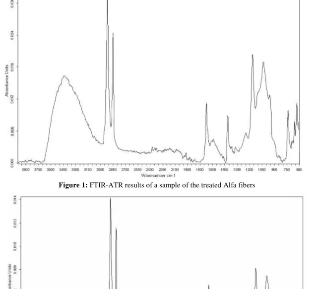
Figure 1: FTIR-ATR results of a sample of the treated Alfa fibers
Figure 2: FTIR-ATR results of a sample of the untreated Alfa fibers
The peak observed at 1733 cm-1 in the spectrogram of the untreated fibers, associated with the stretching vibration of the carbonyl group C=O, proved the presence of pectins, which disappeared after the alkaline extraction with NaOH.
The band observed at 1464 cm-1, corresponding to the bending vibration mode of the CH2 group, and at 1530 cm-1, corresponding to the vibration of the C-H bond in the aromatic ring which is present in the lignin, have decreased considerably.
The peak which lies between 3000 and 3600 cm-1, which is associated with the elongation of the hydroxyl O-H bond, had strongly increased in intensity due to the increase of the proportion of the cellulose in the treated fibers and because of the alkaline treatment. This showed that there are residues of the alkaline sodium hydroxide on the fibers surfaces.
and CH2 group. The peaks at 832 and 724 cm-1, respectively showed the characteristics of the C-O-C stretching vibration and the deformation vibration of the C-OH bond [9-10].
In order to analyze the influence of the pretreatment of the fibers on their crystallinity,
X-ray diffraction analyses were performed. The results of the X-ray diffraction test are shown in the diffraction patterns of figures 3 and 4. These figures show the X-ray diffraction of the untreated and treated Alfa fibers.
Figure 3: Diffraction pattern of the untreated Alfa fibers
Figure 4: Diffraction pattern of the treated Alfa fibers
These diagrams illustrate an intense peak at 2θ= 22,7° corresponding to the crystallographic planes [0 0 2] of the cellulose I. A less intense peak
ISSN : 2248-9622, Vol. 5, Issue 2, ( Part -4) February 2015, pp.54-59
crystallinity Ic was calculated according to the Segal
method [15,16]. The index of crystallinity could be estimated by the values of the diffraction intensities of the crystalline and those of the amorphous structure.
� % =�002−��
�002 × 100 (2) With:
�002: the intensity of the peak of the crystalline phase at 2� = 22,7°
�� : the intensity of diffraction of the amorphous material at 2θ = 18°
These indexes were calculated by using the equation (2) and are presented in Table 1. These results showed that the index of crystallinity becomes more important after the alkaline treatment of the Alfa fibers: 50% for the treated Alfa fibers, 45% for the untreated ones. This confirmed the hypothesis of the gradual elimination of non-cellulosic materials (amorphous part) of the stem of Alfa and it showed that the proportion of the ordered region, namely the cellulose (crystal structure) which is present in the fibers, is increasing.
Alfa fibers 2� (°) hkl
Index of crystallinity
(%)
Treated 22,7 [0 0 2] 50
Untreated 22,7 [0 0 2] 45
Table 1: Main results of the different DRX diffractograms
IV.
Conclusion
The results obtained for the Alfa fibers treated with sodium hydroxide and those in raw state using the technique of FTIR-ATR spectroscopy showed the presence of the cellulose, with an increase in its proportion in those fibers which have undergone an alkaline treatment with NaOH. The existence of the lignin and pectin, as well as their disappearance after the alkaline extraction, has been proven.
The results obtained by using the XRD method showed that the index of crystallinity increases after the treatment of the Alfa fiber. This confirmed the hypothesis of the gradual elimination of non-cellulosic substances.
REFERENCES
[1] Faulstich de Paiva JM., Frollini E., Unmodified and Modified Surface Sisal Fibers as Reinforcement of Phenolic and Lignophenolic Matrices Composites: Thermal Analyses of Fibers and Composites Macromol, Material and Engineering, 291, 405-417, 2006.
[2] Alvarez VA., Vázquez A., Influence of fiber chemical modification procedure on the mechanical properties and water
absorption of MaterBi-Y/sisal fiber composites, Composites: Part A, 37, 1672– 1680, 2006.
[3] Sreekala MS., Kumaran MG., Seena J., Maya J., Oil Palm Fibre Reinforced Phenol Formaldehyde Composites: Influence of Fibre Surface Modifications on the Mechanical Performance, Applied Composite Materials, 7, 295–329, 2000. [4] Al-Khanbashi A., Al-Kaabi K., Hammami
A., Date Palm Fibers as Polymeric Matrix Reinforcement: Fiber Characterization, Polymer Composites, 26 , 486-497, 2005. [5] Wambua P., Jan Ivens, Verpoest. I.,
Natural fibres: Can they replace glass in fibre reinforced plastics, Composites Science and Technology 63,1259-1264, (2003).
[6] Joshi S.V., Drazl L.TL., Mohanty A.K., Arora S., Are natural fiber composites environmentally superior to glass fiber reinforced composite, Composites: Part A 35, 371-376, 2004.
[7] Al-Kaabi K., Al-Khanbashi A., Hammami A., Date Palm Fibers as Polymeric Matrix Reinforcement: DPF/Polyester Composite properties, Polymer Composites, 26, 604-613, 2005.
[8] J.L.Gardette « Caractérisation des polymères par spectrométrie optique », Techniques de l’ingénieur (Décembre 1996).
[9] P.Garside and P.Wyeth « Identification of cellulosic fibres by FTIR spectroscopy: Thread and single fibre analysis by attenuated total reflectance », Studies in Conservation, Vol 48 (4), pp. 269-275 (2003).
[10] P.Garside and P.Wyeth « Identification of cellulosic fibres by FTIR spectroscopy - Differentiation of flax and hemp by polarized ATR FTIR », Studies in Conservation, Vol 51(3), pp. 205-211 (2006)
[11] N.Sgriccia, M.C.Hawley, and M.Misra « Characterization of natural fiber
surfaces and natural fiber composites», Composites Part A Applied Science and Manufacturing 39, (2008), pp. 1632-1637. [12] J.Peng « Détermination des contraintes
résiduelles dans des revêtements par diffraction des rayons X en faible incidence », Thèse de Doctorat, l’Ecole Nationale Supérieure d’Arts et Métiers, Paris (Juillet 2006).
[14] W.H.Bragg « The universe of light », Macmillan, New York (1934).
[15] L.Segal, J.J.Creely, A.E.Martin Jr and C.M.Conrad « An empirical method for estimating the degree of crystallinity of native cellulose using the X-ray diffractometer », Textile Research Journal, Vol 29, pp.786 –794 (1959).