Submitted10 November 2015 Accepted 1 February 2016 Published15 March 2016
Corresponding author Shokoofeh Shamsi, sshamsi@csu.edu.au
Academic editor María Ángeles Esteban
Additional Information and Declarations can be found on page 15
DOI10.7717/peerj.1722
Copyright 2016 Shamsi and Suthar
Distributed under
Creative Commons CC-BY 4.0
OPEN ACCESS
Occurrence of
Terranova
larval types
(Nematoda: Anisakidae) in Australian
marine fish with comments on their
specific identities
Shokoofeh Shamsi and Jaydipbhai Suthar
School of Animal and Veterinary Sciences, Charles Sturt University, Wagga Wagga, NSW, Australia
ABSTRACT
Pseudoterranovosis is a well-known human disease caused by anisakid larvae belonging to the genusPseudoterranova. Human infection occurs after consuming infected fish. Hence the presence ofPseudoterranovalarvae in the flesh of the fish can cause serious losses and problems for the seafood, fishing and fisheries industries. The accurate identification ofPseudoterranovalarvae in fish is important, but challenging because the larval stages of a number of different genera, includingPseudoterranova,Terranova
and Pulchrascaris, look similar and cannot be differentiated from each other using morphological criteria, hence they are all referred to asTerranovalarval type. Given that
Terranovalarval types in seafood are not necessarilyPseudoterranovaand may not be dangerous, the aim of the present study was to investigate the occurrence ofTerranova
larval types in Australian marine fish and to determine their specific identity. A total of 137 fish belonging to 45 species were examined.Terranovalarval types were found in 13 species, some of which were popular edible fish in Australia. The sequences of the first and second internal transcribed spacers (ITS-1 and ITS-2 respectively) of the
Terranovalarvae in the present study showed a high degree of similarity suggesting that they all belong to the same species. Due to the lack of a comparable sequence data of a well identified adult in the GenBank database the specific identity ofTerranovalarval type in the present study remains unknown. The sequence of the ITS regions of the
Terranovalarval type in the present study and those ofPseudoterranovaspp. available in GenBank are significantly different, suggesting that larvae found in the present study do not belong to the genusPseudoterranova, which is zoonotic. This study does not rule out the presence ofPseudoterranovalarvae in Australian fish asPseudoterranova decipiens E
has been reported in adult form from seals in Antarctica and it is known that they have seasonal presence in Australian southern coasts. The genetic distinction ofTerranova
larval type in the present study fromPseudoterranovaspp. along with the presence of more species of elasmobranchs in Australian waters (definitive hosts ofTerranovaspp. andPulchrascaris spp.) than seals (definitive hosts ofPseudoterranova spp.) suggest that Terranovalarval type in the present study belong to either genusTerranova or
Pulchrascaris, which are not known to cause disease in humans. The present study provides essential information that could be helpful to identify AustralianTerranova
larval types in future studies. Examination and characterisation of further specimens, especially adults of Terranova and Pulchrascaris, is necessary to fully elucidate the identity of these larvae.
SubjectsAquaculture, Fisheries and Fish Science, Marine Biology, Parasitology, Taxonomy
Keywords Anisakidae,Terranova, Taxonomy
INTRODUCTION
Psudoterranovosis (Hochberg & Hamer, 2010), the seafood borne parasitic disease, caused by larvae ofPseudoterranova, is another form of anisakidosis, that has caused concern to human beings. The disease is most common in the United States followed by Japan and Europe (Hochberg & Hamer, 2010). With the increased popularity of eating raw or slightly cooked seafood dishes, the number of cases have increased globally (Chai, Darwin Murrell & Lymbery, 2005). The symptoms of the disease vary and may include nausea, severe epigastric pain and other abdominal discomforts, ‘‘tingling throat syndrome’’ from a worm crawling in the upper esophagus or oropharynx, cough and vomiting up live or dead worms (Margolis, 1977). The life cycle of thePseudoterranovaspp. includes crustaceans and fish as their intermediate hosts and marine mammals as their definitive hosts (Anderson, 2000). Human infection occurs after eating infected seafood, therefore the presence ofPseudoterranovalarvae in the flesh of fish can cause serious losses and problems for fish and fisheries industry across the world. For example, up to 36 worms per fish have been reported in cod populations from Norwegian waters (Jensen, Andersen & Desclers, 1994) or Icelandic cod fillets provided by the industry have been reported to be infected with 2.5–17.6 worms per kg fillet (Hafsteinsson & Rizvi, 1987). It has been estimated that detection and removal of the larvae thought to bePseudoterranovafrom the flesh of Atlantic cod (Gadus morhua) and other demersal species, and the resultant downgrading and discard of product, cause an annual loss of $50 million in Atlantic Canada (McClelland, 2002). This implies the need for detection and accurate identifi-cation of these larvae in fish. One of the challenges in diagnosing of parasitic diseases is the specific identification of larval stages of parasites. Larval stages of nematodes cannot be identified reliably using morphological characters alone. This is a consequence of the small size of larval stages and the lack of a sufficient number of characteristic features (Shamsi, Gasser & Beveridge, 2011). Molecular approaches have gained prominence for accurate identification of anisakids, irrespective of developmental stage and sex of the parasite, and for establishing systematic relationships (e.g.,Orecchia et al., 1986). Several studies showed that ITS-1 and ITS-2 are useful genetic markers for specific identifications of nematodes irrespective of their developmental stage or sex and to study their life cycle (e.g.,Shamsi, Gasser & Beveridge, 2011). However, this approach relies on presence of ITS sequences for well identified adults.
In several countries other than Australia, the ability to recognise and diagnose anisakidosis/pseudoterranovosis caused by these larvae has been improved, resulting in progress towards understanding its epidemiology and clinical manifestations of the disease. In Australia, however, little is known about the disease, the causative agent and its epidemiology. Australia is an increasingly multicultural country where seafood prepared in all its forms is very popular. A confirmed case of human anisakidosis was published recently byShamsi & Butcher (2011)and several unpublished cases are on record
(Shamsi, 2014). Therefore, there has been an increasing awareness of anisakidosis in humans and the presence of anisakid larval types in marine fish in Australia (Shamsi, 2014).
A review of the literature shows that Terranovalarval types have been reported quite often in Australian marine fish (e.g.,Cannon, 1977;Doupe et al., 2003;Lester, Barnes & Habib, 1985;Moore et al., 2011) but there is no information on the specific identity of
Terranovalarval types reported in Australia. The dilemma withTerranovalarval types is that it could belong to any of three genera of anisakid nematodes, includingTerranova, PulchrascarisorPseudoterranova,whose adult stages have been reported from Australian waters. Members of TerranovaandPulchrascarisbecome adult in elasmobranchs and are not known to cause harm to humans whereasPseudoterranovaspp. become adult in marine mammals and there are numerous publications about their pathogenicity and human health impacts. The larval stages of all these genera, i.e.,Terranova,Pseudoterranova
andPulchrascarisare morphologically very similar. The typical characteristic of these larvae is the location of the excretory pore at the anterior end of the nematode, presence of a ventriculus without an appendix and having an intestinal caecum (Deardorff, 1987;Gibson & Colin, 1982). Therefore, distinction between larval stages of these genera based solely on morphology can be challenging. With recent increasing awareness about the presence of anisakid larvae in Australian fish as well as the presence of human cases in the country, knowing the specific identity ofTerranovalarval types becomes very important. In the last decade, molecular tools have provided the opportunity for specific identification of larval stages of parasites and there have been several works in the Americas, European countries and Antarctica on specific identification ofTerranovalarval types (Arizono et al., 2011;Paggi et al., 1991). Therefore, the aim of the present study is to employ a combined molecular and morphological approach to investigate the occurrence ofTerranovalarval types in Australian marine fish and to determine their specific identity.
MATERIALS AND METHODS
Parasite collectionA total of 137 fish belonging to 45 species, Abudefduf whitleyi (n=2), Aldrichetta
forsteri (n=1),Atherinomorus vaigiensis(n=1),Caesio cuning (n=8), Carangoides
fulvoguttatus(n=1),Caranx ignobilis(n=2),C. melampygus(n=1),Carcharias taurus (n=1),Chaetodon aureofasciatus(n=1), C. auriga(n=1),C. flavirostris(n=2),C.
lineolatus (n=1), C. melannotus(n=1),Chaetodon sp(n=1),Coryphaena hippurus (n=1),Engraulis australis (n=2),Epinephelus cyanopodus (n=1),Grammatorcynus
bicarinatus(n=3),Haplophryne sp.(n=1),Heniochus monoceros(n=2),H. singularius (n=1),Istiompax indica(n=3),Kajikia audax(n=3),Lutjanus argentimaculatus(n=2),
L. bohar (n=1),L. carponotatus(n=4),L. fulviflamma(n=1),L. sebae(n=4),Makaira
mazara(n=3),Mugil cephalus(n=5),Pastinachus sephen(n=1),Platycephalus laevigatus (n=8),Platycephalus sp.(n=2),Pristipomoides multidens(n=3),Rhombosolea tapirina (n=3),Sardinops sagax neopilchardus(n=8), Scomber australasicus(n=11),Seriola
hippos(n=2),S. lalandi(n=17),Siganus fuscescens(n=1),S. punctatus(n=1),Sillago
flindersi(n=13),Sphyraena novaehollandiae(n=4),Taeniomembras microstomus(n=1),
andThunnus albacares(n=1) were examined for infection with anisakid larval types. Fish were collected off Australian coasts, including Queensland, New South Wales, Victoria, South Australia and Western Australia. No fish were caught or killed for the purpose of this study. All fish were either already euthanized as part of other research projects or were bought from fishermen in various fish markets.
Dead fish were cut open and first examined for presence of larval nematodes in the surface of the internal organs and also for gross pathology. Then the gastro-intestinal tract from mouth to anus was examined for the presence of nematodes. All nematodes found were washed in physiological saline and then preserved in 70% ethanol. A small piece of the mid-body of each nematode was excised for molecular study, and the rest of the nematode were used for microscopy.
Morphological examination
The anterior and posterior parts of each nematode were cleared in lactophenol and examined under a light microscope. Terranovalarvae were identified according to the identification key proposed by Cannon (1977)and were selected for description and further molecular analyses. Illustrations were made using a microscope equipped with camera lucida.
Molecular study
Genomic DNA (gDNA) was isolated from all individual larvae identified morphologically asTerranovalarval type, by sodium dodecyl-sulphate/proteinase K treatment, column-purified (WizardTM DNA Clean-Up; Promega, Madison, WI, USA) and eluted into 45 µl of water. PCR was used to amplify the ITS-1 and ITS-2 regions using primer
sets SS1: 5′-GTTTCCGTAGGTGAACCTGCG-3′ (forward) and NC13R: 5′-GCTGCGTT CTTCATCGAT-3′(reverse) for the former and SS2: 5′-TTGCAGACACATTGAGCACT-3′ (forward) and NC2: 5′-TTAGTTTCTTTTCCTCCGCT-3′(reverse) for the latter region, and cycling conditions, initial 94 ◦C/5′, then 94 ◦C/30′′
, 55 ◦C/40′′
, 72 ◦C/40′′
×30 cycles, 72 ◦C/5′extension and 4 ◦C (Shamsi & Butcher, 2011). An aliquot (4
µl) of each amplicon
was examined on a 1.5% w/v agarose gel, stained with GelRedTM and photographed using a gel documentation system.
Representative samples based on host species and geographical locations were selected for sequencing. Sequences were aligned using the computer program ClustalX (Thompson et al., 1997) and then adjusted manually. Polymorphic sites were designated using International Union of Pure and Applied Chemistry (IUPAC) codes. Pair-wise comparisons of sequence differences (D) were determined using the formulaD=1−(M/L), whereMis the number of alignment positions at which the two sequences have a base in common, andLis the total number of alignment positions over which the two sequences are compared (Chilton, Gasser & Beveridge, 1995).
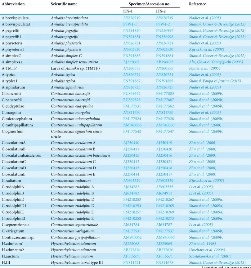
Phylogenetic analysis of the nucleotide sequence data for combined 1 and ITS-2 regions were conducted in PAUP 4.0. Table 1 shows details of the taxa used to build phylogenetic trees. Two tree-building methods, neighbour-joining and maximum parsimony were employed for phylogenetic analysis. The outgroup employed wasHeterakis
Table 1 Scientific name and specimens/accession number of taxa used to build phylogenetic trees in the present study.
Abbreviation Scientific name Specimen/Accession no. Reference
ITS-1 ITS-2
A.brevispiculata Anisakis brevispiculata AY826719 AY826719 Nadler et al. (2005)
A.brevispiculata1 Anisakis brevispiculata PSW4-1 PSW4-2 Shamsi, Gasser & Beveridge (2012)
A.pegreffii Anisakis pegreffii FN391850 FN556997 Shamsi, Gasser & Beveridge (2012)
A.pegreffii1 Anisakis pegreffii FN391851 FN556998 Shamsi, Gasser & Beveridge (2012)
A.physeteris Anisakis physeteris AY826721 AY826721 Nadler et al. (2005)
A.physeteris1 Anisakis physeteris AY603530 AY603530 Kijewska et al. (2008)
A.simplexC Anisakis simplexC FN391883 FN391884 Shamsi, Gasser & Beveridge (2012)
A.simplexs.s. Anisakis simplex sensu stricto AJ225065 AB196672 Abe, Ohya & Yanagiguchi (2005)
A.TMTP Larva ofAnisakissp. (TMTP) AY260555 AY260555 Pontes et al. (2005)
A.typica Anisakis typica AY826724 AY826724 Nadler et al. (2005)
A.typica1 Anisakis typica FN391887 FN391889 Shamsi, Poupa & Justine (2015)
A.ziphidarum Anisakis ziphidarum AY826725 AY826725 Nadler et al. (2005)
C.bancrofti Contracaecum bancrofti EU839572 FM177883 Shamsi et al. (2009b)
C.bancrofti1 Contracaecum bancrofti EU839573 FM177887 Shamsi et al. (2009b)
C.eudyptulae Contracaecum eudyptulae FM177531 FM177562 Shamsi et al. (2009b)
C.margolisi Contracaecum margolisi AY821750 AY821750 Nadler et al. (2005)
C.microcephalum Contracaecum microcephalum FM177524 FM177528 Shamsi et al. (2009b)
C.multipapillatum Contracaecum multipapillatum AM940056 AM940060 Shamsi et al. (2008)
C.ogmorhini Contracaecum ogmorhini sensu stricto
FM177542 FM177547 Shamsi et al. (2009b)
C.osculatumA Contracaecum osculatumA AJ250410 AJ250419 Zhu et al. (2000)
C.osculatumB Contracaecum osculatumB AJ250411 AJ250420 Zhu et al. (2000)
C.osculatumbaicalensis Contracaecum osculatum baicalensis AJ250415 AJ250416 Zhu et al. (2000)
C.osculatumC Contracaecum osculatumC AJ250412 AJ250421 Zhu et al. (2000)
C.osculatumD Contracaecum osculatumD AJ250413 AJ250418 Zhu et al. (2000)
C.osculatumE Contracaecum osculatumE AJ250414 AJ250417 Zhu et al. (2000)
C.radiatum Contracaecum radiatum AY603529 AY603529 Kijewska et al. (2002)
C.rudolphiiA Contracaecum rudolphiiA AJ634782 AY603535 Li et al. (2005)
C.rudolphiiB Contracaecum rudolphiiB AJ634783 AJ634911 Li et al. (2005)
C.rudolphiiD Contracaecum rudolphiiD FM210253 FM210267 Shamsi et al. (2009a)
C.rudolphiiD1 Contracaecum rudolphiiD FM210254 FM210265 Shamsi et al. (2009a)
C.rudolphiiE Contracaecum rudolphiiE FM210257 FM210269 Shamsi et al. (2009a)
C.rudolphiiE1 Contracaecum rudolphiiE FM210258 FM210273 Shamsi et al. (2009a)
C.septentrionale Contracaecum septentrionale AJ634784 AJ634787 Li et al. (2005)
C.variegatum Contracaecum variegatum FM177531 FM177537 Shamsi et al. (2009b)
Contracaecumn.sp. Contracaecum pyripapillatum AM940062 AM940066 Shamsi et al. (2009b)
H.aduncum1 Hysterothylacium aduncum AJ225068 AJ225069 Zhu et al., 1998)
H.aduncum2 Hysterothylacium aduncum AB277826 AB277826 Umehara et al. (2008)
H.auctum Hysterothylacium auctum AF115571 AF115571 Szostakowska et al. (2001)
H.III Hysterothylaciumlarval type III FN811721 FN811678 Shamsi, Gasser & Beveridge (2013) (continued on next page)
Table 1(continued)
Abbreviation Scientific name Specimen/Accession no. Reference
ITS-1 ITS-2
H.III-1 Hysterothylaciumlarval type III FN811723 FN811681 Shamsi, Gasser & Beveridge (2013)
H.IVA Hysterothylaciumlarval type IV Genotype A
FN811724 FN811690 Shamsi, Gasser & Beveridge (2013)
H.IVB Hysterothylaciumlarval type IV Genotype B
FN811730 FN811682 Shamsi, Gasser & Beveridge (2013)
H.IVGA Hysterothylaciumlarval type IV Genotype A
FN811729 FN811690 Shamsi, Gasser & Beveridge (2013)
H.IVGA1 Hysterothylaciumlarval type IV Genotype A
FN811729 FN811691 Shamsi, Gasser & Beveridge (2013)
H.IVGA2 Hysterothylaciumlarval type IV Genotype A
FN811729 FN811692 Shamsi, Gasser & Beveridge (2013)
H.IVGB Hysterothylaciumlarval type IV Genotype B
FN811730 FN811683 Shamsi, Gasser & Beveridge (2013)
H.IVGB1 Hysterothylaciumlarval type IV Genotype B
FN811731 FN811684 Shamsi, Gasser & Beveridge (2013)
H.IVGB2 Hysterothylaciumlarval type IV Genotype B
FN811733 FN811685 Shamsi, Gasser & Beveridge (2013)
H.V Hysterothylaciumlarval type V FN811738 FN811699 Shamsi, Gasser & Beveridge (2013)
H.VI Hysterothylaciumlarval type VI FN811740 FN811701 Shamsi, Gasser & Beveridge (2013)
H.VII Hysterothylaciumlarval type VII FN811749 FN811709 Shamsi, Gasser & Beveridge (2013)
H.VIII Hysterothylaciumlarval type VIII FN811750 FN811710 Shamsi, Gasser & Beveridge (2013)
Heterakisgallinarum Heterakis gallinarum JQ995320 JQ995320 Jimenez et al. (2012)
P.azarasi Pseudoterranova azarasi AJ413973 AJ413974 Zhu et al. (2002)
P.bulbosa Pseudoterranova bulbosa AJ413970 AJ413971 Zhu et al. (2002)
P.cattani Pseudoterranova cattani AJ413982 AJ413984 Zhu et al. (2002)
P.decipiens Pseudoterranova decipiens AJ413979 AJ413980 Zhu et al. (2002)
P.decipiens1 Pseudoterranova decipiens AJ413979 AJ413978 Zhu et al. (2002)
R.acus Raphidascaris acus AY603537 AY603537 Kijewska et al. (2008)
Terranovasp. Terranovasp. LN795828 LN795872 The present study Terranovasp.1 Terranovasp. LN795851 LN795871 The present study
gallinarum(Nematoda: Heteakoidea; GenBank accession numbersJQ995320andJQ995320
for ITS-1 and ITS-2, respectively).
RESULTS
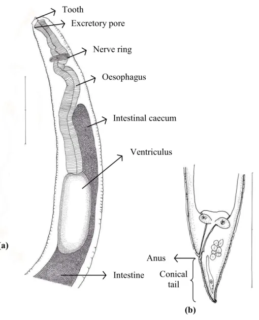
Of 45 species of fish examined in the present study, third stageTerranovatype larvae (n=93) were identified as type II based on the presence of intestinal caecum and ventriculus, absence of developed labia and ventricular appendix, and location of the excretory pore being at the anterior end (Fig. 1). Morphological description of these larvae was summarized inTable 2.
Terranovatype larvae were found in 13 species of fish collected from North-Eastern, Eastern and south eastern coasts of Australia. Material morphologically examined were 10 larvae in good condition fromCaesio cuning (n=3),Caranx ignobilis(n=2),Grammatorcynus
bicarinatus(n=1),Lutjanus argentimaculatus(n=3) andL. carponotatus(n=1) from Heron Island, Queensland.
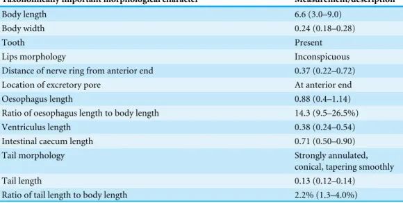
Table 2 Morphological description ofTerranovalarval type found in the present study. All measure-ments are given in millimetres. Mean measuremeasure-ments are given, followed by the range in parentheses.
Taxonomically important morphological character Measurement/description
Body length 6.6 (3.0–9.0)
Body width 0.24 (0.18–0.28)
Tooth Present
Lips morphology Inconspicuous
Distance of nerve ring from anterior end 0.37 (0.22–0.72)
Location of excretory pore At anterior end
Oesophagus length 0.88 (0.4–1.14)
Ratio of oesophagus length to body length 14.3 (9.5–26.5%)
Ventriculus length 0.38 (0.24–0.54)
Intestinal caecum length 0.71 (0.50–0.90)
Tail morphology Strongly annulated,
conical, tapering smoothly
Tail length 0.13 (0.12–0.14)
Ratio of tail length to body length 2.2% (1.3–4.0%)
A total of 93 specimens from various fishes, includingAbudefduf whitleyi, Caesio cuning, Carangoides fulvoguttatus, Caranx ignobilis, Caranx melampygus, Epinephelus cyanopodus, Grammatorcynus bicarinatus, Lutjanus argentimaculatus, L. bohar, L. carponotatusandL. fulviflammaandScomber australasicuswere subjected to PCR amplification. Based on the species of hosts and their geographical locations, 25 and 21 specimens were selected and sequenced for ITS-1 and ITS-2 respectively.
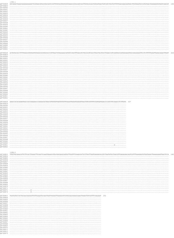
The length of the ITS-1 was 437 bp except for two specimens which were 436 bp long. The difference in length was due to a gap at alignment position 20 in the latter specimens (Fig. 2). Also, sequence polymorphism was detected at alignment position 426 in one specimen (Fig. 2). Sequence variation in the ITS-1 among specimens was 0–0.4% and the G +C content was 47.6–47.9%. The length of the ITS-2 was 252 bp. Sequence polymorphism was detected at alignment position 22 in two specimens. Sequence variation among individuals was 0–0.4% and the G+C content was 46.4–46.8%. ITS-1 and ITS-2 sequences ofTerranovalarval type found in the present study were almost identical among all larvae.
DISCUSSION
Previously, Cannon (1977) described two distinctTerranova larval types, I and II, in Queensland waters which were later reported by other authors from other parts of Australia (e.g., Doupe et al., 2003;Moore et al., 2011). According toCannon (1977), the main difference between larval types I and II is the ratio of intestinal caecum to ventriculus being 1:1 in the former and 2:1 in the latter morphotype. Based on the similarity in the ratio of intestinal caecum to ventriculus and considering the geographical location of larvae and matching it with presence of adult nematodes, he suggestedTerranovalarval type I in his study could beTerranova chiloscyitiandTerranovalarval type II could beT. galeocerdonis
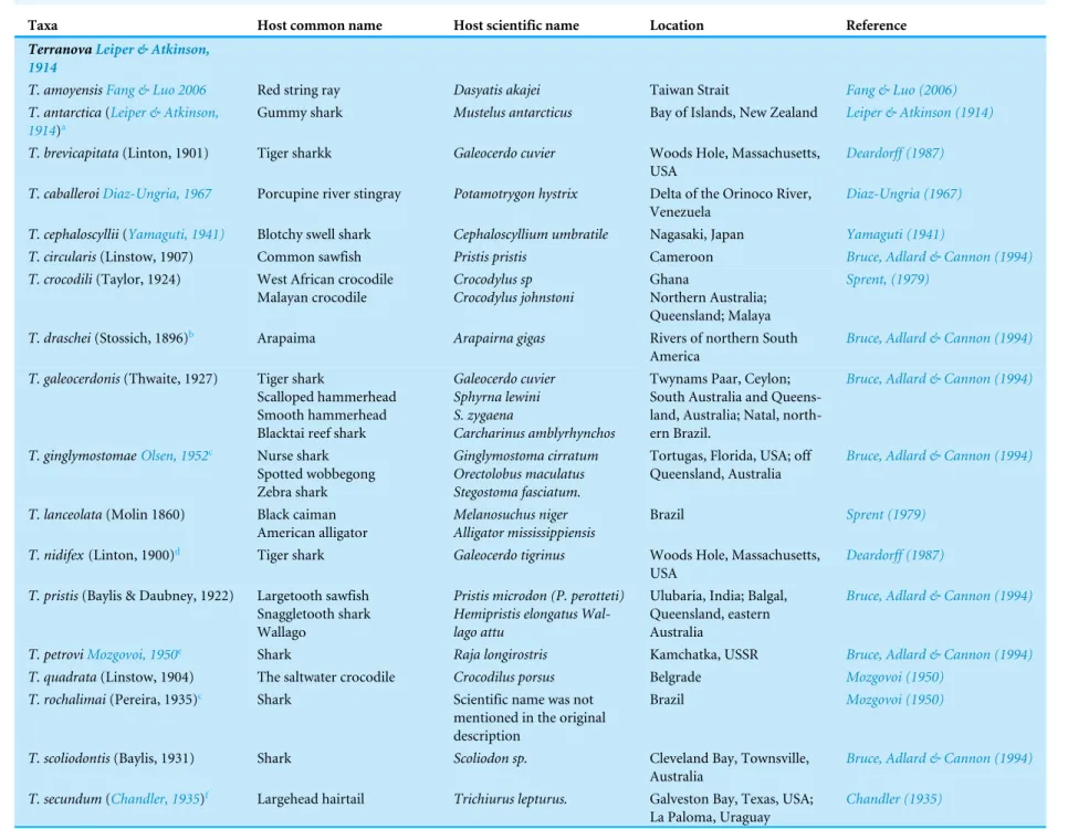
Table 3 Taxa listed under generaTerranova, PulchrascarisandPseudoterranova.
Taxa Host common name Host scientific name Location Reference
TerranovaLeiper & Atkinson, 1914
T. amoyensisFang & Luo 2006 Red string ray Dasyatis akajei Taiwan Strait Fang & Luo (2006)
T. antarctica(Leiper & Atkinson, 1914)a
Gummy shark Mustelus antarcticus Bay of Islands, New Zealand Leiper & Atkinson (1914)
T. brevicapitata(Linton, 1901) Tiger sharkk Galeocerdo cuvier Woods Hole, Massachusetts,
USA
Deardorff (1987)
T. caballeroiDiaz-Ungria, 1967 Porcupine river stingray Potamotrygon hystrix Delta of the Orinoco River, Venezuela
Diaz-Ungria (1967)
T. cephaloscyllii(Yamaguti, 1941) Blotchy swell shark Cephaloscyllium umbratile Nagasaki, Japan Yamaguti (1941)
T. circularis(Linstow, 1907) Common sawfish Pristis pristis Cameroon Bruce, Adlard & Cannon (1994)
T. crocodili(Taylor, 1924) West African crocodile Malayan crocodile Crocodylus sp Crocodylus johnstoni Ghana Northern Australia; Queensland; Malaya Sprent, (1979)
T. draschei(Stossich, 1896)b Arapaima Arapairna gigas Rivers of northern South
America
Bruce, Adlard & Cannon (1994)
T. galeocerdonis(Thwaite, 1927) Tiger shark
Scalloped hammerhead Smooth hammerhead Blacktai reef shark
Galeocerdo cuvier Sphyrna lewini S. zygaena
Carcharinus amblyrhynchos
Twynams Paar, Ceylon; South Australia and Queens-land, Australia; Natal, north-ern Brazil.
Bruce, Adlard & Cannon (1994)
T. ginglymostomaeOlsen, 1952c Nurse shark Spotted wobbegong Zebra shark
Ginglymostoma cirratum Orectolobus maculatus Stegostoma fasciatum.
Tortugas, Florida, USA; off Queensland, Australia
Bruce, Adlard & Cannon (1994)
T. lanceolata(Molin 1860) Black caiman American alligator
Melanosuchus niger Alligator mississippiensis
Brazil Sprent (1979)
T. nidifex(Linton, 1900)d Tiger shark Galeocerdo tigrinus Woods Hole, Massachusetts,
USA
Deardorff (1987)
T. pristis(Baylis & Daubney, 1922) Largetooth sawfish Snaggletooth shark Wallago
Pristis microdon (P. perotteti) Hemipristis elongatus Wal-lago attu
Ulubaria, India; Balgal, Queensland, eastern Australia
Bruce, Adlard & Cannon (1994)
T. petroviMozgovoi, 1950e Shark Raja longirostris Kamchatka, USSR Bruce, Adlard & Cannon (1994)
T. quadrata(Linstow, 1904) The saltwater crocodile Crocodilus porsus Belgrade Mozgovoi (1950)
T. rochalimai(Pereira, 1935)c Shark Scientific name was not
mentioned in the original description
Brazil Mozgovoi (1950)
T. scoliodontis(Baylis, 1931) Shark Scoliodon sp. Cleveland Bay, Townsville,
Australia
Bruce, Adlard & Cannon (1994)
T. secundum(Chandler, 1935)f Largehead hairtail Trichiurus lepturus. Galveston Bay, Texas, USA;
La Paloma, Uraguay
Chandler (1935)
(continued on next page)
Table 3(continued)
Taxa Host common name Host scientific name Location Reference
T. serrata(Drasche, 1896)b Arapaima Arapaima gigas Rivers of northern South
America
Bruce, Adlard & Cannon (1994)
Terranova trichiuri(Chandler, 1935)g
Indian threadfin Polydactylus indicus Trichiurus lepturus
Galveston Bay, Texas, USA; Khulna, Pakistan
Bruce, Adlard & Cannon (1994)
PulchrascarisVicente and dos Santos, 1972
P. caballeroiVicente and dos San-tos, 1972
Angelshark Squatina squatinah Rio de Janeiro, Brazil Bruce, Adlard & Cannon (1994)
P. chiloscyllii(Johnston and Maw-son, 1951)
Brownbanded bambooshark Blacktip reef shark
Gummy shark Scalloped hammerhead Smooth hammerhead Whitetip reef shark
Chiloscyllium punctatum Carcharinus melanopterus Mustelus antarcticus Sphyrna lewini S. zygaena Triaenenodon obesus
Halfway Island, Australia; Hawaii, Alabama, USA; South Africa
Bruce, Adlard & Cannon (1994)
P. secunda(Chandler, 1935) Largehead hairtail Trichiurus lepturus. Galveston Bay, Texas, USA; La Paloma, Uraguay
Bruce, Adlard & Cannon (1994)
PseudoterranovaMozgovoi, 1951 Pseudoterranova azarasi (Yam-aguti & Arima, 1942)
Steller’s sea lion Californian sea lion Harbor seal Bearded seal
Eumetopias jubatus Zalophus californianus Phoca vitulina richardsii Erignathus barbatus
Japanese and Sakhalinese waters of the North Pacific Ocean
Mattiucci & Nascetti (2008)
P. bulbosa(Cobb, 1888) Bearded seal Erignathus barbatus Barents and Norwegian Seas,
the Canadian Atlantic and the Sea of Japan,
Mattiucci & Nascetti (2008)
P.cattaniGeorge-Nascimento and Urrutia, 2000
South American sea lion Otaria byronia South-East Pacific, Chilean coast
Mattiucci & Nascetti (2008)
P. decipiens(Krabbe, 1868) (sensu stricto)
Californian sea lion Harbor seal Harbor seal Grey seal Hooded seal
Norhern elephant seal
Zalophus californianus Phoca vitulina richardsii Phoca vituline
Halichoerus grypus Cystophora cristata Mirounga angustirostris
North-East and North-West Atlantic
Mattiucci & Nascetti (2008)
P. krabbeiPaggi, Mattiucci et al., 2000
Harbor seal Grey seal
Phoca vituline Halichoerus grypus
North-East Atlantic; Faeroe Islands
Mattiucci & Nascetti (2008)
P. decipiensE ofBullini et al., 1997 Antarctic Weddell seal Leptonychotes weddellii Antarctica Mattiucci & Nascetti (2008)
Notes.
aThe species has been described based on a single female and should be redescribed.
bMozgovoi (1953) lists this species asTerranova serrata(Drasche 1884) whileBruce, Adlard & Cannon (1994)listed it asPorrocaecurn draschei(Stossich, 1896) and noted that there is some doubt as to
which name has priority for this species.
cThis taxon was considered as junior synonym ofT. galeocerdonisbyTanzola & Sardella (2006). dAccording toJohnston & Mawson (1945)T. nidifexmay be identical toT. galeocerdonis. eThis taxon was regarded as species inquirenda byGibson & Colin (1982).
fNow is known asPulchrascaris secunda(Deardorff, 1987).
gThis species was considered as a synonym ofT. secundum(Chandler, 1935) byOlsen (1952). hAccording toDeardorff (1987)this is a misidentification of host.
TMIn the original descriptionCação pananwas stated as type host which could not be assigned to any specific elsamobranch.
Figure 1 Diagram ofTerranovalarval type found in the present study indicating taxonomically im-portant features (scale-bars=0.3 mm).
or T. scoliodontis.Although some species within Pseudoterranova(e.g.,P. cattani) have the same ratio of intestinal caecum to ventriculus and although Pulchrascarishas been reported from the same general location (Table 3), the possibility of these larvae being
Pulchrascarisspp. orPseudoterranovaspp. was not discussed in Cannon’s work. In addition, assigning larval type to adults based on the ratio of intestinal caecum to ventriculus has been considered to be unreliable.Huizinga (1967)showed that the length of the intestinal caecum is shorter in smaller/younger larvae and increases as the larvae grow in length. This can affect the ratio of intestinal caecum to other organs, such as ventricular appendix or ventriculus. As a result the specific identity of Terranovalarval types remains unknown. For the same reasons, despite of morphological resemblance betweenTerranovalarval type in the present study and those described byCannon (1977)there is no certainty that they
Figure 2 Alignment of the sequences of the ITS-1 and ITS-2 regions ofTerranovalarval type II of Can-non, 1977c found in the present study. The left column indicates the GenBank accession number of spec-imens. Numbers to the right of alignment indicate the alignment position. Polymorphic sites were desig-nated using IUPAC codes.
are genetically similar or belong to the same species due to lack of comparable molecular data for Cannon’s specimens.
In an attempt to specifically identifyTerranovalarval type in the present study, we genetically characterised allTerranovalarval type found in the present study from broad geographical region as well as a broad variety of fish species, based on their ITS-1 and ITS-2 sequences followed by phylogenetic analyses.
The nucleotide variation withinTerranovalarval type in the present study was very low (0–0.4% for both ITS-1 and ITS-2), and was within the range for nucleotide variation (0–0.2% and 0–0.4% for ITS-1 and ITS-2 respectively) calculated for members of the same species in the family Anisakidae (Shamsi et al., 2009b). This suggests they all should be the same genotype/species.
To reveal the specific identity of theTerranovalarval type found in the present study comparable ITS sequences from well identified adults must be available. To date, there is no such sequence in the GenBank database. Among reliably identified species whose ITS-1 and ITS-2 sequences were available in the GenBank database, there was no identical or highly similar sequence to ITS-1 and ITS-2 sequences found in the present study. Alignment of ITS-1 and ITS-2 sequences ofTerranova larval type in the present study with those available in GenBank database did not result in finding identical or highly similar sequences. Although the closest ITS sequences in the GenBank database belonged toPseudoterranova azarasi, P. bulbosa, P. cattaniandP. decipiens sensu strict the nucleotide difference between ITS sequences of the larvae in the present study and those ofPseudoterranovaspp. in the GenBank was too great (38.9–39.8% and 46.7–48.4% for ITS-1 and ITS-2, respectively) to be considered within the genus Pseudoterranova(Table 4). The distinction between
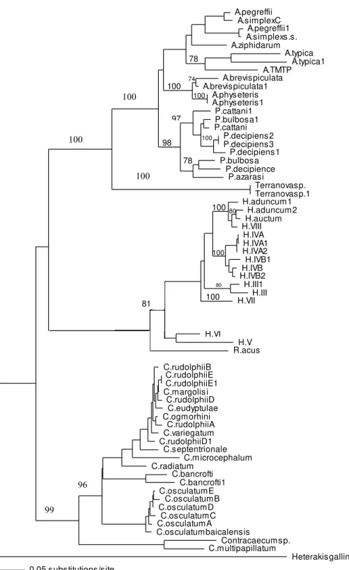
Terranovalarval type in the present study andPseudoterranovaspp. was also supported by phylogenetic analyses (Fig. 3).
ITS-1 and ITS-2 sequences of well identified closely related taxa were selected to build the phylogenetic tree to investigate the association of larvae in the present study with other taxa within family Anisakidae. Both neighbour joining and maximum parsimony (the latter is not shown) trees had similar profile and grouping of taxa were the same among both trees. In the neighbour joining phylogenetic tree (Fig. 3),Terranovalarval type found in the present study were resolved as a distinct clade with strong bootstrap support of 100%. None of the anisakid species (Pseudoterranovaspp.,Anisakisspp. andContracaecum
spp. becoming adult in marine mammals) with similar morphology toTerranovaspp. (i.e., having excretory pore opened at the base of the labia) were grouped in the same clade asTerranovalarval type found in the present study. Closely related species becoming adult in teleost fishes (Hysterothylaciumspp. andRaphidascaris acus) were also included in the phylogentic tree, although the excretory system in this group has a different feature to
Terranovaspp. These anisakids also resolved as a distinct clade toTerranovalarval type. In both phylogenetic trees produced in the present study based on the combined ITS-1 and ITS-2 sequences, the Terranova larval type was resolved separately from
Pseudoterranovaspp. suggesting they do not belong to the genusPseudoterranova.
As reviewed in the Introduction, AustralianTerranova larval types could potentially be larval stages of Pseudoterranovaspp.,Terranova spp., orPulchrascarisspp. Species
A.pegreffii A.simplexC A.pegreffii1 A.simplexs.s. A.ziphidarum A.typica A.typica1 A.TMTP A.brevispiculata A.brevispiculata1 A.physeteris A.physeteris1 P.cattani1 P.bulbosa1 P.cattani P.decipiens2 P.decipiens3 P.decipiens1 P.bulbosa P.decipience P.azarasi Terranovasp. Terranovasp.1 H.aduncum1 H.aduncum2 H.auctum H.VIII H.IVA H.IVA1 H.IVA2 H.IVB1 H.IVB H.IVB2 H.III1 H.III H.VII H.pelagicum H.tetrapteri H.sp. H.VI H.V R.acus Masonascaris C.rudolphiiB C.rudolphiiE C.rudolphiiE1 C.margolisi C.rudolphiiD C.eudyptulae C.ogmorhini C.rudolphiiA C.variegatum C.rudolphiiD1 C.septentrionale C.microcephalum C.radiatum C.bancrofti C.bancrofti1 C.osculatumE C.osculatumB C.osculatumD C.osculatumC C.osculatumA C.osculatumbaicalensis Contracaecumsp. C.multipapillatum Heterakisgallinarum 0.05 substitutions/site 100 78 74 100 100 98 80 78 80 100 100 97 100 81 100 100 100 99 96
Figure 3 Phylogenetic analysis of the combined ITS-1 and ITS-2 sequence data for members of the Anisakidae withHeterakis gallinarumas outgroup, using the neighbour-joining method. Bootstrap support values are indicated. SeeTable 1for detailed abbreviations. Note that Terranovasp and Terra-novasp1 both belong to the same taxon and only different in polymorphic sites as shown inFig. 2. They are representative of 93Terranovalarval type examined in the present study.
Table 4 Pairwise comparisons of the nucleotide differences (%) in the consensus sequences of ITS-1 and ITS-2 betweenTerranovalarval type found in the present study andPseudoterranova spp.(the only taxa with closest ITS sequence similarity available in GenBank database).
Terranovalarval type in the present study
ITS-1 ITS-2
P. azarsi 39.8 46.7
P. bulbosa 38.9 48.4
P. cattani 39.3 47.2
P. decipiens sensu tricto 39.0 48.3
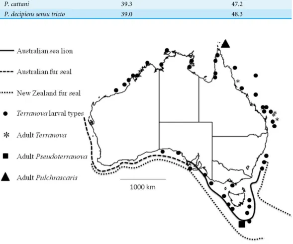
Figure 4 Map shows reported cases ofTerranovalarval types (circles), AdultTerranovaspp (asterisk), adultPseudoterranovaspp (square), adultPulchrascaris(triangle), distribution of Australian sea lion (solid line), Australian fur seal (square dots) and New Zealand fur seal (round dot).
associated with these genera have been listed inTable 3. Comparison of ITS sequence of Terranovalarval type found in the present study with those ofPseudoterranovaspp. available in GenBank (Table 4) shows a considerable nucleotide difference of 38.9–39.8% in both ITS-1 and ITS-2 regions. This is greater than nucleotide difference found for distinct species within a genus of family Anisakidae (Shamsi et al., 2009b) suggestingTerranova
larval type in the present study does not belong to the genusPseudoterranova.
To date, four species of Terranovahave been reported from Australian sharks,T. galeocerdonis, T. ginglymostomae, T. pristisandT. scoliodontis(Bruce & Cannon, 1990). In addition, T. crocodyliwas found in Australian crocodiles (Sprent, 1979). They all have
a similar relationship between length of the intestinal caecum and ventriculus to the
Terranovalarval type in the present study.Pulchrascarisis a small genus in terms of number of species under family Anisakidae. Like members of the genusTerranova, Pulchrascaris
spp. become adult in elasmobranches. There is intra/inter specific variation in the ratio of the intestinal caecum to ventriculus of Pulchrascarisspp. (Bruce & Cannon, 1990). Since there is no ITS sequences available forTerranovaspp. orPulchrascarisspp. in the GenBank database, the specific identity of the Terranovalarval type found in the present study remains unknown and we are not able to associate these larvae to anyTerranovaspp. or
Pulchrascarisspp. however, the present study, particularly the ITS sequence data, provides the essential information for future studies when the adult form is found and characterised. This is the first report of aTerranovalarval type fromAbudefduf whitleyi, Carangoides fulvoguttatus, Caranx ignobilis, C. melampygus, Chaetodon flavirostris, Lutjanus argenti-maculatus, L. bohar, Pristipomoides multidens, Scomber australasicus.Some of these fish, such as Australian mackerel (Scomber australasicus)are popular edible fish. Infection of those fish species that are not edible is also very important due to their role in the survival and transmission ofTerranovalarval type in the ecosystem.
Although the present study could not specifically identify theTerranova larval type in Australian waters, it could rule out the possibility of them being Pseudoterranova
larvae which would have different implications for seafood and consumers’ safety and policy development in the country. It should be emphasized that it is very likely that
Pseudoterranovalarvae exist in Australian waters, infect some fish and await discovery. Their definitive hosts, Australian sea lion, Australian fur seal and New Zealand fur seal are found in southern coast of Australia (Fig. 4) and have been found to be infected withPseudoterranova decipiens E (Bullini et al., 1997). However, given that in Australian waters the diversity of elasmobranch species is considerably higher (approximately 200 species,www.fishbase.net) than that of marine mammals (3 species of seals) our suggestion is that Terranovalarval type in Australian waters is more likely to be aTerranovaor a
Pulchrascaris. To date there is no evidence that larval stage ofTerranovaorPulchrascaris
can cause infection in humans.
ACKNOWLEDGEMENTS
Authors are grateful to Prof. Tom Cribb (University of Queensland, Australia) and his research group for their generous cooperation in collecting specimens from Heron Island.
ADDITIONAL INFORMATION AND DECLARATIONS
Funding
This work had partial financial support by School of Animal and Veterinary Sciences, Charles Sturt University and University of Melbourne. Shokoofeh Shamsi received support from the Graham Centre for Agricultural Innovations. The funders had no role in study design, data collection and analysis, decision to publish, or preparation of the manuscript.
Grant Disclosures
The following grant information was disclosed by the authors: School of Animal and Veterinary Sciences.
Charles Sturt University. University of Melbourne.
Graham Centre for Agricultural Innovations.
Competing Interests
The authors declare there are no competing interests. Shokoofeh Shamsi is a member of the Graham Centre for Agricultural Innovations.
Author Contributions
• Shokoofeh Shamsi conceived and designed the experiments, performed the experiments, analyzed the data, contributed reagents/materials/analysis tools, wrote the paper, prepared figures and/or tables, reviewed drafts of the paper, drawings of the parasite. • Jaydipbhai Suthar performed the experiments, analyzed the data, prepared figures and/or
tables, reviewed drafts of the paper.
DNA Deposition
The following information was supplied regarding the deposition of DNA sequences: Materials examined morphologically were deposited in South Australian Natural History Museum. The isolate numbers are as below: 301-1, 304-18, 304-2, 316-2, 324-4, 333-10, 336-4, 336-6, 340-4 and 341-3.
Nucleotide sequence data reported in this paper are available in the GenBank database under the accession numbersLN795828–LN795873.
Data Availability
The following information was supplied regarding data availability: The research in this article did not generate any raw data.
REFERENCES
Abe N, Ohya N, Yanagiguchi R. 2005.Molecular characterization ofAnisakis pegreffii
larvae in Pacific cod in Japan.Journal of Helminthology79:303–306
DOI 10.1079/JOH2005290.
Anderson RC. 2000.Nematode parasites of vertebrates: their development and transmission. Wallingford: CABI Publishing, 284.
Arizono N, Miura T, Yamada M, Tegoshi T, Onishi K. 2011.Human infection with
Pseudoterranova azarasiroundworm.Emerging Infectious Diseases17:555–556
DOI 10.3201/eid1703.101350.
Bruce NL, Adlard RD, Cannon LRG. 1994.Synoptic checklist of ascaridoid parasites (Nematoda) from fish hosts.Invertebrate Taxonomy8:583–674
DOI 10.1071/IT9940583.
Bruce NL, Cannon LRG. 1990.Ascaridoid nematodes from sharks from Australia and the Solomon Islands, Southwestern Pacific Ocean.Invertebr. Taxon.4:763–783
DOI 10.1071/IT9900763.
Bullini L, Arduino P, Cianchi R, Nascetti G, D’Amelio S, Mattiucci S, Paggi L, Orecchia P, Plotz J, Berland B, Smith JW, Brattey J. 1997. Genetic and ecological research on anisakid endoparasites of fish and marine mammals in the Antarctic and Arctic -Boreal regions. In:Antarctic communities. Cambridge: Cambridge University Press, 39–44.
Cannon LRG. 1977.Some larval ascaridoids from south-eastern Queensland marine fishes.International Journal for Parasitology7:233–243
DOI 10.1016/0020-7519(77)90053-4.
Chai J-Y, Darwin Murrell K, Lymbery AJ. 2005.Fish-borne parasitic zoonoses: status and issues.International Journal for Parasitology35:1233–1254
DOI 10.1016/j.ijpara.2005.07.013.
Chandler AC. 1935.Parasites of fishes in Galveston Bay.Proceedings of the United States National Museum83:123–157DOI 10.5479/si.00963801.83-2977.123.
Chilton NB, Gasser RB, Beveridge I. 1995.Differences in a ribosomal DNA sequence of morphologically indistinguishable species within theHypodontus macropi complex
(Nematoda: Strongyloidea).International Journal for Parasitology25:647–651
DOI 10.1016/0020-7519(94)00171-J.
Deardorff TL. 1987.Redescription ofPulchrascaris chiloscyllii(Johnston and Mawson, 1951) (Nematoda: Anisakidae), with comments on species inPulchrascarisand
Terranova.Proceedings of the Helminthological Society of Washington54:28–39.
Diaz-Ungria C. 1967.Tres especies de nematodes de peces venezolanos, con descriptcion de Terranova caballeroi, n. sp. (Nematoda), vol. 22. Maracay: Revista de Medicina Veterinaria y Parasitologia, 1–8.
Doupe RG, Lymbery AJ, Wong S, Hobbs RP. 2003.Larval anisakid infections of some tropical fish species from North-West Australia.Journal of Helminthology 77:363–365DOI 10.1079/JOH2003193.
Fang WZ, Luo DM. 2006.Description of a new ascarid species in elasmobranchs from Taiwan Strait.Journal of Parasitology92:822–825DOI 10.1645/GE-694R1.1.
Gibson DI, Colin JA. 1982.TheTerranovaenigma.Parasitology 85:R36–R37.
Hafsteinsson H, Rizvi SSH. 1987.A review of the sealworm problem—biology, implica-tions and soluimplica-tions.Journal of Food Protection50:70–84.
Hochberg NS, Hamer DH. 2010.Anisakidosis: perils of the deep.Clinical Infectious Diseases51:806–812DOI 10.1086/656238.
Huizinga HW. 1967.The life cycle ofContracaecum multipapillatum(Von Drasche, 1882) Lucker, 1941 (Nematoda: heterocheilidae).The Journal of Parasitology 53:368–375DOI 10.2307/3276593.
Jensen T, Andersen K, Desclers S. 1994.Sealworm(Pseudoterranova decipiens)in dem-ersal fish from 2 areas in Norway.Canadian Journal of Zoology-Revue Canadienne De Zoologie72:598–608 DOI 10.1139/z94-082.
Jimenez FA, Gardner SL, Navone G, Orti G. 2012.Four events of host switching in Aspidoderidae Nematoda involve convergent lineages of mammals.Journal of Parasitology 98:1166–1175DOI 10.1645/GE-3045.1.
Johnston TH, Mawson PM. 1945.Parasitic nematodes. British, Australian and New Zealand Antarctic research expedition.Reports, Series B5:73–159.
Kijewska A, Czarna A, Fernandez M, Zdzitowiecki K, Rokicki J, Wrobel B. 2008.
Analysis of 5.8S rDNA and internal transcribed spacer 1 (ITS1) sequences of
ascaridoid nematodes: phylogenetic signal and hypothesis testing.Genes & Genomics 30:291–306.
Kijewska A, Rokicki J, Sitko J, Wegrzyn G. 2002.Ascaridoidea: a simple DNA assay for identification of 11 species infecting marine and freshwater fish, mammals, and fish-eating birds.Experimental Parasitology101:35–39
DOI 10.1016/S0014-4894(02)00031-0.
Leiper RT, Atkinson EL. 1914.Helminthes of the British Antarctic Expedition 1910– 1913.Proceedings of the Zoological Society of London1914:222–226.
Lester RJG, Barnes A, Habib G. 1985.Parasites of skipjack tuna,Katsuwonus pelamis:
fishery implications.Fishery Bulletin83:343–356.
Li A, D’Amelio S, Paggi L, He F, Gasser RB, Lun Z, Abollo E, Turchetto M, Zhu X. 2005.Genetic evidence for the existence of sibling species withinContracaecum rudolphii(Hartwich, 1964) and the validity ofContracaecum septentrionale
(Kreis, 1955) (Nematoda: Anisakidae).Parasitology Research96:361–366
DOI 10.1007/s00436-005-1366-y.
Margolis S. 1977.Public health aspects of codworm infection: a review.Journal of the Fisheries Research Board of Canada34:887–898DOI 10.1139/f77-140.
Mattiucci S, Nascetti G. 2008.Advances and trends in the molecular systematics of anisakid nematodes, with implications for their evolutionary ecology and host—parasite co-evolutionary processes.Advances in Parasitology66:47–148
DOI 10.1016/S0065-308X(08)00202-9.
McClelland G. 2002.The trouble with sealworms (Pseudoterranova decipiens species complex,Nematoda): a review.Parasitology124:s183–s203.
Moore BR, Stapley J, Allsop Q, Newman SJ, Ballagh A, Welch DJ, Lester RJG. 2011.
Stock structure of blue threadfinEleutheronema tetradactylumacross north-ern Australia, as indicated by parasites.Journal of Fish Biology78:923–936
DOI 10.1111/j.1095-8649.2011.02917.x.
Mozgovoi AA. 1950. Key to parasitic nematodes. In: Skrjabin KI,ed.Oxyurata and Ascaridata, vol 2. New Delhi: Academiya NAUK SSSR. Washington, D.C.: United States Department of Agriculture and the National Science Foundation, 1982.
Nadler SA, D’Amelio S, Dailey MD, paggi L, Siu S, Sakanari JA. 2005.Molecular phylogenetics and diagnosis ofAnisakis,Pseudoterranova, andContracaecum
from Northern Pacific marine mammals.Journal of Parasitology91:1413–1429
DOI 10.1645/GE-522R.1.
Olsen LS. 1952.Some nematodes parasitic in marine fishes. Vol. 2. Port Aransas: Institute of Marine Science of the University of Texas, 173–215.
Orecchia P, Paggi L, Mattiucci S, Smith JW, Nascetti G, Bullini L. 1986.Electrophoretic identification of larvae and adults ofAnisakis(Ascaridida: Anisakidae).Journal of Helminthology60:331–339DOI 10.1017/S0022149X00008580.
Paggi L, Nascetti G, Cianchi R, Orecchia P, Mattiucci S, D’Amelio S, Berland B, Brattey J, Smith JW, Bullini L. 1991.Genetic evidence for three species within
Pseudoterranova decipiens(nematoda, ascaridida, ascaridoidea) in the north atlantic and norwegian and barents seas.International Journal for Parasitology21:195–212
DOI 10.1016/0020-7519(91)90010-5.
Pontes T, D’Amelio S, Costa G, Paggi L. 2005.Molecular characterisation of larval anisakid nematodes from marine fishes of Madeira by a PCR-based approach, with evidence for a new species.Journal of Parasitology 91:1430–1434
DOI 10.1645/GE-565R1.1.
Shamsi S. 2014.Recent advances in our knowledge of Australian anisakid nematodes.
International Journal for Parasitology: Parasites and Wildlife3:178–187.
Shamsi S, Butcher AR. 2011.First report of human anisakidosis in Australia.Medical Journal of Australia194:199–200.
Shamsi S, Gasser RB, Beveridge I. 2011.Mutation scanning-coupled sequencing of nuclear ribosomal DNA spacers (as a taxonomic tool) for the specific identification of differentContracaecum(Nematoda: Anisakidae) larval types.Molecular and Cellular Probes25:13–18DOI 10.1016/j.mcp.2010.09.003.
Shamsi S, Gasser R, Beveridge I. 2012.Genetic characterisation and taxonomy of species of Anisakis (Nematoda:Anisakidae) parasitic in Australian marine mammals.
Invertebrate Systematics26:204–212DOI 10.1071/IS11019.
Shamsi S, Gasser R, Beveridge I. 2013.Description and genetic characterisation of Hys-terothylacium(Nematoda: Raphidascarididae) larvae parasitic in Australian marine fishes.Parasitology International 62:320–328DOI 10.1016/j.parint.2012.10.001.
Shamsi S, Gasser R, Beveridge I, Shabani AA. 2008.Contracaecum pyripapillatum
n. sp. and a description ofC. multipapillatum(von Drasche, 1882) from the Australian pelican,Pelecanus conspicillatus.Parasitology Research103:1031–1039
DOI 10.1007/s00436-008-1088-z.
Shamsi S, Norman R, Gasser R, Beveridge I. 2009a.Genetic and morphological evi-dences for the existence of sibling species withinContracaecum rudolphii(Hartwich, 1964) (Nematoda: Anisakidae) in Australia.Parasitology Research105:529–538
DOI 10.1007/s00436-009-1424-y.
Shamsi S, Norman R, Gasser R, Beveridge I. 2009b.Redescription and genetic character-ization of selectedContracaecumspp. (Nematoda: Anisakidae) from various hosts in Australia.Parasitology Research104:1507–1525DOI 10.1007/s00436-009-1357-5.
Shamsi S, Poupa A, Justine J-L. 2015.Characterisation of Ascaridoid larvae from marine fish off New Caledonia, with description of new Hysterothylacium larval types XIII and XIV.Parasitology International 64:397–404DOI 10.1016/j.parint.2015.05.014.
Sprent JFA. 1979.Ascaridoid nematodes of amphibians and reptiles:Terranova.Journal of Helminthology53:265–282DOI 10.1017/S0022149X00006088.
Szostakowska B, Myjak P, Kur J, Sywula T. 2001.Molecular evaluation of Hysterothy-lacium auctum (Nematoda, Ascaridida, Raphidascarididae) taxonomy from fish of the southern Baltic.Acta Parasitologica46:194–201.
Tanzola RD, Sardella NH. 2006.Terranova galeocerdonis(Thwaite, 1927) (Nematoda: Anisakidae) fromCarcharias taurus(Chondrichthyes: Odontaspididae) off Ar-gentina, with comments on some related species.Systematic Parasitology64:27–36
DOI 10.1007/s11230-005-9015-5.
Thompson JD, Gibson TJ, Plewniac F, Jeanmougin F, Higgins DG. 1997.The Clustal X windows interface:flexible strategies for multiple sequence alignment aided by quality analysis tools.Nucleic Acids Research24:4876–4882.
Umehara A, Kawakami Y, Araki J, Uchida A. 2008.Multiplex PCR for the identification of Anisakis simplex sensu stricto,Anisakis pegreffiiand the other anisakid nematodes.
Parasitology International 57:49–53DOI 10.1016/j.parint.2007.08.003.
Yamaguti S. 1941.Studies on the helminth fauna of Japan. Part 33. Nematodes of fishes, II.Japanese Journal of Zoology9:343–396.
Zhu X, D’Amelio S, Paggi L, Gasser RB. 2000.Assessing sequence variation in the internal transcribed spacers of ribosomal DNA within and among members of theContracaecum osculatumcomplex (Nematoda: Ascaridoidea: Anisakidae).
Parasitology Research86:677–683DOI 10.1007/PL00008551.
Zhu X, Gasser RB, Podolska M, Chilton NB. 1998.Characterisation of anisakid nema-todes with zoonotic potential by nuclear ribosomal dna sequences.International Journal for Parasitology28:1911–1921DOI 10.1016/S0020-7519(98)00150-7.
Zhu XQ, D’amelio S, Palm HW, Paggi L, George-Nascimento M. 2002.SSCP-based identification of members within thePseudoterranova decipienscomplex (Nematoda: Ascaridoidea: Anisakidae) using genetic markers in the internal transcribed spacers of ribosomal DNA.Parasitology124:615–623.