INTRODUCTION
Celadon glaze constituted with (SiO2–Al2O3–CaO–
MgO–K2O–Na2O) baic raw ceramics shows characteristic
bright color, which strongly depends on the oxidizing and deoxidizing thermal treatment at higher-temperature of about 1200 to 1300 °C and transition-metal ions included naturally and artificially in the complex raw ceramics. Larid (1918) [1] and Hunghan [2] reported for the Chinese celadons that the blue-green color of the celadon glazes is induced by chemical reaction from Fe2O3 to FeO in the
celadon glazes under the deoxidizing thermal treatment.
The celadon glaze changes gradually its color from yellow-brown to black color, as increasing the iron oxides under the deoxidizing thermal treatment [3]. Recently, we have been studying the fascinating and mysterious coloration-mechanism of the celadon glaze for the Hizen celadons, which were produced at Arita, Hasami and Imari areas in 1630’s to 1790’s (Edo period, Japan) by means of the X-ray diffraction and the X-ray absorption spectra (XAS) near the Fe-K edge by using synchrotron radiation, in addition to the ordinary X-ray fluorescence analysis and the Mössbauer spectrum [4, 5]. We found that that the glaze colors of the Hizen celadons depend on the material properties of the
Structural and electronic properties of iron oxides in the
celadon glazes (II)
(Propriedades estruturais e eletrônicas de óxidos
de ferro em esmaltes celadon (II))
M. Hidaka1, K. Takeuchi2, R. P. Wijesundera1,6, L. S. R. Kumara1, S. Sugihara3, N. Momoshima3,
S. Kubuki4, Nark Eon Sung5
1Department of Physics, Graduate School of Science, Kyushu University, Fukuoka 812-8581, Japan 2Ceramic Research Center of Nagasaki, Nagasaki, 859-3726, Japan
3Radioisotope Center, Kyushu University, Fukuoka 812-8581, Japan
4Department of Chemical and Biological Engineering, Ube National College of Technology,
Yamaguchi, 755-8555, Japan
5Beamline Department, Pohang Accelerator Laboratory, Pohang University of Science and Technology,
Pohang, 790-784, Korea
6Department of Physics, University of Kelaniya, Kelaniya, Sri Lanka
hidaka@phys.kyushu-u.ac.jp
Abstract
Celadon glazes have been investigated by means of an X-ray absorption spectrum (XAS) near a Fe-K edge by using synchrotron radiation and a Mössbauer spectrum. High-temperature treatments under CO-deoxidizing and oxidizing till about 1300 °C show the different glaze-color each other. The XAS analyses suggest that the deoxidized celadon glaze (Seiji A) and the oxidized one (Seiji C) have a modified α-Fe2O3 structure and an ideal one, respectively, but not FeO structure. The Mössbauer spectra also suggest that the celadon glaze-color depends on the hybridized 3d5L and 3d6L bands near an electronic Fermi level (E
F), where the hybridization is induced by an electronic exchange interaction between 3d orbitals of Fe ions and 2p orbitals of surrounding O ions in the celadon glaze of glass-state.
Keywords: glass ceramics, color, traditional ceramics, X-ray methods.
Resumo
Esmaltes celadon foram investigados por meio do espectro de absorção de raios X (XAS) próximo da borda Fe-K usando radiação síncrotron e espectro Mössbauer. Tratamentos térmicos a altas temperaturas sob atmosfera desoxidante (CO) e oxidante até ~ 1300 °C apresentam diferentes cores dos esmaltes. As análises XAS sugerem que o esmalte celaton desoxidado
(Seiji A) e o oxidado (Seiji C) apresentam uma estrutura α-Fe2O3 modificada e uma ideal, respectivamente, mas não a estrutura
FeO. Os espectros Mössbauer também sugerem que a cor dos esmaltes celadon depende das bandas híbridas 3d5L and 3d6L
próximas do nível de Fermi eletrônico (EF), onde a hibridização é induzida por uma interação de troca eletrônica entre orbitais
3d dos íons Fe e orbitais 2p dos íons de oxigênio próximos no esmalte celadon do estado vítreo.
used raw ceramics and the transition-metal ions of Cr, Cu, Zn (very small amounts) and Fe (small amounts). It was also found that the celadon glaze includes microscopically the fine iron oxides of Fe2O3, but not FeO, under the deoxidizing
thermal treatment of high-temperature, even if the celadon glaze is macroscopically in glass-state of short-range order.
More recently, in order to reduce the intrinsic parameters related to the appearance of the glaze color, we tentatively made some celadon glazes under the oxidizing and deoxidizing thermal treatment and studied the structural and electronic properties of the glazes by means of the X-ray diffraction and the X-ray absorption spectra in our first paper [6]. The results suggested that the characteristic color of blue-green, white-green-brown, and white-blue-green result from the complex hybridized 3d5L and 3d6L bands of
Fe ions. The 3d6L hybridization is induced by an electronic
exchange interaction between the empty 3d6 orbitals of Fe
ions and the occupied 2p orbitals of surrounding O ions in the (SiO2–Al2O3–CaO) basic complex ceramics of
glass-state under the deoxidizing thermal treatment. In order to confirm the results, we studied the local structure around Fe ions and the electron valence of Fe ions in the present celadon glaze. In this paper, we will report the structural and electronic properties of iron oxides in the tentative celadon glazes by means of the X-ray absorption spectrum (XAS) near the K-edge of Fe ions and the Mössbauer spectrum.
MATERIALS AND METHODS
In the present investigations, the tentative celadon glazes were made by mixing raw celadon materials of Masuda feldspar, limestone, Hadong kaolin, quartz, and extra-added Fe2O3 of about 1wt.%. The material properties of the used
celadon glazes (Seiji A, B, C, and D) were already shown [6]. For comparison, Table I also lists a red-color overglaze of the Kakiemon-style porcelain, produced at Arita in 1670-80’s [7 ].
We measured the X-ray absorption spectrum (XAS) near the Fe-K edge for Seiji A and C, in addition to those of the red-color overglaze of the Kakiemon-style porcelain and the marketed fine powders of FeO, by using monochromatic incident X-ray beams of synchrotron radiations at the Pohang
Light Source. A double crystal monochrometor of Si (111) gave a relative energy resolution ΔE to be less than about 0.2 eV at the respective monochromatic incident X-ray beams. The incident X–ray photons (Io) were detected with an ionic
chamber set before the specimens, which were already shown in Fig. 1 of our first paper [6], while the X–ray fluorescence photons (I) emitted from the surface of the specimens were simultaneously detected with an X–ray fluorescence detector. The flat surface of specimen was always set with about 45.0° to the incident X-ray beam. The used beam size was about 3 mm in horizontal and 1 mm in vertical on the specimen surface. Since the incident synchrotron X-ray gradually loses intensity, we always monitored the incident X–ray photons (Io) at the respective measurement. In the present
investigations, we firstly normalized the observed XAS by subtracting background counts at the pre-edge region far from the X-ray absorption Fe-K edge. Secondly, we analyzed the normalized XAS with software programs of Artemis and
Athena, which was developed by Booth and Bridges [8].
The local structure around Fe ions can be obtained from the XAS in the region of about 50 to 700 eV around its X-ray absorption threshold (Eo), of which the spectrum is called an
EXAFS spectrum.
To study an electronic valence of Fe ions in the present celadon glazes we also measured the Mössbauer spectrum at room temperature. The Mössbauer effect involves a resonant absorption of gamma rays by atoms of the same isotope. The source of gamma rays is a radioactive isotope of an element which decays into an excited state of the isotope, which returns to its ground state by the emission of a gamma ray or electron. In the present measurements, we used the gamma ray emitted from the isotope of 57Co, which undergoes the
nuclear decay to 57Fe of nuclear spin I=5/2 excited state.
The decay via gamma ray or conversion electrons is in the Mössbauer spectrum of iron system.
RESULTS
To study the long-range ordering of the Fe2O3 and FeO
structures, we first measured the XAS around the Fe-K edge for the red-color overglaze of the Kakiemon-style porcelain and the marketed fine powders of FeO. The red-color
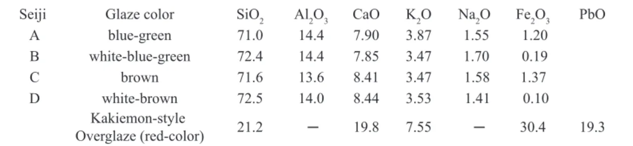
Seiji Glaze color SiO2 Al2O3 CaO K2O Na2O Fe2O3 PbO
A blue-green 71.0 14.4 7.90 3.87 1.55 1.20
B white-blue-green 72.4 14.4 7.85 3.47 1.70 0.19
C brown 71.6 13.6 8.41 3.47 1.58 1.37
D white-brown 72.5 14.0 8.44 3.53 1.41 0.10
Kakiemon-style
Overglaze (red-color) 21.2 ─ 19.8 7.55 ─ 30.4 19.3
Table I - Oxide composition (wt.%) in the tentative celadon glazes (Seiji A, B, C, D) and the red-color overglaze of the Kakiemon-style porcelain.
Figure1: X-ray absorption spectra around the Fe-K edge for the red-color overglaze of the Kakiemon-style porcelain and the fine powders of FeO.
[Figura 1: Espectros de absorção de raios X próximos da borda Fe-K do esmalte vermelho da porcelana de estilo Kakiemon e pós finos de FeO.]
1.2
1.1
1.2 1.05
I/Io I/Io
I/Io I/Io
E (eV) E (eV)
E (eV) E (eV)
0.8 1.0
0.8 0.4
0.4
1.00
0.95
0.0 0.9
0.0 7100 7100
7300 7300
7300 7300
7500 7500
7500 7500
7200 7200
7200 7200
7400 7400
7400 7400
7600 7600
7600 7600
Figure2: X-ray absorption spectra around the Fe-K edge for the celadon glazes of Seiji A and C.
[Figura 2: Espectros de absorção de raios X próximos da borda Fe-K para os esmaltes celadon de Seiji A e C.] 1.2
1.2
1.1
1.1
I/Io
I/Io
I/Io
I/Io
E (eV)
E (eV)
E (eV)
E (eV) 0.8
0.8
1.0
1.0 0.4
0.4 0.0
0.0
0.9
0.9 7100
7100 7000
7000
7300
7300
7300
7300 7500
7500
7500
7500 7200
7200
7200
7200 7400
7400
7400
7400 7600
7600
7600
overglaze includes Fe2O3 of about 30.4 wt.% in Table I, and
its X-ray diffraction showed some reflections belonging to the α−Fe2O3 structure, in addition to the complex patterns of
the (SiO2−CaO−PbO) glaze ceramics [7].This suggests that in the red-color overglaze there are the fine crystals of α− Fe2O3 to be in long-range order. Fig. 1 shows the Fe-K XAS
of the Kakiemon-style red-color overglaze and the FeO fine powders. Figs. 1 a2 and b2 represent the EXAFS spectrum in the region of 7150 to 7600 eV, respectively. Thus, the large oscillating amplitude of the EXAFS spectrum suggests that the Fe2O3 fine particles make a considerably large cluster
of long-range order in the red-color overglaze. The clear difference of the XAS between the Kakiemon-style red-color overglaze and the FeO fine powders results from the Fe2O3 structure and the FeO one.
On the other hand, Fig. 2 shows the Fe-K XAS for the celadon glazes of Seiji A and C, where Figs. 2 a2 and b2 represent the EXAFS spectrum in the region of 7150 to 7600 eV of Seiji A and C, respectively. Thus, the smoothly small oscillating amplitude of the EXAFS spectra suggests that Fe ions are in glassy state of short-range order. We already confirmed that Seiji A, B, C, and D show the hallo-like X-ray diffraction pattern [6].Thus, we are interested whether Fe ions in the celadon glaze are still in the micro-crystal state of Fe2O3 or FeO structure or in non-crystal state to be perfectly
complex free ions, because the EXAFS spectrum gives the structural information of the local circumstance around the X-ray absorbing ions in a material.
To study the local structure around Fe ions in the celadon glaze, we carried out to analyze the EXAFS spectra of Seiji A and C. We firstly obtained an observed Fourier transformation spectrum │Fobs (R)│ to the observed oscillating EXAFS
spectrum Хobs(K), after replacing an energy E of X-ray
photons with a wave-number K{= 8π2m
e(E-Eo)/h2}, where
me and h are an effective mass of electron and Plank constant,
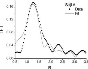
respectively. Fig. 3 shows │Fobs(R)│ of the EXAFS spectrum
for Seiji A, where the abscissa refers to a radial distance (R(Å)) from the X-ray absorbing Fe ion, as a coordinate origin, to its surrounding shells including cations or anions in the celadon glaze. The peaks of the observed │Fobs(R)│ approximately
indicate the existence of some shells being away from the X-ray absorbing Fe ions by Rj (Ǻ). Each shell includes the equivalent surrounding ions. Since the X-ray diffraction of Seiji A shows the hallo-like pattern, it is considered that, in the celadon glaze, the surrounding O and Fe ions are in short-range order to the X-ray absorbing Fe ion. The similar feature is also confirmed for the celadon glaze of Seiji C. From the│Fobs(R)│of Seiji A and C, we assume that the short-range
order of Fe ions is microscopically in a crystal-state of the Fe2O3 or FeO structure, but not perfectly isolated free ions.
After obtaining the Хobs(K), we calculated a theoretical
oscillating EXAFS spectrum Хcal(K) based on an optical
interference theory for the X-ray photoelectron waves emitted from the X-ray absorbing Fe ions and its back-scattering waves induced by the surrounding ions. For the refinement of the Хobs(K) with the Хcal(K), we used the
software programs of Artemis and Athena [8]. The best fitting between the Хobs(K) and the Хcal(K) is usually done
by a least squares method with several refined parameters, which are an equivalent ion-number (Nj) on the shell having the same radial distance Rj, a Debye-Waller factors (σj), a characteristic temperature (QDj), a passive
electron reduction factor (S2
o). The refinements are always
monitored by a convergence factor RF (={Σ|Хobs(K)–
kХcal(K)|2}/Σ|Хobs(K)|2 ), where k is a scale factor. The
details were already reported [9].
Figure 3: Observed Fourier transformation spectrum │Fobs(R)│of the Fe-K EXAFS spectrum for Seiji A in Fig. 2b.
[Figura 3: Espectro da transformada de Fourier │Fobs(R)│do
espectro Fe-K EXAFS para Seiji A na Fig. 2b.] 0.20
0.15
0.10
0.05
0.00 0.5
R
I Fobs (R)I
2.5
1.5 3.5
1.0 2.0 3.0 4.0
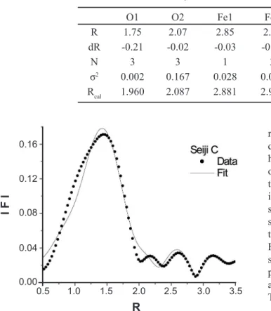
Figure 4: Theoretical |Fcal(R)| (solid line-fit) of the EXAFS spectrum, taken by the least squares refinement to the observed │Fobs(R)│ (solid circles) for the Fe ions in the α−Fe2O3 structure of Seiji A in Fig. 3.
[Figura 4: Espectro teórico |Fcal(R)| EXAFS (ajuste-linha sólida),
obtido por ajuste de mínimos quadrados do espectro │Fobs(R)│
observado (círculos sólidos) para os íons Fe da estrutura α−Fe2O3
de Seiji A na Fig. 3.] 0.16
0.12
0.08
0.04
0.00 0.5
R
I F I
2.5
1.5 3.5
Fig. 4 shows the refinement of the EXAFS spectra for the Fe ions due to the α-Fe2O3 structure of Seiji A in Figs. 2 and
3. The abscissa refers to the radial distance (R(Å)) from the X-ray absorbing Fe ion to its surrounding shells including the cations or anions of the α-Fe2O3 structure. The solid line
and the solid circles represent the theoretical and observed Fourier transformation spectrum │F(R)│, respectively. The theoretical calculation was based on the ideal α-Fe2O3
structure, which has a hexagonal symmetry with its lattice constants of a=5.035 Ǻ and b=13.72 Ǻ. In the present analyses the equivalent ion-numbers (Nj) were fixed to those of the ideal α-Fe2O3 structure. The RF-values were about 0.06,
and the refined value S2
o=0.83. The present spectral analysis
is roughly satisfied. However, the calculated peak amplitudes are not so consistent with the observed ones, in contrasted to those of the peak positions. This suggests that the α-Fe2O3
structure is slightly modified in the celadon glaze of Seiji A. The refined results are listed in Table II. In the table, dR(=R− Rcal,) represents a radial difference between the
refined radial distance (R) and the calculated one (Rcal) due to the ideal α-Fe2O3 structure to each surrounding shell having Oj or Fej. It is found that the most nearest neighbor of O ions O1 shows a large difference of dR. This means that the octahedron of FeO6 is largely distorted to the ideal α-Fe2O3 structure in celadon glaze of Seiji A. When studying the local structure by the refinement of the EXAFS spectrum, it is important to regard an optical phase between the photoelectron waves emitted from the X-ray absorbing Fe ions and the backscattering waves produced by the surrounding shells. Thus, we should also refine the phase parameter in the present investigations. The R values in the abscissa of Fig. 4 are slightly different to those in Table II. The difference results from the optical phase.
We also carried the refinement of the EXAFS spectra for the Fe ions of the α-Fe2O3 structure of Seiji C in Fig. 2. The result is shown in Fig. 5. The RF-values were about 0.03, and S2
o=1.26. The peak amplitudes and the peak position
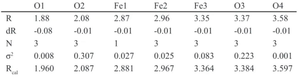
are considerably consistent for the observed and calculated │F(R)│. Thus, the present refinement is sufficiently satisfied. This suggests that Seiji C shows the ideal α-Fe2O3 structure. Table III shows the refined parameters for the celadon glaze of Seiji C. The most nearest neighbor of O ions O1 shows a small difference of dR. This suggests that the octahedron of FeO6 is not so distorted to the ideal α-Fe2O3 structure in celadon glaze of Seiji C. In the present investigations, we also tried to refine the EXAFS supectra of Seiji A and C with the FeO structure [1, 2].However, we could not succeed the FeO refinements of both EXAFS spectra of Seiji A and C. We will describe the reason in a discussion.
To study the electronic valence of Fe ions in the present celadon glazes we measured the Mössbauer spectrum at room temperature. The Mössbauer spectrum is affected by temperature and three other factors of isomer shift, quadruple splitting, and magnetic splitting. In the present investigations, we regard only the effects of the isomer shift (δ) and the quadruple splitting (Δ) because of the measurement at room temperature and no applied magnetic field. The δ results from the difference in the electron densities at the nuclear sites in the emitting and absorbing atoms. The δ is sensitive to the oxidation state and can therefore be used to study valence electrons of Fe ions in the celadon glaze. If the nuclei do not have a Figure 5: Theoretical |Fcal(R)| (solid line-fit) of the EXAFS
spectrum, taken by the least squares refinement to the observed │Fobs(R)│ (solid circles) for the Fe ions in the α−Fe2O3 structure of Seiji C in Fig. 2.
[Figura 5: Espectro teórico |Fcal(R)| EXAFS (ajuste-linha sólida),
obtido pelo ajuste de mínimos quadrados do espectro observado
│Fobs(R)│ (círculos sólidos) para os íons Fe na estrutura α−Fe2O3
de Seiji C na Fig. 2.]
Table II - Refined radial distance Rj (Ǻ) of the surrounding ions (oxygen ions Oj and Fe ions Fej) around the X-ray absorbing Fe ion of the α-Fe2O3 structure in the celadon glaze of Seiji A.
[Tabela II - Distância radial refinada Rj (Ǻ) dos íons vizinhos (íons de oxigênio Oj e íons Fe Fej) em torno dos íons Fe da absorção de raios X da estrutura Rj (Ǻ) do esmalte celadon de Seiji A.]
O1 O2 Fe1 Fe2 Fe3 O3 O4
R 1.75 2.07 2.85 2.94 3.33 3.35 3.56
dR -0.21 -0.02 -0.03 -0.03 -0.03 -0.03 -0.04
N 3 3 1 3 3 3 3
σ2 0.002 0.167 0.028 0.029 0.119 0.184 0.013
Rcal 1.960 2.087 2.881 2.967 3.364 3.384 3.597
0.16
0.12
0.08
0.04
0.00 0.5
R
I F I
2.5
1.5 3.5
charge distribution of spherically symmetric, the nucleus will possess an electric nuclear quadruple moment. This moment interacts with an asymmetric electronic charge distribution splits the degeneracy of the excited state into two levels, which are separated by the Δ. The Mössbauer spectrum is a doublet. The Δ can be broken down into two contributions of a valence contribution from the atom itself and a lattice contribution from neighboring atoms. Fe3+
(3d5) ions have no contribution to the electric field gradient
from the 3d electron orbitals. This means that Fe3+ ions
have the relatively low Δ. On the hand, Fe2+ (3d6) ions have
the large electric field gradient contribution from the 6th 3d-electron, and give the large Δ of about 1.5-3 mm/s (with a maximum of > 4 mm/s). Thus, the δ and Δ are useful tools to determine whether Fe ion has the valence electrons of +3 (Fe2O3) or +2 (FeO). If Fe2O3 and FeO particles coexist
in the glaze, we can observe a mixed Mössbauer spectrum induced by Fe3+ and Fe2+ ions.
Table IV lists the refined parameters of the isomer shift δ, the quadruple splitting ∆, and the line width H for the observed Mössbauer spectra of the present tentative celadon glazes, Seiji A, B, C, and D. We could not observe any Mössbauer spectrum of Seiji D. The reason comes from the very small amount of Fe2O3 of about 0.10 wt.%. The
Mössbauer spectroscopy for Fe oxides and oxyhydroides were reported [10, 11]. From their analyses the present Mössbauer spectra suggest that the celadon glaze of Seiji A and B include only Fe2+ ions, while Seiji C includes
only Fe3+ ions. Thus, we suspect that the high-temperature
treatment under the oxidizing and the CO-deoxidizing in the used kiln induce the different electronic property of Fe ions in the celadon glaze.
DISCUSSION
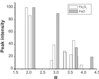
To study the structural and electronic properties of iron oxides in the celadon glazes we carried out the measurements of the X-ray absorption spectra near the Fe-K edge by using synchrotron radiation and the Mössbauer spectra. We suspected from the analyses of the EXAFS spectra that the CO-deoxidized celadon glaze (Seiji A) and the oxidized one (Seiji C) have the slightly modified α-Fe2O3 structure and the ideal one, respectively, even if the celadon glazes are macro-structurally in the glass-state of short-range order. However, we could not succeed the refinements of the observed EXAFS spectrum Хobs(K) with the theoretical one Хcal(K) of the FeO structure for Seiji A and C. Fig. 6 shows theoretical peak intensities of |Fcal(R)| for the α-Fe2O3 structure and the FeO one. When we consider the FeO structure, there should be only two separate high peaks within the radial distance of about 3.5 Å.
Thus, it was impossible to refine the observed Хobs(K) of Seiji A and C with the FeO structure. In fact, the monitoring factor RF was quickly diverged at the first cycle of the refinements. Thus, we considered that the characteristic glaze color of blue-green (Seiji A) and white-blue-color (Seiji B) results from the slight modulation of the ideal α-Fe2O3 structure induced by the CO-deoxidizing thermal treatment at high-temperature of about 1200 to 1250 °C. This suggests that there is no chemical reaction from Fe2O3 to FeO in the celadon glazes under the deoxidizing thermal treatment, as reported [1, 2]. On the other hand, the Mössbauer spectra suggest that the electron valence of Fe ions is 2+ and 3+ in Seiji A and Seiji C, respectively. The results are not consistent with those taken by the EXAFS refinements, where the iron
O1 O2 Fe1 Fe2 Fe3 O3 O4
R 1.88 2.08 2.87 2.96 3.35 3.37 3.58
dR -0.08 -0.01 -0.01 -0.01 -0.01 -0.01 -0.01
N 3 3 1 3 3 3 3
σ2 0.008 0.307 0.027 0.025 0.083 0.223 0.001
Rcal 1.960 2.087 2.881 2.967 3.364 3.384 3.597
Table III - Refined radial distance Rj (Ǻ) of the surrounding ions (oxygen ions Oj and Fe ions Fej) around the X-ray absorbing Fe ion of the α-Fe2O3 structure in the celadon glaze of Seiji C.
[Tabela III - Distância radial refinada Rj (Ǻ) dos íons vizinhos (íons oxigênio Oj e íons ferro
Fej) em torno do íon Fe da estrutura α-Fe2O3 no esmalte celadon de Seiji C.]
Table IV - Isomer shift δ, quadruple splitting ∆, and line width H with a unit mm/s for the Mössbauer spectrum of Seiji A, B, C, and D in Table I.
[Tabela IV - Desvio isomérico δ, desdobramento quadrupolar ∆, e largura de linha H de unida de mm/s do espectro Mössbauer de Seiji A, B, C, e D na Tabela I.]
Seiji Glaze color δ ∆ H
A blue-green 1.00(1) 1.82(2) 0.69(2)
B white-blue-green 0.99(3) 1.78(5) 0.62(7)
C brown 0.30(4) 0.99(5) 0.90(5)
oxide should be Fe2O3, but not FeO, in the celadon glaze of Seiji A. The modified and ideal Fe2O3 structures are not so largely different each other, as in Tables II and III. Thus, it is considered that the difference of δ and Δ in Table IV results mainly from the electric field gradient of the 3d electronic orbitals around the Fe nucleus, if the iron oxides in Seiji A and C have almost the Fe2O3-type structure.
More recently, from the pre-edge XANES near the X-ray absorption threshold Eo, that is the electronic Fermi level EF, we found that Seiji A shows the complex hybridized 3d5L
and 3d6L bands of Fe ions, while Seiji C shows only the
hybridized 3d5L one.16 The hybridization is induced by the
electronic exchange interaction between empty 3d orbitals of an X-ray absorbing Fe ion and full-occupied 2p orbitals of its surrounding O ions [12 ]. Thus, we suspect that, in a solid state, the hybridized Fe-3d6L band induces the asymmetric
electronic charge distribution at the Fe-nuclear sites. This gives the large quadruple splitting (Δ), in contrast with the symmetric Fe-3d5L band. In Tables II and III, the radial
distance between the X-ray absorbing Fe ion and the most nearest neighbor O ions, O1, of Seiji A is slightly shorter than that of Seiji C. This suggests that the octahedron FeO6 of Seiji A is distorted slightly larger than that of Seiji C for the crystallographic symmetry, in addition to the increase of the electronic overlapping between the Fe-3d and O-2p orbitals. This also gives that the large isomer shift (δ) of Seiji A, in contrast with that of Seiji C.
We consider that the present Mössbauer spectra give the electronic and structural information of Fe2+-like and
Fe3+-like ions, but not pure Fe2+ or Fe3+ ions, in the celadon
glaze of Seiji A and C, respectively, due to the hybridized 3d5L and 3d6L bands of Fe ions. The considerations are not
inconsistent with the modified α-Fe2O3 structure in Seiji A and the ideal α-Fe2O3 in Seiji C, taken by the EXAFS spectrum analyses. The CO deoxidizing thermal treatment of the celadon glaze affects electronically the hybridization of the Fe-3d band states, which are localized just below and
above the Fermi level [13], and structurally the distortion of the FeO6 octahedra. The orbital hybridization between
the Fe ions and the surrounding O ions sensitively affects the glaze-color emission in the celadon glaze, of which the constructive ions are in the glass-state of short-range order. It is therefore deduced that the high-temperature deoxidization of the celadon glaze burn and remove the O ions of Fe2O3,
and that the surrounding O ions of the (SiO2-Al2O3-CaO)
complex ceramics are rearranged into the empty sites of O ions in the Fe2O3 structure. Thus, the rearrangement gives
the slight distortion of FeO6 and shorts the radial distance
between Fe ion and its surrounding O ions. The feature contributes to the color brightness of the celadon glaze.
CONCLUSIONS
The EXAFS spectra suggested that the CO-deoxidized celadon glaze (Seiji A and B) have the slightly modified α-Fe2O3 structure, while the oxidized glaze (Seiji C) has
the ideal α-Fe2O3 structure. Thus, we considered that the
characteristic glaze color of blue-green (Seiji A) and white-blue-color (Seiji B) results from the slight modulation of the ideal α-Fe2O3 structure induced by the CO-deoxidizing
thermal treatment at high-temperature of about 1200 to 1250 °C. This suggests that there is no chemical reaction from Fe2O3 to FeO in the celadon glazes under the deoxidizing
thermal treatment, as reported [1, 2]. The Mössbauer spectra also suggested the electronic valence of Fe2+-like (3d6L ) and
Fe3+-like (3d5L ) of the Fe ions in the celadon glaze of Seiji A
and C, respectively, due to the hybridization induced by the electronic exchange interaction between the 3d orbitals of Fe ions and the 2p orbitals of surrounding O ions in the celadon glaze of glass-state. We suspected that the CO deoxidizing thermal treatment of the celadon glaze affects electronically the hybridization of the Fe-3d band states, localized just below and above the Fermi level, and structurally the distortion of the FeO6 octahedra. Thus, the orbital hybridization between
the Fe ions and the surrounding O ions of the (SiO2-Al2O3
-CaO) complex ceramics sensitively affects the glaze-color emission in the celadon, of which the constructive ions are in glass-state of short-range order.
REFERENCES
[1] J. S. Larid, The composition of Chinese celadon pottery, J. Am. Ceram. Soc. 1 (1918) 675.
[2] R. R. Hunghan, Early Chinese ceramics glazes,Ceram. Age 56 (1950) 40.
[3] V. P. Shvaiko, A black glaze for porcelain, Steklo i Keramika 18 (1961) 33
[4] M. Hidaka, K. Ohashi, R. P. Wijesundera, L. S. R. Kumara, M. Watanabe, K. Koga, J. Y. Choi, N. E. Sung, Y. J. Park, Correlation between glaze-colors and structural properties of the HIZEN celadons produced in the Edo
period of Japan by means of X-ray diffraction (І), Cerâmica
57 (2011) 106. Figure 6: Theoretical peak intensity of |Fcal(R)| for the α-Fe2O3
structure (empty) and the FeO one (filled).
[Figura 7: Intensidade do pico teórico de |Fcal(R)| para a estrutura
α-Fe2O3 (vazio) e para a estrutura do FeO (preenchido).]
100
80
60
40
20
0 1.5
R
Peak intensity
2.5 3.5
2.0 3.0 4.0 4.5
[5] M. Hidaka, K. Ohashi, R. P. Wijesundera, L. S. R Kumara, S. Sugihara, N. Momoshima, S. Kubuki, N. E. Sung, Local structures and electronic band states of α−
Fe2O3 polycrystalline particles in the glazes of the HIZEN
celadons produced in the Edo period of Japan by means of
X-ray absorption spectra (II), Cerâmica 57 (2011) 155.
[6] M. Hidaka, K. Takeuchi, R. P. Wijesundera, L. S. R. Kumara, M. Watanabe, Jae-Young Choi, Nark Eon Sung,
Correlation between the green-like coloration and the
structural and electronic properties of Celadon glazes (I),
Cerâmica 58, 347 (2012) 328-337.
[7] M. Hidaka, K. Ohashi, S. Kajihara, R. P. Wijesundera, L. S. R. Kumara, M. Watanabe, J. Y. Choi, N. E. Sung,
Structural properties of the red-color overglaze for the HIZEN porcelains produced in the early Edo period of
Japan, Ceram. Int. 35 (2009) 875-886.
[8] C. H. Booth, F. Bridges, Phys. Scr. T115 (2005) 202. [9] M. Hidaka, H. Horiuchi, K. Ohashi, R. P. Wijesundera,
L. S. R. Kumara, N. E. Sung, Local structures and electronic band states of α−Fe2O3 polycrystalline particles included in the red-color overglazes and the transparent glazes of the Kakiemon-style porcelains by means of X-ray absorption
spectra,Cerâmica 55 (2009) 223-232.
[10] E. De Grave, A. Van Alboom, Evaluation of ferrous and
ferric Mössbauer fractions, Phys. Chem. Minerals 18 (1991)
337-342.
[11] M. Darby Dyar, Mössbauer spectroscopy (2009). http://serc.carleton.edu/research_education/geochemsheets/ techniques/mossbauer.html
[12] J. Zaanen, G. A. Sawatzky, J. W. Allen, Band Gaps and
Electronic Structure of Transition-Metal Compounds, Phys.
Rev. Lett. 55 (1985) 418-421.
[13] W. C. Mackrodt, F. Jollet, M. Gautier-Soyer, A first Hatree-Fock interpretation of the X-ray oxygen K-edge of haematite (α−Fe2O3), Phil. Mag. B 79 (1999) 25-36.