Crystalline Silver Nanoparticles Biosynthesis by
Morganella
spp.
Rasesh Y. Parikh1, Rajesh Ramanathan2, Peter J. Coloe2, Suresh K. Bhargava2, Milind S. Patole1, Yogesh S. Shouche1*, Vipul Bansal2*
1National Centre for Cell Science, Pune University Campus, Pune, India,2School of Applied Sciences, RMIT University, Melbourne, Victoria, Australia
Abstract
This study was performed to determine whether extracellular silver nanoparticles (AgNPs) production is a genus-wide phenotype associated with all the members of genusMorganella, or onlyMorganella morganii RP-42 isolate is able to synthesize extracellular Ag nanoparticles. To undertake this study, all the availableMorganellaisolates were exposed to Ag+ ions, and the obtained nanoproducts were thoroughly analyzed using physico-chemical characterization tools such as transmission electron microscopy (TEM), UV-visible spectrophotometry (UV-vis), and X-ray diffraction (XRD) analysis. It was identified that extracellular biosynthesis of crystalline silver nanoparticles is a unique biochemical character of all the members of genusMorganella, which was found independent of environmental changes. Significantly, the inability of other closely related members of the family Enterobacteriaceae towards AgNPs synthesis strongly suggests that AgNPs synthesis in the presence of Ag+ions is a phenotypic character that is uniquely associated with genusMorganella.
Citation:Parikh RY, Ramanathan R, Coloe PJ, Bhargava SK, Patole MS, et al. (2011) Genus-Wide Physicochemical Evidence of Extracellular Crystalline Silver Nanoparticles Biosynthesis byMorganellaspp.. PLoS ONE 6(6): e21401. doi:10.1371/journal.pone.0021401
Editor:Dimitris Fatouros, Aristotle University of Thessaloniki, Greece
ReceivedOctober 20, 2010;AcceptedJune 1, 2011;PublishedJune 21, 2011
Copyright:ß2011 Parikh et al. This is an open-access article distributed under the terms of the Creative Commons Attribution License, which permits unrestricted use, distribution, and reproduction in any medium, provided the original author and source are credited.
Funding:This work was supported in part by the Australian Research Council (ARC) Discovery grant and an APD Fellowship to V.B. (DP0988099), and an Endeavour Research Award to R.Y.P. from the Department of Education, Employment and Workplace Relations (DEEWR), Commonwealth of Australia to pursue this work at RMIT University, Australia. The funders had no role in study design, data collection and analysis, decision to publish, or preparation of the manuscript.
Competing Interests:The authors have declared that no competing interests exist.
* E-mail: [email protected] (YSS); [email protected] (VB)
Introduction
As nanotechnology is emerging as an interdisciplinary field with potential to influence various aspects of human life through a myriad of applications, biological synthesis of nanomaterials is gaining particular attention as a rapidly growing discipline of Bio-nanotechnology with an enormous application potential in the coming future [1,2]. There has been a strong interest in developing environmentally benign protocols for biological synthesis of nanomaterials that do not involve toxic chemicals in synthesis process. As demonstrated previously by our group and others [1–4], this has been successfully achieved by biological synthesis of various metal (Au [5–7], Ag [8–12], and Pt [13]), metal oxide (silica [14–18], titania [17], zirconia [19], magnetite [20–21] and barium titanate [22]), and metal sulphide (CdS [23], and Fe2S3 [21]) nanoparticles by using prokaryotic as well as eukaryotic organisms including bacteria [4–5,9–10,12,21,23], fungi [7–8,14–17,19–20,22], and plants [6,11,13]. However, among various organisms studied until to date, prokaryotes remain the choice of organism for biological synthesis of nanomaterials [4–5,9–10,12,21,23]. This is predominantly be-cause prokaryotes offer well-defined advantages over eukaryotic organisms such as easy handling, ease of downstream processing and ease of genetic manipulation. However, the full potential of prokaryotic organisms for biological synthesis of nanoparticles can only be realized when plausible biochemical mechanism of nanoparticle synthesis is clearly understood. Among synthesis of
different nanoparticles by various microorganisms, bacterial synthesis of silver nanoparticles (AgNPs) is particularly attractive from microbiology perspective due to existence of well-known silver resistance machinery in few silver resistant bacterial species, thus making their study significantly important for biomedical applications [10]. Moreover, silver nanoparticles have remained an attractive choice of nanomaterial because of their ability of encompassing broad application area from electronics to medicine to food technology [3–4,24–30].
Recently, in an attempt to understand the biomolecular mechanism of extracellular AgNPs synthesis, we demonstrated that Morganella morganii strain RP-42 isolate [10] was capable of synthesizing AgNPs extracellularly, and explored the phenotypic and genotypic characters of putative silver resistant machinery in
capability of all the knownMorganellabiogroups till date. We have also investigated the presence of gene homologue of putative gene of the silver binding protein (silE) from all strains ofMorganellaspp. that were tested for AgNPs synthesis. To achieve this, we have carried out a genus-wide characterization of AgNPs synthesis using all the members of genus Morganella isolated from different environments.
Results and Discussion
On exposure to 5 mM colourless AgNO3 solutions, all
Morganellabiogroups formed dark brown coloured solutions within 20 h of reaction, except forM. psychrotoleransthat formed greenish brown colloidal solution, indicating formation of extracellular AgNPs by all the biogroups. The color of the solutions did not significantly change from that point onward (except in intensity), even after continuing the reaction for up to 5 days. The AgNPs solutions remained stable for at least 8 weeks without any visible aggregation or precipitation. To understand the nature of nanoparticles, detailed physico-chemical characterization of
ex-tracellular AgNPs formed by allMorganellastrains was carried out using UV-Vis absorbance spectroscopy, transmission electron microscopy (TEM), and X-ray diffraction (XRD) studies as described in the materials and methods section [10].
Figure 1 shows the time-dependent UV-vis absorbance spectra of colloidal solutions obtained after reaction of all Morganella
biogroups with 5 mM AgNO3for 1, 3, 8, 16, 20, 48 and 120 h. The presence of a characteristic Ag surface plasmon resonance (SPR) between 400 and 500 nm is clearly evident in all the samples, thus confirming the formation of extracellular AgNPs by allMorganella biogroups [10]. The differences in the position of absorbance maxima of SPR features of AgNPs synthesized by different biogroups is notable, which is most likely due to the difference in the size and/or shape of Ag nanocrystals synthesized by these biogroups [12,31,]. It is also interesting to note that most of theMorganellabiogroups started synthesizing AgNPs as early as within 1 h of reaction and the yield of AgNPs by different biogroups increased as the reaction progresses over a period of time. However, the amount of AgNPs produced by different biogroups reached to a saturation state somewhere between 20 h
Figure 1. UV-vis spectra of culture supernatants from different biogroups ofMorganellashowing the extracellular synthesis of AgNPs in a time-dependent manner.
and 120 h of reaction, which varied from one biogroup to another. This suggests that although all Morganella biogroups have the capability to reduce Ag+ ions to form AgNPs (Ag0
), the rate of AgNPs formation may vary among them. To compare the rate of AgNPs formation by different biogroups, the maximum absor-bance intensity (Amax) of the Ag SPR feature of different biogroups was plotted with respect to different time points of the biosynthesis reaction (Figure 2). It is clearly evident from Figure 2 that different biogroups indeed followed different reaction kinetics in terms of AgNPs formation, among which M. psychrotolerans showed the fastest activity towards AgNPs biosynthesis, followed byM. morganii
strain RP-42 (compare absorbance intensities at 1 h). However, after 8 h of reaction, AgNPs production byM. morganiistrain RP-42 superseded that from M. psychrotolerans, thus RP-42 strain showing largest overall AgNPs production capability within 120 h time frame. In a control experiment, SPR signatures correspond-ing to Ag nanoparticles were found absent in the media control wherein no bacteria were inoculated, thus ruling out the possibility of direct role of media components on AgNPs synthesis, and affirming that AgNPs synthesis resulted as a whole function of extracellular micro-environment created by different strains of
Morganella.
It is also interesting to note that AgNPs production by most of theMorganellabiogroups (except biogroups RP-42 and A) reached to a saturation state within 20 h of reaction, after which no further increase in AgNPs synthesis was observed. Therefore, in this study, although UV-Vis analysis was performed up to 5 days to follow the reaction kinetic, we performed TEM and XRD analysis on AgNPs obtained after 20 h of reaction. In our opinion, 20 h time point provides a better representation than 120 h time point for comparison between AgNPs synthesized by different biogroups, predominantly because at 20 h time point AgNPs biosynthesis is in its log (growth) phase, which enables to capture the state of as-formed particles, rather than a possibility of their further modification while AgNPs stay in the bacterial growth media up to 120 h. It should also be noted that when we previously performed a detailed precursor concentration-dependent experi-ment on M. morganiistrain RP-42, the rate of AgNPs formation was found to be maximum at 5 mM AgNO3concentration, and was reduced by increasing the precursor concentration [10]. This
motivated us to perform all the experiments reported in the current study at 5 mM AgNO3concentration. However, consid-ering the differences in the rate of AgNPs biosynthesis by different
Morganellabiogroups, it is likely that optimal precursor concentra-tion for maximum AgNPs synthesis rate may vary from one biogroup to another. This will require separate detailed investi-gations concerning each of the biogroups in the future, wherein influence of various parameters such as precursor concentration, solution pH, reaction time and temperature should be studied in detail to obtain more insights about each system.
To understand the morphology of AgNPs formed by different biogroups ofMorganella, the TEM analysis of AgNPs synthesized by all biogroups of Morganella was performed after 20 h of biosynthesis (Figure 3). It is evident from TEM images that AgNPs formed by all biogroups were quasi-spherical in shape, ranging 10–50 nm in diameter (Figure 3a–r). The particle size distribution of AgNPs formed by different Morganellabiogroups was assessed using TEM micrographs for at least 200 particles in each biogroup, which revealed that the average particle diameters with the standard error of mean of quasi-spherical AgNPs synthesized by different biogroups were 39.961.1 nm (A), 1961 nm (B), 12.360.7 nm (C), 10.260.2 nm (D), 32.961.3 nm (E), 15.160.9 nm (F), 8.361.1 nm (G1), 14.860.8 nm (G2), 32.861 nm (RP-42), and 46.361.2 nm (M. psychrotolerans).
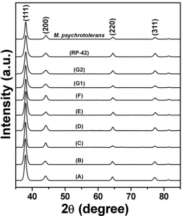
The crystallography of AgNPs formed by different biogroups of
Morganellaafter 20 h of reaction was investigated by XRD. As is evident from XRD patterns in Figure 4, extracellular AgNPs synthesized by all the biogroups are highly crystalline in nature, that could be perfectly indexed to the {111}, {200}, {220} and {311} Bragg reflections of the face centered cubic (fcc) form of crystalline silver [12,24,31–32]. XRD analysis thus provided a clear indication of formation of high quality crystalline AgNPs using a Morganella mediated biosynthesis process by all its type strains. Additionally, the crystallinity of AgNPs was further confirmed by performing SAED analysis of AgNPs formed by
M. morganii strain RP-42 and M. psychrotolerans during TEM imaging (Figure 5). The SAED patterns from both the samples revealed well-defined diffraction spots in the form of rings, which are indicative of polycrystalline silver.
The UV-vis, TEM, XRD and SAED results presented in this study clearly demonstrate that formation of AgNPs is a genus-wide characteristic phenotype of all reported type strains ofMorganellato date. Further experiments were performed to explore whether AgNPs formation is a characteristic phenotype restricted to genus
Morganella, or whether other taxonomically related genera of Enterobacteriaceae family also show this feature. To obtain this insight, when comparative analysis of AgNPs synthesis using laboratory strains ofEscherichia coli,Salmonella typhimurium,Kelebisella pneumoniaeandSerratia marcescenswas performed in the presence of 5 mM AgNO3, no AgNPs formation was observed in any of these closely related organisms. Additionally, distant taxonomic relatives of Morganella such as Firmicutes and Actinobacter did not lead to formation of detectable AgNPs in solutions. This strongly suggests that AgNPs synthesis in the presence of Ag+ions is a phenotypic character that is uniquely associated with genusMorganella.
It has been previously established that in silver resistant bacteria, silver resistance mechanism involves the gene (SilE) which encodes a periplasmic silver binding protein (silE). This macromolecule plays a major role in highly specific uptake of Ag+ions from surrounding environment by providing histidine sites as primary candidate for Ag+ ions binding [33]. Similarly, in our previous study, we established that silver resistance machinery in Morganella morganii
RP-42 is associated with AgNPs synthesis capability of this particular strain [10]. Since, in the current study, all reported
Figure 2. Comparative production of AgNPs by different biogroups of Morganella represented in terms of maximum absorbance SPR intensities of AgNPs plotted against biosyn-thesis reaction time.
strains of genus Morganella were found to exhibit phenotype of AgNPs synthesis in the presence of Ag+ions, we found it important to associate the presence of silver resistance (SilE gene) in all these strains with their AgNPs synthesis capability [10]. Therefore, to determine whether all strains of Morganella spp. exhibit silver resistance, further efforts were made to identify gene homologue of SilE in all the members of genusMorganella. As can be seen from Figure 6, all Morganellastrains showed the presence of SilE gene homologue. This observation further strengthens the probable role of silver resistance genes and gene products in AgNPs synthesis in
Morganella. Detailed investigation of functional role of silver resistance genes in AgNPs synthesis is currently being pursued.
In the present study, we have demonstrated that the phenotype of extracellular AgNPs synthesis inMorganellais not just restricted to an isolate pertaining to one environment, but it is indeed a unique biochemical character associated with all the members of this genus isolated from different environment [34–37]. This clearly establishes that AgNPs synthesis by genusMorganella is a phenotype independent of environmental influence. Although AgNPs synthesis has been previously reported by other
microor-Figure 3. Transmission electron microscopy (TEM) images of extracellular AgNPs formed by different biogroups ofMorganella. Biogroup A (panels a and b), biogroup B (panels c and d), biogroup C (panels e and f), biogroup D (panels g and h), biogroup E (panels i and j), biogroup F (panels k and l), biogroup G1 (panels m and n), biogroup G2 (panels o and p),M. morganiistrain RP-42 (panels q and r), andM. psychrotolerans(panels s and t). Scale bar in each panel corresponds to 100 nm.
ganisms, this is for the first time that extracellular synthesis of AgNPs by all the members a particular Genus (Morganella) has been established, and their AgNPs synthesis capability has been followed in a time-dependent manner. The observation that members of other genera of the same Enterobacteriaceafamily are incapable of AgNPs synthesis, establishes AgNPs synthesis as a unique phenotypic character of genusMorganella. These observa-tions might, in future, not only provide a complementary tool for
easy detection and purification of Morganella in the presence of other members ofEnterobacteriaceaefamily, but might also establish new evolutionary links between different microorganisms by comparing their metal ion reducing capabilities.
Materials and Methods
Growth and identification ofMorganellastrains
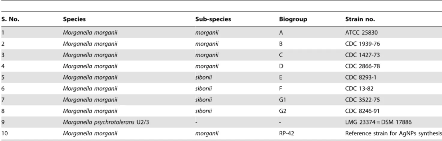
The genus Morganella comprises of two species viz. Morganella morganii with two sub-species morganii and sibonii, and Morganella psychrotolerans. On the basis of biochemical and taxonomic profiling, two sub-species morganii and sibonii have been further divided into a total of eight biogroups (Table 1) [34–37]. All strains of Morganella were routinely maintained on LB agar slants and preserved at280uC. The identity of all strains was confirmed by
16S rRNA gene sequencing as shown earlier [10], and sequences were submitted to GenBank with accession numbers HM122047, HM122048, HM122049, HM122050, HM122051, HM122052, HM122053, HM122054 and HM122055.
Extracellular biosynthesis of silver nanoparticles by
Morganellaspp. and nanoparticles characterization
All reported type strains of Morganella were used for AgNPs synthesis in the present study (Table 1). All strains of Morganella
were initially grown at 37uC for 24 h in a 500-mL Erlenmeyer flask that contained LB broth (100 mL) without added NaCl in a shaker incubator set at 200 rpm, as reported previously forM. morganiistrain RP-42 [10], except forM. psychrotolerans, wherein the bacteria were grown at 20uC, while maintaining all other synthesis conditions similar to those for other Morganella strains. For M. psychrotolerans, 20uC was used as the growth temperature, because this being a psychrotolerant species, 15–20uC is its optimum growth temperature and M. psychrotolerans could not be grown successfully at 37uC even after 5 days of incubation [12]. For all other Morganella biogroups, the optimum growth temperature being 37uC, they were grown at 37uC. Following bacterial growth, all the culture suspensions were incubated with aqueous 5 mM solutions of AgNO3at 37uC in a shaker incubator at 200 rpm in the dark, and the reactions were carried out in a time-dependent manner for up to 120 h (5 days). TheM. morganiistrain RP-42 that was earlier reported by us to synthesize extracellular AgNPs was considered as a positive control in all the experiments [10]. The
Figure 4. X-ray diffraction (XRD) patterns of extracellular AgNPs formed by different biogroups ofMorganella.Each XRD pattern has been labelled with respective biogroup, and the Bragg reflections corresponding to (111), (200), (220), and (311) planes have been indicated that are characteristic of crystalline silver.
doi:10.1371/journal.pone.0021401.g004
Figure 5. Selected area electron diffraction (SAED) patterns obtained from AgNPs formed by (A)M. morganiistrain RP-42, and (B)M. psychrotolerans.
extracellular synthesis of AgNPs was initially detected by visual inspection of the culture flask for a change in color of culture medium from clear light-yellow to brown/green. Extracellular AgNPs were separated from bacterial cells by centrifuging aliquots of culture supernatants (1.5 mL) at 3000 rpm for 6 min at 25uC. The supernatants thus obtained were clear brown/green homog-enous suspensions of AgNPs, which were analyzed in a time-dependent fashion using UV-vis spectroscopy (Cary 50 Bio-spectrophotometer) at a spectral resolution of 2 nm, transmission electron microscopy (Jeol 1010 TEM) at an accelerating voltage of 100 keV, selected area electron diffraction (SAED) coupled with TEM instrument, and X-ray diffraction (XRD - Bruker AXS D8 Discover) using Cu Ka radiation and a General Area Detector Diffraction System (GADDS). For UV-vis analysis, the AgNPs suspensions were diluted 10 times using MilliQ deionized water at every time point and UV-vis spectra were obtained. For TEM analysis, AgNPs samples obtained after 20 h of reaction were prepared by drop casting the colloidal suspensions of AgNPs onto carbon-coated Cu grids followed by drying under air for 24 hours.
For XRD analysis, the samples were prepared by precipitating AgNPs obtained after 20 h of biosynthesis at 10,000 rpm for 15 min, followed by three washings with MilliQ deionised water, and drop casting the samples onto a glass substrate.
Acknowledgments
Authors gratefully acknowledge the generous contribution of Caroline Mohr O’Hara, CDC, USA, for the gift ofM. morganii biogroupsand Jette Emborg, Danish Institute for Fisheries Research, Denmark, for theM. psychrotolerans strain. We also acknowledge RMIT Microscopy and Microanalysis Facility (RMMF) for providing access to the electron microscopes.
Author Contributions
Conceived and designed the experiments: RYP PJC SKB MSP YSS VB. Performed the experiments: RYP RR VB. Analyzed the data: RYP VB. Contributed reagents/materials/analysis tools: VB. Wrote the paper: RYP VB.
References
1. Niemeyer CM, Mirkin CA (2005) Nanobiotechnology – concepts, applications and perspectives: Wiley-VCH Verlag GmbH & Co.
2. Bansal V, Ramanathan R, Bhargava SK (2011) Fungus-mediated biological approaches towards ‘green’ synthesis of oxide nanomaterials. Aus J Chem 64: 279–293.
3. Thakkar KN, Mhatre SS, Parikh RY (2010) Biological synthesis of metallic nanoparticles. Nanomedicine 6: 257–262.
4. Klaus -JT, Joerger R, Olsson E, Granqvist C (2001) Bacteria as workers in the living factory: metal-accumulating bacteria and their potential for materials science. Trends Biotechnol 19: 15–20.
5. Nangia Y, Wangoo N, Goyal N, Shekhawat G, Suri CR (2009) A novel bacterial isolate Stenotrophomonas maltophilia as living factory for synthesis of gold nanoparticles. Microb Cell Fact 8: 39.
6. Shankar SS, Rai A, Ankamwar B, Singh A, Ahmad A, et al. (2004) Biological synthesis of triangular gold nanoprisms. Nat Mater 3: 482–488.
7. Ahmad A, Senapati S, Khan MI, Kumar R, Sastry M (2003) Extracellular biosynthesis of monodisperse gold nanoparticles by a novel extremophilic actinomycete,Thermomonosporasp. Langmuir 19: 3550–3553.
8. Ahmad A, Mukherjee P, Senapati S, Mandal D, Khan MI, et al. (2003) Extracellular biosynthesis of silver nanoparticles using the fungus Fusarium oxysporum. Colloids Surf B 28: 313–318.
9. Klaus -JT, Joerger R, Olsson E, Granqvist CG (1999) Silver-based crystalline nanoparticles, microbially fabricated. Proc Natl Acad Sci USA 96: 13611–13614.
10. Parikh RY, Singh S, Prasad BLV, Patole MS, Sastry M, et al. (2008) Extracellular synthesis of crystalline silver nanoparticles and molecular evidence
Table 1.List of existing members of genusMorganella.
S. No. Species Sub-species Biogroup Strain no.
1 Morganella morganii morganii A ATCC 25830
2 Morganella morganii morganii B CDC 1939-76
3 Morganella morganii morganii C CDC 1427-73
4 Morganella morganii morganii D CDC 2866-78
5 Morganella morganii sibonii E CDC 8293-1
6 Morganella morganii sibonii F CDC 13-82
7 Morganella morganii sibonii G1 CDC 3522-75
8 Morganella morganii sibonii G2 CDC 8246-91
9 Morganella psychrotoleransU2/3 - - LMG 23374 = DSM 17886
10 Morganella morganii morganii RP-42 Reference strain for AgNPs synthesis
doi:10.1371/journal.pone.0021401.t001
of silver resistance from Morganella sp.: towards understanding biochemical synthesis mechanism. ChemBioChem 9: 1415–1422.
11. Song JY, Kim BS (2009) Rapid biological synthesis of silver nanoparticles using plant leaf extracts. Bioprocess Biosyst Eng 32: 79–84.
12. Ramanathan R, O’Mullane AP, Parikh RY, Smooker PM, Bhargava SK, et al. (2011) Bacterial kinetics-controlled shape-directed biosynthesis of silver nano-plates usingMorganella psychrotolerans. Langmuir 27: 714–719.
13. Song JY, Kwon EY, Kim BS (2010) Biological synthesis of platinum nanoparticles using Diopyros kaki leaf extract. Bioprocess Biosyst Eng 33: 159–164.
14. Bansal V, Ahmad A, Sastry M (2006) Fungus-mediated biotransformation of amorphous silica in rice husk to nanocrystalline silica. J Am Chem Soc 128: 14059–14066.
15. Bansal V, Ahmad A, Sastry M (2007) Fungus-mediated selective bioleaching of silica as a means of enrichment of zirconia in zircon sand. Langmuir 23: 4993–4998.
16. Bansal V, Sanyal A, Rautaray D, Ahmad A, Sastry M (2005) Bioleaching of sand by the fungus,Fusarium oxysporumas a means of producing extracellular silica nanoparticles. Adv Mater 17: 889–892.
17. Bansal V, Rautaray D, Bharde A, Ahire K, Sanyal A, et al. (2005) Fungus-mediated biosynthesis of silica and titania particles. J Mater Chem 15: 2583–2589.
18. Ramanathan R, Campbell J, Soni SK, Bhargava SK, Bansal V (2011) Cationic amino acids specific biomimetic silicification in ionic liquid: a quest to understand the formation of 3-D structures in diatoms. PLoS ONE 6: e17707. 19. Bansal V, Rautaray D, Ahmad A, Sastry M (2004) Biosynthesis of zirconia nanoparticles using the fungusFusarium oxysporum. J Mater Chem 14: 3303–3305. 20. Bharde A, Rautaray D, Bansal V, Ahmad A, Sarkar I, et al. (2006) Extracellular
biosynthesis of magnetite using fungi. Small 2: 135–141.
21. Bharde A, Parikh RY, Baidakova M, Jouen S, Hannoyer B, et al. (2008) Bacteria-mediated precursor-dependent biosynthesis of superparamagnetic iron oxide and iron sulfide nanoparticles. Langmuir 24: 5787–5794.
22. Bansal V, Poddar P, Ahmad A, Sastry M (2006) Room-temperature biosynthesis of ferroelectric barium titanate nanoparticles. J Am Chem Soc 128: 11958–11963.
23. Sweeney RY, Mao C, Gao X, Burt JL, Belcher AM, et al. (2004) Bacterial biosynthesis of cadmium sulfide nanocrystals. Chem Biol 11: 1553–1559. 24. Bansal V, O’Mullane AP, Bhargava SK (2009) Galvanic replacement mediated
synthesis of hollow Pt nanocatalysts: significance of residual Ag in H2evolution reactions. Electrochem Commun 11: 1639–1642.
25. Egger S, Lehmann RP, Height MJ, Loessner MJ, Schuppler M (2009) Antimicrobial properties of a novel silver-silica nanocomposite material. Appl Environ Microbiol 75: 2973–2976.
26. Galeano B, Korff E, Nicholson WL (2003) Inactivation of vegetative cells, but not spores, ofBacillus anthracis,B. cereus, andB. subtilison stainless steel surfaces coated with an antimicrobial silver- and zinc-containing zeolite formulation. Appl Environ Microbiol 69: 4329–4331.
27. Schultz S, Smith DR, Mock JJ, Schultz DA (2000) Single-target molecule detection with nonbleaching multicolor optical immunolabels. Proc Natl Acad Sci USA 97: 996–1001.
28. Yang HL, Lin JC, Huang C (2009) Application of nanosilver surface modification to RO membrane and spacer for mitigating biofouling in seawater desalination. Water Res 43: 3777–3786.
29. Brenner DJ, Farmer JJ, Fanning GR, Steigerwalt AG, Klykken P, et al. (1978) Deoxyribonucleic acid relatedness of Proteus and Providencia species. Int J Syst Bacteriol 28: 269–282.
30. Pearson A, O’Mullane AP, Bansal V, Bhargava SK (2010) Galvanic replacement mediated transformation of Ag nanospheres into dendritic Au-Ag nanostructures in the ionic liquid [BMIM][BF4]. Chem Commun 46: 731–733.
31. Bansal V, Li V, O’Mullane AP, Bhargava SK (2010) Shape dependent electrocatalytic behaviour of silver nanoparticles. Cryst Eng Commun 12: 4280–4286.
32. Selvakannan PR, Swami A, Srisathiyanarayanan D, Shirude PS, Pasricha R, et al. (2004) Synthesis of aqueous Au core-Ag shell nanoparticles using tyrosine as a pH-dependent reducing agent and assembling phase-transferred silver nanoparticles at the air-water interface. Langmuir 20: 7825–7836.
33. Gupta A, Matsui K, Lo JF, Silver S (1999) Molecular basis for resistance to silver cations inSalmonella. Nature Med 5: 183–188.
34. Emborg J, Dalgaard P, Ahrens P (2006)Morganella psychrotolerans sp. nov., a histamine-producing bacterium isolated from various seafoods. Int J Syst Evol Microbiol 56: 2473–2479.
35. Fulton M (1943) The identity of bacteriumColumbensis Castellani. J Bacteriol 46: 79–82.
36. Jensen KT, Frederiksen W, Hickman-Brenner FW, Steigerwalt AG, Riddle CG, et al. (1992) Recognition ofMorganellasubspecies, with proposal ofMorganella morganiisubsp.morganiisubsp. nov. andMorganella morganiisubsp.siboniisubsp. nov. Int J Syst Bacteriol 42: 613–620.