A total laparoscopic technique for endovascular
thoracic stent graft deployment
Ricardo Yoshida, MD,aRalf R. Kolvenbach, MD,bZhidong Ye, MD,b and Winston Yoshida, MD, PhD,aDusseldorf, Germany; and Botucatu, Brazil
Background:Limitations of endovascular thoracic aneurym treatment include small, tortuous, or severely calcified iliac arteries. We present our experience with a total laparoscopic access to deploy thoracic endografts.
Methods:A total laparoscopic left retrocolic approach was used in all cases. A Dacron conduit was laparoscopically sutured to either the iliac artery or to the aorta directly. The endograft was inserted through this conduit. After graft deployment, the Dacron prosthesis was tunneled to the groin and anastomosed with the femoral artery.
Results:The laparoscopic procedure could successfully be performed in 11 patients. In six cases, the aorta was used as an access and in five patients, the iliac arteries were preferred. In one of these cases, the right iliac artery was used for deployment of the endograft. After successful aorto- or ileo-femoral bypass grafting, all patients had an improvement of their ankle brachial index postoperatively. The mean operative time was almost four hours, including laparoscopy, laparoscopic anastomosis, endograft deployment, and femoral artery anastomosis or profundaplasty.
Conclusion:Totally laparoscopic assisted graft implantation in aorta or iliac arteries provides a safe and effective access for the endovascular delivery system. However, further evaluation and long follow-up are necessary to ensure the potential advantages of this technique. It is a less invasive option to overcome access-related problems with thoracic endograft deployment, giving the patient the advantage of a totally minimal invasive procedure. ( J Vasc Surg 2010;51:504-8.)
Thoracic aortic aneurysms (TAAs) are successfully treated by endovascular techniques (TEVAR),1-4
with the benefit of aneurysm exclusion without the invasiveness of prolonged proximal aortic clamping and direct surgical exposure. However, not all patients with TAAs are eligible for these less invasive endovascular procedures.2,3,5
Limitations include the anatomic morphology of the aneurysm and tortuous or stenotic access arteries.5-7
The femoral artery is the most commonly used access vessel, but the common iliac artery or abdominal aorta can be used in case of anatomic problems.5
The 21-22 F introducer sheaths make deployment in patients with aorto-iliac occlu-sive disease often difficult or impossible.2,8 An
extraana-tomic exposure of the aorta or iliac arteries significantly increases the invasiveness of the procedure.6
In the following, we describe our experience with a laparoscopic technique to deploy thoracic endografts. The video endoscopic technique is presented in detail.
MATERIALS AND METHODS
Using a regular bypass graft as a conduit, we wanted to obtain a safe and durable access to the iliac arteries or to the aorta as well as a bypass graft to treat the occlusive disease. This could easily be accomplished by tunneling the Dacron graft to the groin and subsequently performing an anasto-mosis with the femoral artery after introducing the stent graft.
Laparoscopic exposure. For all procedures involving the left iliac artery and the infrarenal aorta, the patient was positioned on a vacuum bag and could be tilted at a 70° slope (Fig 1). As previously described, we prefer a transperi-toneal left retrocolic approach similar to the one originally described by Dion as the “Apron technique.”9
Mobiliza-tion of the left hemicolon, as originally described by Mat-tox, was modified by Coggia to facilitate laparoscopic ex-posure of the aorta.10Mobilization of the left hemicolon
was initiated using the line of Toldt as a landmark. A retractor was inserted from the right side to retract the small bowel and the left kidney medially (Fig 2). The surgeon, first assistant, and second assistant, holding the
From the Department of Surgery and Orthopedics, Botucatu School of Medicine, São Paulo State Universityaand the Department of Vascular
Surgery and Endovascular Therapy, Augusta Hospital and Catholic Clin-ics.b
Competition of interest: none.
Reprint requests: Ralf Kolvenbach, MD, PhD, Department of Vascular Surgery, Augusta Hospital and Union of Catholic Clinics Duesseldorf, Amalien Str. 9, 40472 Duesseldorf (e-mail. [email protected]).
The editors and reviewers of this article have no relevant financial relation-ships to disclose per the JVS policy that requires reviewers to decline review of any manuscript for which they may have a competition of interest.
0741-5214/$36.00
Copyright © 2010 by the Society for Vascular Surgery. doi:10.1016/j.jvs.2009.06.060
camera, all stood on the right side of the patient during the laparoscopic portion of the procedure (small diagram in Fig 3). A maximum number of six ports were required to expose the aorta or the iliac artery (Fig 3). As can be seen in
Fig 1, the Dacron graft was exteriorized through one of the
lower ports (F or A). Tunneling to the groin, particularly on the left side, is associated with gas loss and compromised exposure. Therefore, we preferred the technique described though the angle of introduction of the graft into the artery is steeper.
In all cases, an end-to-side anastomosis with a 10 mm preclotted Dacron Graft was performed (Fig 4). Two run-ning sutures secured at the end with a felt pledged as described by Coggia were used to perform the anastomo-sis.10
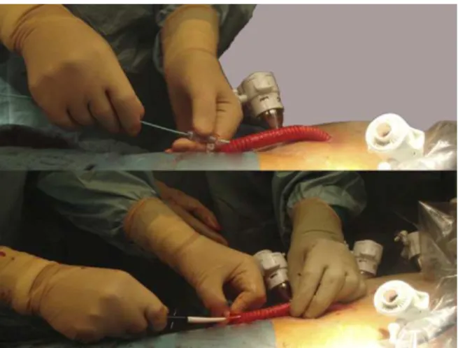
At the end of the procedure after graft deployment, the Dacron graft was tunneled to the groin and anasto-mosed with the femoral artery. For thoracic endograft deployment, the Dacron graft was accessed with a 5F sheath after direct puncture and a super stiff wire was placed in the ascending aorta. The patient was repositioned and C-Arm fluoroscopy was used to obtain multiple oblique angles of the proximal landing zone. Without removing the trocars, the endograft was inserted through the Dacron graft and deployed. Simultaneously, passage of the graft through the anastomosis was controlled with the laparo-scopic camera (Fig 5). After deployment of the thoracic endograft, the Dacron prosthesis was cut to length and after tunneling to the groin, the distal anastomosis with the femoral artery was performed.
RESULTS
The laparoscopic procedure could successfully be per-formed in all 11 patients. In six cases, the aorta was used as an access and in five patients, the iliac arteries were pre-ferred (Fig 6). In one of these cases, the right iliac artery was used for deployment of the endograft. In four patients, a transbrachial wire was placed and pulled out through the prosthesis to facilitate passage of the endograft through a
Fig 1. Intraoperative photo of ports and Dacron conduit.
Fig 2. Intraoperative picture and schematic depiction of laparo-scopic exposure.
Fig 3. Schematic drawing of placement of trocars.
kinked aorta and iliac arteries. After successful aorto- or ileo-femoral bypass grafting, all patients had an improve-ment of their ankle brachial index postoperatively (Table I). The mean operative time was almost four hours includ-ing laparoscopy, laparoscopic anastomosis, endograft de-ployment, and femoral artery anastomosis or profunda-plasty. Laparoscopy time was dependent on the access site. The mean period of time to expose the aorta laparoscopi-cally was 136.8 minutes (range, 70-195 minutes) com-pared with 62.6 minutes (range, 48.0-85.0 minutes) when the anastomosis had to be performed with the external or common iliac artery (Table II).
DISCUSSION
Unobstructed transluminal arterial access to the tho-racic aorta remains an important necessity for endoluminal repair of thoracic aneurysms and accounts for a substantial amount of technical failures.1,2,5,6 Extensive iliac artery
calcification and occlusive disease often precludes femoral access for TEVAR. In an international survey of thoracic aortic stent-grafting, primary access other than the com-mon femoral artery was required in approximately 15% of TEVAR cases.1,7
A recent retrospective study showed that 26% of TAAs were unsuitable for endovascular repair be-cause of anatomic limitations and, in 33%, access limitations were a major reason.2
Inadequate iliofemoral access can be overcome by us-ing an appropriately sized Dacron conduit.2
Retroperito-neal dissection and placement of a prosthetic iliac conduit is the most commonly used option to overcome the limita-tions of iliac disease, but this requires major surgical expo-sure associated with significant trauma and side effects.1As
a consequence, the patient cannot fully benefit from the reduced invasiveness of the endovascular procedure.2
Retroperitoneal dissection accounts for a significant morbidity. Lee et al found a 2.6-fold greater blood loss, 82% longer procedure time, 1.5-day longer hospital length of stay, and 1.8-fold higher risk of perioperative complica-tions when retroperitoneal procedures were performed rather than standard femoral exposure.6Data on incisional Fig 5. Insertion of stent graft into Dacron conduit.
Fig 6. Different sites of end-site anastomosis with the Dacron graft.
Table I. Patient demographics
Mean age (yrs) 70.9 (59-81)
Male 7
Female 4
Diameter AA (cm) 6.1 (5.0-8.1)
Iliac access 5
Aortic access 6
Brachial wire 4
ABI preop. 0.67 (0.5-0.9)
ABI postop. 0.84 (0.7-1.0)
Aortic access,Number of patients with laparoscopic aortic access;ABI preop., preoperative ankle brachial Index;ABI postop.,postoperative ankle brachial index;Brachial wire,number of patients where a transbrachial wire was placed;Diameter AA,diameter of the thoracic aortic aneurysm;Iliac access, number of patients with laparoscopic iliac access.
Table II. Perioperative data
Mean value Minimum Maximum
Total operative time (min) 236.1 190 298
Laparoscopy time (min) 120.3 74 180
x. clamp time (min) 55.6 30 110
ICU stay (days) 0.9 0.0 2.0
Hospital stay (days) 6.0 5.0 9.0
pain, lumbosacral neuritic pain, incisional hernia, and de-forming abdominal bulge after retroperitoneal exposure have been published. In a retrospective study, Honig et al reported a 23% incidence of abdominal bulge, a 7% inci-dence of incisional hernia, and, more importantly, a 37% incidence of prolonged disabling pain.11In a more recent
paper, the authors could show that retroperitoneal incisions were associated with severe rectus abdominis muscle atro-phy.12
A less invasive alternative can be percutaneous translu-minal angioplasty of the iliac arteries or serial dilatation of the iliac arteries with hydrophilic dilators.1 These often
allow passage of delivery sheaths used during TEVAR, but an aggressive angioplasty is often needed for these large sheaths to pass, which can lead to iliac artery dissection or even iliac artery rupture.1,7
As a minimally invasive technique, laparoscopic aortic surgery was shown to be safe and effective in abdominal aortic reconstructions.13-19
Aortic and iliac artery exposure and control are safe and effective during the entire proce-dure when dedicated laparoscopic vascular instruments are used, preventing any inadvertent bleeding.5,16The
learn-ing curve of a laparoscopic anastomosis with the iliac artery is less steep compared with a total laparoscopic aneurysm resection or with the anastomosis of a bifurcated graft with the infrarenal aorta.14-17Exposure of the aorta where
nec-essary can be performed in a more limited way; in some cases, the laparoscopic anastomosis can be performed just proximal to the aortic bifurcation without the need for further extensive dissection and mobilization of the left kidney.9,10
Preoperative angio computed tomography or magnetic resonance angiography determined in combina-tion with the clinical examinacombina-tion whether the aorta or the iliac artery were chosen as an access for TEVAR. When in doubt the aorta was preferred. There was a short learning curve of two cases with this laparoscopic procedure until the laparoscopic equipment as well as the C-arm and the angio operating table were positioned in an optimal way. Similar to a conventional approach to the right iliac artery from the left side, laparoscopic exposure of the right iliac artery was more challenging and took longer compared with the left iliac artery. In cases where the left iliac artery is unsuitable, we now prefer the aorta close to the bifurcation, which, in our experience, permits more expeditious expo-sure and allows for a faster laparoscopic anastomosis.
The use of a special conduit as described by Fearn et al would greatly facilitate the procedure.20
In a porcine model, they used a curved hollow needle, a partially stented Dacron conduit, an airtight laparoscopic port, and a sealing sheath and valve specifically developed for percutaneous access through the abdominal wall. The conduit was in-serted over the guide wire after needle removal and de-ployed under fluoroscopy. The distal end of the conduit was secured by the sealing sheath and valve, enabling wire and catheter exchange thereafter. Yet this elegant and simple solution requires a strong interest from industry to manufacture such a device before wider use can be antici-pated.
The operating time in our series is still significantly longer compared with the time required for an extraperi-toneal approach and an anastomosis under direct vi-sion.13,14,19In this subset of patients, the laparoscopic
approach described seemed to be a more straightforward procedure than performing femoral and iliac artery throm-bendarterectomy or a hybrid open/endovascular proce-dure.
Totally laparoscopic-assisted graft implantation in aorta or iliac arteries provides a safe and effective access for the endovascular delivery system. However, further evaluation and long follow-up are necessary to ensure the potential advantages of this technique. A prospective comparison of laparoscopic assisted TEVAR versus a standard retroperito-neal access will be required to assess the role of this tech-nique as an adjunct to endovascular aneurysm exclusion, though we know from several studies that the immunolog-ical consequences and late sequelae of multiple ports are significantly less compared with a conventional often mus-cle splitting incision. The retroperitoneal approach as such does not necessarily offer advantages over a transperitoneal exposure.21-23
Laparoscopic-assisted TEVAR is a less invasive option to overcome access related problems with thoracic en-dograft deployment, giving the patient the advantage of a totally minimal invasive procedure.
AUTHOR CONTRIBUTIONS
Conception and design: RK, ZY Analysis and interpretation: RY Data collection: RY, ZY Writing the article: RK, RY, ZY Critical revision of the article: RK, WY Final approval of the article: RK, RY Statistical analysis: RY, ZY
Obtained funding: N/A Overall responsibility: RK
REFERENCES
1. Peterson BG, Matsumura JS. Internal endoconduit: an innovative tech-nique to address unfavorable iliac artery anatomy encountered during thoracic endovascular aortic repair. J Vasc Surg 2008;47:441-5. 2. Jackson BM, Carpenter JP, Fairman RM, Moser GW, Pochettino A,
Woo EY, Bavaria JE. Anatomic exclusion from endovascular repair of thoracic aortic aneurysm. J Vasc Surg 2007;45:662-6.
3. Cambria RP, Brewster DC, Lauterbach SR, Kaufman JL, Geller S, Fan CM, et al. Evolving experience with thoracic aortic stent graft repair. J Vasc Surg 2002;35:1129-36.
4. Criado FJ, Clark NS, Barnatan MF. Stent graft repair in the aortic arch and descending thoracic aorta: a 4-year experience. J Vasc Surg 2002; 36:1121-8.
5. Fukui S, Gigou F, Daneshvar M, Marteau V, Soury P, Petit MD, Laurian C. Totally laparoscopic assisted thoracic aorta endograft deliv-ery by direct sheath placement into the aorta. J Vasc Surg 2006;43: 1274-7.
6. Lee WA, Berceli SA, Huber TS, Ozaki CK, Flynn TC, Seeger JM. Morbidity with retroperitoneal procedures during endovascular ab-dominal aortic aneurysm repair. J Vasc Surg 2003;38:459-63; discus-sion 464-5.
phase II multicenter trial of the GORE TAG thoracic endoprosthesis. J Vasc Surg 2005;41:1-9.
8. Criado FJ, Barnatan MF, Rizk Y, Clark NS, Wang CF. Technical strategies to expand stent-graft applicability in the aortic arch and proximal descending thoracic aorta. J Endovasc Ther 2002;9 Suppl 2:II32-8.
9. Dion YM, Thaveau F, Fearn SJ. Current modifications to totally lapa-roscopic “apron technique.” J Vasc Surg 2003;38:403-6.
10. Coggia M, Di Centa I, Javerliat I, Colacchio G, Goeau-Brissonniere O. Total laparoscopic aortic surgery: transperitoneal left retrorenal ap-proach. Eur J Vasc Endovasc Surg 2004;28:619-22.
11. Honig MP, Mason RA, Giron F. Wound complications of the retroperitoneal approach to the aorta and iliac vessels. J Vasc Surg 1992;15:28-34.
12. Yamada M, Maruta K, Shiojiri Y, Takeuchi S, Matsuo Y, Takaba. Atrophy of the abdominal wall muscles after extraperitoneal approach to the aorta. J Vasc Surg 2003;38:346-53.
13. Kolvenbach R. Total laparoscopic aortic aneurysm surgery. Acta Chir Belg 2006;106:36-9.
14. Kolvenbach R, Puerschel A, Fajer S, Lin J, Wassiljew S, Schwierz E, Pinter L. Total laparoscopic aortic surgery versus minimal access techniques: review of more than 600 patients. Vascular 2006;14: 186-92.
15. Wassiljew S, Kolvenbach R, Puerschel A, Schwierz E. Total laparo-scopic iliac artery aneurysm repair using endolaparo-scopic techniques and endovascular balloon occlusion. Eur J Vasc Endovasc Surg 2006;32: 270-2.
16. Kolvenbach R, Ferrari M, Shifrin EG. Laparoscopic assisted aortic surgery. A review. J Cardiovasc Surg (Torino) 2006;47:547-56. 17. Yoshida R, Yoshida W, Kolvenbach R, Rollo H, Lorena S. Laparoscopic
aortic surgery learning curve: experimental study in pigs. J Vasc Br 2008;7:231-8.
18. Ferrari M, Adami D, Del Corso A, Berchiolli R, Pietrabissa A, Romag-nani F, Mosca F. Laparoscopy-assisted abdominal aortic aneurysm repair: early and middle-term results of a consecutive series of 122 cases. J Vasc Surg 2006;43:695-700.
19. Kolvenbach R, Da Silva L, Deling O, Schwierz E. Video-assisted aortic surgery. J Am Coll Surg 2000;190:451-7.
20. Fearn SJ, Burke K, Hartley DE, Semmens JB, Lawrence-Brown MM. A laparoscopic access technique for endovascular procedures: surgeon training in an animal model. J Endovasc Ther 2006;13:350-6. 21. Cambria RP, Brewster DC, Abbott WM, Freehan M, Megerman J,
LaMuraglia G, et al. Transperitoneal versus retroperitoneal approach for aortic reconstruction: a randomized prospective study. J Vasc Surg 1990;11:314-24; discussion 324-5.
22. Bouvy ND, Marquet RL, Jeekel J. Laparoscopic surgery is associated with less tumor growth stimulation than conventional surgery.: An experimental study. Br J Surg 1997;84:358-61.
23. Sheen-Chen SM, Chen HS, Eng HL, Chen WJ, Jawan B. Systemic immune response after laparoscopic and open cholecystectomy. World J Surg 2002;26:1418-22.