Ventilator-associated pneumonia: epidemiology
and impact on the clinical evolution of ICU patients*
Pneumonia associada à ventilação mecânica: epidemiologia e impacto na evolução clínica de pacientes em uma unidade de terapia intensiva
Pedro Mendes de Azambuja Rodrigues, Edgard do Carmo Neto, Luiz Rodrigo de Carneiro Santos, Marcos Freitas Knibel
Abstract
Objective: Although ventilator-associated pneumonia (VAP) is a major cause of nosocomial infection, its role in the prognosis of patients remains undefined. The objective of this study was to evaluate the impact of VAP on the clinical evolution of patients. Methods: This was a prospective cohort study involving 233 patients on mechanical ventilation (VAP group, n = 64; control group, n = 169). Primary outcomes were time on mechanical ventilation (TMV), time in ICU (TICU), overall length of hospital stay (LHS) and in-ICU mortality. Secondary outcomes were in-hospital mortality, microbiological profile, prior use of antibiotics and risk factors for VAP acquisition. Results: Control and VAP group outcomes were, respectively, as follows: median TMV (days), 9 (interquartile range [IQR]: 5-15) and 23 (IQR: 15-37; p < 0.0001); median TICU (days), 12 (IQR: 8-21) and 27 (IQR: 17-42; p < 0.0001); median LHS (days), 33 (IQR: 18-64) and 46 (IQR: 25-90; p = 0.05); and in-ICU mortality, 38% (95% CI: 31-45) and 55% (95% CI: 42-67; p = 0.02). VAP was a predictor of in-ICU mortality (OR = 3.40; 95% CI: 1.54-7.48). TMV (OR = 2.27; 95% CI: 1.05-4.87) and prior use of antibiotics (OR = 1.07; 95% CI: 1.04-1.10) were risk factors for VAP. VAP did not affect in-hospital mortality. Acinetobacter spp. was the most common isolate (28%). Inappropriate empirical antibiotic therapy was administered in 48% of cases. Conclusions: In this study, there was a high incidence of infection with resistant bacteria and inappropriate initial antibiotic therapy. Long TMV and prior use of antibiotics are risk factors for VAP.
Keywords: Pneumonia, ventilator-associated; Cross infection; Intensive care units; Hospital mortality; Risk factors.
Resumo
Objetivo: Apesar de representar uma das principais causas de infecção nosocomial, o papel da pneumonia asso-ciada à ventilação mecânica (PAVM) no prognóstico ainda permanece indefinido. O objetivo deste estudo foi avaliar o impacto dessa doença na evolução clínica dos pacientes. Métodos: Estabeleceu-se uma coorte prospectiva de 233 pacientes sob ventilação mecânica (grupo PAV, n = 64; grupo controle, n = 169). Os desfechos primários foram tempo de ventilação mecânica (TVM), tempo de permanência na UTI (TUTI), tempo de permanência hospi-talar (TH) e mortalidade na UTI. Os desfechos secundários foram mortalidade hospihospi-talar, perfil microbiológico, uso prévio de antibióticos e fatores de risco para PAVM. Resultados: Os desfechos dos grupos controle e PAVM foram, respectivamente, os seguintes: mediana do TVM (dias), 9 (intervalo interquartílico [II]: 5-15) e 23 (II: 15-37; p < 0,0001); mediana do TUTI (dias), 12 (II: 8-21) e 27 (II: 17-42; p < 0,0001); mediana do TH (dias), 33 (II: 18-64) e 46 (II: 25-90; p = 0,02); e mortalidade na UTI, 38% (IC95%: 31-45) e 55% (IC95%: 42-67; p = 0,02). A PAVM foi um preditor de mortalidade na UTI (OR = 3,40; IC95%: 1,54-1,78). O TVM (OR = 2,27; IC95%: 1,05-4,87) e o uso prévio de antibióticos (OR = 1,07; IC95%: 1,04-1,10) foram fatores de risco para PAVM. A PAVM não afetou a mortalidade hospitalar. Acinetobacter spp. foi o isolado mais frequente (28%). Antibioticoterapia empí-rica inadequada foi administrada em 48% dos casos. Conclusões: No presente estudo, houve uma alta incidência de bactérias resistentes e de antibioticoterapia inicial inadequada. TVM longo e o uso prévio de antibióticos são fatores de risco para PAVM.
Descritores: Pneumonia associada à ventilação mecânica; Infecção hospitalar; Unidades de terapia intensiva; Mortalidade hospitalar; Fatores de risco.
* Study carried out in the Intensive Care Unit of the São Lucas Copacabana Hospital, Rio de Janeiro, Brazil.
Correspondence to: Pedro Mendes de Azambuja Rodrigues. Unidade de Cuidados Intensivos, Hospital São Lucas Copacabana, Travessa Frederico Pamplona, 32, Copacabana, CEP 22061-080, Rio de Janeiro, RJ, Brasil.
Tel 55 21 2545-4152. E-mail: [email protected] Financial support: None.
having been transferred from another hospital with a permanent artificial airway; having been prescribed end-of-life care by the attending physician; and falling outside the inclusion age bracket. For the purpose of data analysis, the cohort was divided into two groups: VAP group and control group.
In patients who were admitted to the ICU more than once, only the first admission was included in the cohort.
ICU follow-up was performed for up to 48 h after extubation or complete withdrawal of ventilatory support (for tracheostomized patients), continued until death or continued for 60 days on invasive mechanical ventilation. Late (in-hospital) follow-up continued until the outcome (discharge or death) was known or until the end of the six-month study period.
Cases of VAP were defined as those in which there was new or progressive infiltrate on chest X-ray accompanied by fever (> 38°C) or changes in leukocyte counts (≥ 12,000 or < 4,000 cells/mm3) and at least one of the
following findings: purulent tracheal secre-tions; isolation of a likely pulmonary pathogen in a sample from the lower respiratory tract; or PaO2/FiO2 ratio < 240.(9) Circulatory shock was
defined as that which was dependent on vasoac-tive amines (noradrenaline > 0.1 µg/kg/min or dobutamine > 5 µg/kg/min) for more than 12 h and was acquired after the first 24 h of enroll-ment.(10)
Quantitative BAL cultures were used to analyze the etiologic profile of VAP. The cut-off point for infection was set at 104 UFC/mL.(9,11)
The antibiotic therapy initially prescribed was considered appropriate when the pathogens isolated were sensitive to at least one drug in the regimen.
The data collection scenario consisted of a private ICU, with 46 clinical-surgical beds (ratio, 3:1), in a hospital without a long-term respira-tory care unit, located in Copacabana, in the city of Rio de Janeiro, Brazil. In this ICU, the density rate of use of mechanical ventilation remains at approximately 27.5% (according to the registries of the Quality in Intensive Care System of the Brazilian Association of Intensivists).
The variables evaluated included those determined at admission—age, gender, Acute Physiology and Chronic Health Evaluation II (APACHE II) and Sequential Organ Failure
Introduction
Pneumonia is the leading cause of nosoco-mial infection in ICUs, occurring primarily (in more than 90% of cases) in patients submitted to endotracheal intubation and mechanical ventilation (MV).(1,2) Due to its clinical relevance
and epidemiological profile, ventilator-associ-ated pneumonia (VAP) is studied as a clinical entity distinct from other nosocomial pneumo-nias, representing one of the major challenges faced by intensivists in daily practice.
Various studies have sought to charac-terize VAP in terms of prognosis, predisposing features, microbiology and costs.(3-5) In addition
to showing the magnitude of the problem, such studies point out characteristics to be subse-quently examined in strategies for diagnosis, prevention and treatment. Despite the advances in the understanding of the epidemiology of VAP, some fundamental aspects, such as its influence on in-ICU mortality and time in ICU, remain a matter of controversy, and conflicting data have been observed in the literature.(5-8)
The present study, through the prospective follow-up of patients submitted to MV in the ICU of a private hospital in the city of Rio de Janeiro, Brazil, can be included in this context. Comparing the subgroup of patients who devel-oped VAP with the remaining patients, we sought to determine the impact of VAP on the prognosis of the patients submitted to MV by assessing the following primary outcomes: in-ICU mortality rate; time on MV; time in ICU; and overall length of hospital stay. The secondary outcomes assessed were in-hospital mortality, risk factors for VAP acquisition, microbiological profile and prior use of antibiotics.
Methods
dence of tracheostomy, indication for MV and diagnostic category. The potential risk factors for the occurrence of the outcomes were included in the multivariable models based on previ-ously reported studies on risk.(10,12-14) The severity
scores were included in the regression model separately in order to avoid the phenomenon of autocorrelation. The level of significance was set at p < 0.05.
Assessment (SOFA) scores, admission type (clinical or surgical), admission diagnosis and indica-tion for MV—as well as those determined over the course of hospitalization—in-ICU mortality, in-hospital mortality, time on MV, time in ICU, overall length of hospital stay, incidence of VAP, hemodialysis (as a marker of severe acute renal failure),(12) shock and tracheostomy.
The indications for MV were divided into three categories: exacerbation of chronic pulmonary disease; acute cardiorespiratory disease (pneumonia, shock, acute pulmonary edema, trauma, cardiopulmonary arrest, aspira-tion, ARDS, immediate postoperative care); and primarily neurological disease (a drop in the level of consciousness or neuromuscular disease).(13)
Continuous and ordinal variables were expressed as median and interquartile range (IQR), whereas categorical variables were expressed as proportion and 95% CI.
Hypothesis testing of continuous and ordinal variables was performed using the Wilcoxon test, whereas hypothesis testing of categorical vari-ables was performed using the chi-square test.
Susceptibility bias was controlled by multi-variate (stepwise) logistic regression adjustment. The independent variables included in the model for the evaluation of mortality outcomes were as follows: age; gender; diagnostic category; indication for MV; APACHE II score and SOFA score at admission; as well as incidence of VAP; hemodialysis, circulatory shock and tracheos-tomy. The independent variables tested in the models for the VAP acquisition outcome were time on MV, prior use of antibiotics, age,
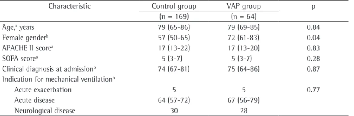
inci-Table 1 - Comparison of the two study groups in terms of baseline characteristics.
Characteristic Control group VAP group p
(n = 169) (n = 64)
Age,a years 79 (65-86) 79 (69-85) 0.84
Female genderb 57 (50-65) 72 (61-83) 0.04
APACHE II scorea 17 (13-22) 17 (13-20) 0.83
SOFA scorea 5 (3-7) 5 (3-7) 0.28
Clinical diagnosis at admissionb 74 (67-81) 75 (64-86) 0.87
Indication for mechanical ventilationb
Acute exacerbation 5 5 0.77
Acute disease 64 (57-72) 67 (56-79)
Neurological disease 30 28
VAP: ventilator-associated pneumonia; APACHE: Acute Physiology and Chronic Health Evaluation; and SOFA: Sequential
Organ Failure Assessment. aOrdinal variables expressed as median (interquartile range). bCategorical variables expresses as
percentage (95% CI).
Negativ
e
Figure 1 - Distribution of the 53 BAL culture results of 51/64 patients (80%) with ventilator-associated pneumonia. ACNB 0: carbapenem-sensitive
Acinetobacter baumannii; ACNB 1:
carbapenem-resistant A. baumannii; PSEUDO 0: carbapenem-sensitive Pseudomonas aeruginosa; PSEUDO 1: carbapenem-resistant P. aeruginosa; MRSA: methicillin-resistant Staphylococcus aureus; MSSA: methicillin-sensitive S. aureus; ESBL 1: extended-spectrum beta-lactamase-producing
Klebsiella pneumoniae; ESBL 0: extended-spectrum
beta-lactamase-nonproducing K. pneumoniae; STENO: Stenotrophomonas maltophilia; SER:
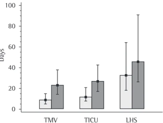
ICU (median = 27 days; IQR: 17-42 vs. median = 12 days; IQR: 8-21; p < 0.0001) and overall length of hospital stay (median = 46 days; IQR: 25-90 vs. median = 33 days; IQR: 18-64; p = 0.02; Figure 3).
There was no significant difference in mortality between VAP group patients (67%; 95% CI: 56-79) and control group patients (59%; 95% CI: 51-66). However, in-ICU mortality was significantly higher in the VAP group (55%; 95% CI: 42-67 vs. 38%; 95% CI: 31-45; p = 0.02; Figure 4), and VAP was found to be an independent risk factor for in-ICU mortality, as evidenced by multiple logistic regression (OR = 3.01; 95% CI: 1.36-6.66; p = 0.006). Other independent risk factors for in-ICU mortality were acute renal failure (OR = 3.99; 95% CI: 2.10-7.59; p < 0.0001); shock (OR = 4.66; 95% CI: 2.08-10.41; p = 0.0002); tracheostomy (OR = 0.17; 95% CI: 0.07-0.36; p < 0.0001); and male gender (OR = 0.36; 95% CI: 0.18-0.71; p < 0.0034).
Discussion
The results of the present study demonstrate that the occurrence of VAP was positively related to longer time on invasive ventilatory support (approximately 15 additional days), as well as significantly prolonging time in ICU (in approxi-mately 15 additional days) and overall length The study methodology was approved by the
local research ethics committee.
Results
During the study period, 233 patients were monitored, after 87 exclusions. Of those patients, 143 (61%) were female (95% CI: 55-68). The median age was 79 years (IQR: 66-86). Regarding admission diagnosis, 173 patients (74%) fell into the clinical category (95% CI: 69-80). The median baseline APACHE II score and the median baseline SOFA score were 17 (IQR: 13-22) and 5 (IQR: 3-7), respectively.
Of the 133 patients, 64 developed VAP during the study follow-up period (incidence: 16.79‰ days on MV). The median time in ICU and the median time on MV prior to the diagnosis of VAP were 12 days (IQR: 7-20) and 10 days (IQR: 5-17), respectively. The group of patients who developed VAP was found to be homogeneous in relation to the control group in terms of the baseline characteristics assessed, with the excep-tion of gender—there was a predominance of females in the VAP group (72%; 95% CI: 61-83; p = 0.04; Table 1). Of the patients diagnosed with VAP, 51 (80%) were submitted to BAL. The micro-biological profile associated with the etiology of VAP is represented in Figure 1. The most common pathogens isolated were Acinetobacter baumannii (in 28%), Pseudomonas aeruginosa (in 19%) and Staphylococcus aureus (in 20%). In addition, 16% of the BAL cultures were nega-tive. Based on the culture results, we found that 48% (95% CI: 36-61) of the patients received empirical antibiotic therapy that was inappro-priate for the etiologic agent in question.
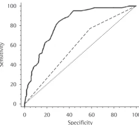
Multiple logistic regression analysis demon-strated that time on MV (OR = 1.07; 95% CI: 1.05-1.10, p < 0.0001) and prior use of antibi-otics (OR = 2.36; 95% CI: 1.12-4.99, p = 0.023) were independent risk factors for the develop-ment of VAP. The analysis of the performance of these variables using the ROC curve revealed that the area under the curve was 0.82 (95% CI: 0.76-0.87) and 0.59 (95% CI: 0.52-0.65), respectively, for time on MV and for prior use of antibiotics (Figure 2).
The comparison of the VAP group with the control group revealed that, in the former, time on MV was significantly longer (median: 23 days; IQR: 15-37 vs. median = 9 days; IQR: 5-15; p < 0.0001), as were time in
Sen
sitivity
Specificity
Figure 2 - ROC curves evaluating the performance of time on mechanical ventilation (solid line) and
discrepancies might be due to the variety of populations, as well as to the enrollment criteria and the data analysis used.(16) Therefore, it is
important to consider the various characteristics of the studies when comparing and extrapo-lating their results.
The incidence of VAP in the cohort was 16.79‰ days on MV, this rate being higher than the 90th percentile (8.9‰ days on MV) from the 2004 National Nosocomial Infections Surveillance System report, which is used as a reference by various authors and whose criteria for defining VAP are similar to those used in the present study.(17) However, since compliance with
this surveillance system is voluntary, the system is subject to a selection bias, since some centers obtain better results.
The population in the two study groups was found to be homogeneous in terms of baseline characteristics. The only demographic datum that revealed a significant difference was gender, with a predominance of females in the VAP group. It is important to emphasize that most patients in both groups were older than 79 years of age, which is likely a reflection of the demo-graphic characteristics of the region in which the hospital is located, namely Copacabana, where there is the highest concentration of elderly individuals in the city of Rio de Janeiro—24.8% of the population is over 60 years of age.(18)
Regarding admission diagnosis, more than 60% of the patients belonged to the clinical category, and there was no difference between the groups in terms of distribution.
These characteristics are in contrast to those of other study populations, and the potential of these variables to have an impact on prog-nosis should be taken into consideration. One group of authors(8) demonstrated the absence
of attributable risk of death in patients with VAP in a population consisting exclusively of patients hospitalized for trauma. Another group of authors(6) found similar results in a
prospec-tive cohort study in which only 26% of the patients belonged to the clinical category. Those results were in contrast to those observed in populations with a higher prevalence of clin-ical patients, such as that analyzed in another study(7) (44%) and that of the present study. Of
the patients from a large database used by one group of authors,(3) those in the VAP group were
predominantly male and significantly younger of hospital stay (in approximately 13 additional
days). Similarly, there was an increase in in-ICU mortality. In contrast, in-hospital mortality was not affected by the incidence of this infection. The multivariate analysis confirmed VAP as an independent risk factor for in-ICU mortality.
There is a significant discrepancy in the results of epidemiological studies in terms of the contribution of VAP to mortality. Some studies have demonstrated that the rate of survival is similar between patients with and without VAP,(6,8) whereas others have established VAP
as a risk factor for mortality.(7,15) In part, these
Figure 3 - Differences in time on mechanical ventilation (TMV), time in ICU (TICU) and overall length of hospital stay (LHS) between the control group (light bars) and the ventilator-associated pneumonia group (dark bars). Ordinate values expressed as medians and interquartile ranges (p < 0.0001; p< 0.0001; and p = 0.02, respectively).
Day
s
TMV TICU LHS
Figure 4 - Differences in in-ICU mortality and late (in-hospital) mortality between the control group (light bars) and the ventilator-associated pneumonia group (dark bars; p = 0.02 and p = 0.30, respectively).
Proportion of ev
ents
. . . . . . . . . .
In-ICU mortality
the sample in the present study did not allow the analysis of this topic through the use of etio-logic subgroups.
In the present study, methods to control bias were used, which made it possible to properly weigh the prognostic effect of VAP, adjusting the model for the interference of other extrinsic variables, whose contribution to clinical outcomes has been pointed out in previous studies.(10,12,14,24,25) The prospective nature of the
study design made it possible to collect variables based on the study objective, not limiting data collection to a review of historical records stored for various purposes, a practice that can produce selection bias. Enrollment procedures followed a strict restriction (exclusion) protocol, based on the presence of AIDS, advanced neoplastic disease, prior cognitive impairment (GCS ≤ 8) and advanced neuromuscular disease. However, restriction criteria affect the generalization of the findings to other populations. Multiple logistic regression adjustment was used in the data analysis, and the proportional weight of each explanatory variable was evaluated. The impossibility of stratifying the results by comor-bidities, of which no prospective records were obtained, was a limitation of the study design.
In the multivariate analysis, admission severity scores were not risk factors for mortality, in contrast to previous data. Conversely, the organ dysfunctions assessed during the course of hospitalization, that is, shock and acute renal failure, were found to be important prognostic factors.
In conclusion, the results of the present study point out that VAP is an independent risk factor for in-ICU mortality. However, in-hospital mortality in the VAP group was not signifi-cantly different from that in the control group. In addition, VAP correlated with longer time on MV, longer overall length of hospital stay and longer time in ICU. The etiology of VAP in this population was associated with pathogens at high risk for antimicrobial resistance. The initial antibiotic therapy was found to be inappropriate in a substantial proportion of cases. Time on MV and prior use of antibiotics were independent risk factors for VAP. The characteristics of the population studied should be taken into consid-eration when attempting to generalize these results to other groups of patients.
than were those in the control group, and there was no difference between the groups in terms of incidence of mortality.
In the present study, the independent risk factors for the development of VAP identified using multivariate analysis were time on MV and prior use of antibiotics. Based on the analysis of the ROC curve, we can conclude that, of these two variables, although prior use of antibiotics has an OR that demonstrate a closer relation-ship with VAP acquisition, time on MV has greater reproducibility, with higher sensitivity and higher specificity, and this is revealed by the area under the curve above 0.8, for the cut-off point of 12 days.
Late-onset VAP (which develops from the fifth day on MV onward) can be associated with a higher risk of morbidity and mortality, and this has been attributed to the higher prevalence of resistant microorganisms in this subgroup.(4) In
the present study, the median time on MV until VAP acquisition was 10 days, and late-onset VAP accounted for 75% of the cases. Even among the 25% who developed early-onset VAP, most of the BAL culture results revealed pathogens at high risk for antimicrobial resistance. The most prevalent etiologic agent in our sampling unit was Acinetobacter baumannii (in 28%), half of its strains being resistant to carbapenems. Although the distribution of the proportions of causal agents varies among studies, the preva-lence of infection with this pathogen is usually low.(3,5,19) In 48% of the cases, this disparity
reflected in the inappropriateness of the initial empirical antibiotic therapy administered to the patients with VAP, confirmed by the BAL culture results. This emphasizes the importance of conducting periodic microbiological studies in each facility in order to develop an appropriate empirical therapeutic strategy, since previous studies underscore the fact that an inappropriate initial antimicrobial regimen is an independent risk factor for mortality(4,20) and that even its
subsequent correction based on culture results seems to be unable to counteract this effect.(21-23)
To that end, in addition to BAL culture results, tracheal aspirate culture results and protected bronchial brush culture results can be consid-ered.(2) Although the presence of pathogens that
13. Frutos-Vivar F, Esteban A, Apezteguía C, Anzueto A, Nightingale P, González M, et al. Outcome of mechanically ventilated patients who require a tracheostomy. Crit Care Med. 2005;33(2):290-8. 14. Estenssoro E, González F, Laffaire E, Canales H, Sáenz
G, Reina R, et al. Shock on admission day is the best predictor of prolonged mechanical ventilation in the ICU. Chest. 2005;127(2):598-603.
15. Cunnion KM, Weber DJ, Broadhead WE, Hanson LC, Pieper CF, Rutala WA. Risk factors for nosocomial pneumonia: comparing adult critical-care populations. Am J Respir Crit Care Med. 1996;153(1):158-62. 16. Cook DJ, Kollef MH. Risk factors for ICU-acquired
pneumonia. JAMA. 1998;279(20):1605-6.
17. National Nosocomial Infections Surveillance System. National Nosocomial Infections Surveillance (NNIS) System Report, data summary from January 1992 through June 2004, issued October 2004. Am J Infect Control. 2004;32(8):470-85.
18. Instituto Brasileiro de Geografia e Estatística [homepage on the Internet]. Brasília: Ministério do Planejamento, Orçamento e Gestão [cited 2009 Abr 17]. População residente censo 2000; 2000. Available from: http:// www.ibge.gov.br/
19. Vincent JL, Bihari DJ, Suter PM, Bruining HA, White J, Nicolas-Chanoin MH, et al. The prevalence of nosocomial infection in intensive care units in Europe. Results of the European Prevalence of Infection in Intensive Care (EPIC) Study. EPIC International Advisory Committee. JAMA. 1995;274(8):639-44.
20. Alvarez-Lerma F. Modification of empiric antibiotic treatment in patients with pneumonia acquired in the intensive care unit. ICU-Acquired Pneumonia Study Group. Intensive Care Med. 1996;22(5):387-94. 21. Luna CM, Vujacich P, Niederman MS, Vay C, Gherardi C,
Matera J, et al. Impact of BAL data on the therapy and outcome of ventilator-associated pneumonia. Chest. 1997;111(3):676-85.
22. Sandiumenge A, Diaz E, Bodí M, Rello J. Therapy of ventilator-associated pneumonia. A patient-based approach based on the ten rules of “The Tarragona Strategy”. Intensive Care Med. 2003;29(6):876-83. 23. Carmo Neto E, Souza PC, Azevedo F, Lugarinho ME.
Pneumonia Associada à Ventilação Mecânica: Análise de Fatores Epidemiológicos na Confecção de Profilaxia e Terapêutica. Rev Bras Ter Intensiva. 2006;18(4):344-50.
24. Guidet B, Aegerter P, Gauzit R, Meshaka P, Dreyfuss D; CUB-Réa Study Group. Incidence and impact of organ dysfunctions associated with sepsis. Chest. 2005;127(3):942-51.
25. Ibrahim EH, Ward S, Sherman G, Kollef MH. A comparative analysis of patients with early-onset vs late-onset nosocomial pneumonia in the ICU setting. Chest. 2000;117(5):1434-42.
References
1. Cook DJ, Walter SD, Cook RJ, Griffith LE, Guyatt GH, Leasa D, et al. Incidence of and risk factors for ventilator-associated pneumonia in critically ill patients. Ann Intern Med. 1998;129(6):433-40.
2. American Thoracic Society; Infectious Diseases Society of America. Guidelines for the management of adults with hospital-acquired, ventilator-associated, and healthcare-associated pneumonia. Am J Respir Crit Care Med. 2005;171(4):388-416.
3. Rello J, Ollendorf DA, Oster G, Vera-Llonch M, Bellm L, Redman R, et la. Epidemiology and outcomes of ventilator-associated pneumonia in a large US database. Chest. 2002;122(6):2115-21.
4. Kollef MH, Silver P, Murphy DM, Trovillion E. The effect of late-onset ventilator-associated pneumonia in determining patient mortality. Chest. 1995;108(6):1655-62.
5. Heyland DK, Cook DJ, Griffith L, Keenan SP, Brun-Buisson C. The attributable morbidity and mortality of ventilator-associated pneumonia in the critically ill patient. The Canadian Critical Trials Group. Am J Respir Crit Care Med. 1999;159(4 Pt 1):1249-56.
6. Papazian L, Bregeon F, Thirion X, Gregoire R, Saux P, Denis JP, et al. Effect of ventilator-associated pneumonia on mortality and morbidity. Am J Respir Crit Care Med. 1996;154(1):91-7.
7. Fagon JY, Chastre J, Hance AJ, Montravers P, Novara A, Gibert C. Nosocomial pneumonia in ventilated patients: a cohort study evaluating attributable mortality and hospital stay. Am J Med. 1993;94(3):281-8.
8. Baker AM, Meredith JW, Haponik EF. Pneumonia in intubated trauma patients. Microbiology and outcomes. Am J Respir Crit Care Med. 1996;153(1):343-9. 9. Calandra T, Cohen J; International Sepsis Forum
Definition of Infection in the ICU Consensus Conference. The international sepsis forum consensus conference on definitions of infection in the intensive care unit. Crit Care Med. 2005;33(7):1538-48.
10. Vincent JL, Moreno R, Takala J, Willatts S, De Mendonça A, Bruining H, et al. The SOFA (Sepsis-related Organ Failure Assessment) score to describe organ dysfunction/ failure. On behalf of the Working Group on Sepsis-Related Problems of the European Society of Intensive Care Medicine. Intensive Care Med. 1996;22(7):707-10. 11. Campbell GD Jr. Blinded invasive diagnostic procedures
in ventilator-associated pneumonia. Chest. 2000;117(4 Suppl 2):207S-211S.
About the authors
Pedro Mendes de Azambuja Rodrigues
Full-time Attending Physician. Intensive Care Unit, São Lucas Copacabana Hospital, Rio de Janeiro, Brazil.
Edgard do Carmo Neto
Full-time Attending Physician. Intensive Care Unit, São Lucas Copacabana Hospital, Rio de Janeiro, Brazil.
Luiz Rodrigo de Carneiro Santos
Full-time Attending Physician. Intensive Care Unit, São Lucas Copacabana Hospital, Rio de Janeiro, Brazil.
Marcos Freitas Knibel