Zebrafish Egg Infection Model for Studying
Candida albicans
Adhesion Factors
Yin-Zhi Chen1, Yun-Liang Yang2,3, Wen-Li Chu1, May-Su You4, Hsiu-Jung Lo1,5*
1National Institute of Infectious Diseases and Vaccinology, National Health Research Institutes, Miaoli, Taiwan,2Department of Biological Science and Technology, National Chiao Tung University, Hsinchu, Taiwan,3Institute of Molecular Medicine and Bioengineering, National Chiao Tung University, Hsinchu, Taiwan,4Institute of Molecular and Genomic Medicine, National Health Research Institutes, Miaoli, Taiwan,
5School of Dentistry, China Medical University, Taichung, Taiwan
Abstract
Disseminated candidiasis is associated with 30–40% mortality in severely
immunocompro-mised patients. Among the causal agents,Candida albicansis the dominant one. Various animal models have been developed for investigating gene functions inC.albicans. Zebra-fish injection models have increasingly been applied in elucidatingC.albicanspathogenesis because of the conserved immunity, prolific fecundity of the zebrafish and the low costs of care systems. In this study, we established a simple, noninvasive zebrafish egg bath infec-tion model, defined its optimal condiinfec-tions, and evaluated the model with variousC.albicans
mutant strains. The deletion ofSAP6did not have significant effect on the virulence. By con-trast, the deletion ofBCR1,CPH1,EFG1, orTEC1significantly reduced the virulence under current conditions. Furthermore, all embryos survived when co-incubated withbcr1/bcr1,
cph1/cph1 efg1/efg1,efg1/efg1, ortec1/tec1mutant cells. The results indicated that our novel zebrafish model is time-saving and cost effective.
Introduction
The prevalence of invasive fungal infections has increased substantially because the size of pop-ulations at risk have increased [1,2]. The use of antifungal drugs and the incidence of drug resistance have increased concurrently [3,4]. The limited variety of antifungal drugs and the emergence of drug resistance emphasize the importance of novel antifungal agents. Dissemi-nated candidiasis is associated with 30–40% mortality in severely immunocompromised patients andCandida albicansis one of frequently isolated fungal pathogenic species in humans [5–7].
Mouse models are predominantly used for studyingC.albicanspathogenesis, but are lim-ited by difficulties in performing large-scale studies, high costs, and being time-consuming. To overcome these limitations, investigators have developed several invertebrate models, such as fruit flies (Drosophila melanogaster), nematodes (Caenorhabditis elegans), and larvae of wax moths (Galleria mellonella) [8–13] models. They feature conserved innate immunity and inex-pensive care systems and enable experiments to be performed on a large scale [14].
a11111
OPEN ACCESS
Citation:Chen Y-Z, Yang Y-L, Chu W-L, You M-S, Lo H-J (2015) Zebrafish Egg Infection Model for StudyingCandida albicansAdhesion Factors. PLoS ONE 10(11): e0143048. doi:10.1371/journal. pone.0143048
Editor:Joy Sturtevant, Louisiana State University, UNITED STATES
Received:December 30, 2014
Accepted:October 30, 2015
Published:November 16, 2015
Copyright:© 2015 Chen et al. This is an open access article distributed under the terms of the
Creative Commons Attribution License, which permits unrestricted use, distribution, and reproduction in any medium, provided the original author and source are credited.
Data Availability Statement:All relevant data are within the paper.
Zebrafish (Danio rerio) are used in biomedical research because manual experimentation and drug administration are easy and the fish exhibits prolific fecundity. In addition, the opti-cal transparency of zebrafish embryos enables real-time visualization of host-pathogen interac-tions. Chao et al. demonstrated thatC.albicanscould colonize and invade zebrafish at multiple anatomical sites, and caused mortality after being injected into the peritoneal cavities of adult fish or the yolks of embryos [15]. Brothers et al. subsequently observed that bath infections of zebrafish were not associated with mortality or fungal invasion, and developed a hindbrain ventricle infection model by using larvae [16]. The disadvantages of an injection model include unavoidable damage caused by injection, stringent technical requirements, and the time-con-suming procedures. Gratacap et al. developed a noninvasive model of mucosal candidiasis of the swimbladder, natural infection site forC.albicans. [17]. However, this model did not cause mortality, which is a limitation for investigating virulence even though it is an appropriate model for studying immunity.
In this study, we established a zebrafish egg bath infection model as a simple noninvasive model for investigatingC.albicanspathogenesis.Candida albicansswitches between the uni-cellular yeast and the filamentous forms to adapt to various conditions. This switch is induced by many environmental cues. The induction by serum or by macrophages may be the most critical ones for the pathogenicity ofC.albicans[18,19]. Thecph1/cph1 efg1/efg1double mutant fails to form filamentsin vitroand does not cause lethal infections in a mouse model [20,21]. These findings suggest thatC.albicansstrains possessing the ability to switch between the yeast and filament forms are those capable of penetrating vital organs and prolif-erating sufficiently to cause lethal infections. Even though the null mutation ofEFG1but not
CPH1affected the virulence ofC.albicansin a mouse systemic infection model [21], thecph1/ cph1mutant cells exhibited decreased virulence in a fly infection model [22]. In addition to
cph1/cph1,efg1/efg1,cph1/cph1 efg1/efg1mutants, several other mutants were applied in the present study to evaluate this newly established model. Sap6p is a member of the secreted aspartyl protease family [23] and is highly up-regulated in biofilms [24]. However, the dele-tion ofSAP6exerted no significant effects on germ tube and hyphal formation as well as on virulence in a mouse model [25]. Hyphal formation was defective intec1/tec1mutant cells in liquid media but not solid ones [21,26].BCR1deletion caused defects in adhesion and biofilm formation but not in hyphal growth [27–29]. Our results show that this model enables the observation of hyphal/biofilm formation on the chorion and identification of the genes involved in adhesion.
Materials and Methods
Strains and media
TheC.albicansstrains used were the SC5314 wild type (WT) [30]; JKC19,cph1/cph1[31]; HLC52,efg1/efg1[21]; HLC54,cph1/cph1 efg1/efg1[21]; CAY3672,bcr1/bcr1[32]; DSY346,
sap6/sap6[33]; CAY2504,tec1/tec1[32]; CAF2-dTomato [34] and OG1[15]. Yeast peptone dextrose (1% yeast extract, 2% peptone, and 2% dextrose), Roswell Park Memorial Institute 1640 medium (RPMI) (31800–022, GibcoBRL), and 10% fetal bovine serum (FBS) (GibcoBRL, US-628531) were prepared as described previously [35]. The compounds supplementing the media were obtained from Difco unless otherwise stated.Candida albicanscells were grown on YPD agar medium at 30°C for 1 day. Then,C.albicanswere grown overnight with shaking in YPD broth at 30°C to stationary phase and the cell density of the inoculum of each strain was determined by OD600and confirmed by plating.
Competing Interests:The authors have declared
Zebrafish egg bath infection model
Wild-type zebrafish (Danio rerio), aged approximately 8–15 months were maintained in the zebrafish core facility at National Health Research Institutes (NHRI) ( http://www.zebrafish-nthu-nhri.org/tzcf/) at 28°C in a 10-h dark 14-h light cycle. The fish were maintained accord-ing to maintenance and culturaccord-ing procedures described previously [36]. Embryos were obtained from natural mating and staged according to the procedure described by Kimmel et al. [37]. One day post-fertilization, the embryos were sterilized using 0.028% chlorine bleach containing 0.0017% sodium hypochlorite to reduce the possibility of contamination. Data were derived from2 repeated experiments unless otherwise stated.
The effect of 0.028% chlorine bleach to chorion structure was determined by adding fluores-cein (CAS No. 2321-07-5) to a final concentration of 0.1 mg/mL after the bleach treatment. After one additional day of incubation, representative embryos were anesthetized in 0.2 mg/ mL Tris-buffered tricaine methane sulfonate (Fluka A5040, China) and further immobilized in a mixture of 1% low-melting point agarose (Sigma A9414, St. Louis, MO, USA) in egg water. A Leica TCS SP5 II inverted microscope was used for confocal imaging. The green fluorescein was detected by optical filters for excitation/emission at 500 nm/550 nm. The distance between two slices was approximately 5μm.
In general, after co-incubation, non-adheredC.albicanscells were removed from embryos by washing with egg water 3 times. Embryos incubated in 1 mL of egg water in 24-well plates at 30°C were imaged daily under an inverted microscope, with a beating heart used to indicate viability.
For the investigation of optimal conditions for conducting the egg infection model, embryos were placed in a 125 mL flask with 10 mL media containing 1 × 10⁶or 1 × 10⁷cells/mL of SC5314. The media used in the evaluations were egg water (0.03% sea salt), egg water contain-ing 10% FBS (egg water/serum), RPMI, and RPMI containcontain-ing 10% FBS (RPMI/serum). The embryos andC.albicanscells were co-incubated at 30°C for 1 h or 4 h with or without shaking at 80 or 180 repetitions per minute (rpm). After non-adheredC.albicanscells were removed, the embryos were incubated in egg water for additional 2 days. Since the laboratory standard wild-type SC5314 strain was used for identifying the conditions for conducting the infection model, we used approximately 10 embryos for each treatment.
To determine whetherC.albicanshyphae were inside larvae, we co-incubated embryos with 1 × 10⁶cells/mL of OG1C.albicanscells in 6-well plates containing 4 mL of RPMI/serum with shaking at 80 rpm and 30°C for 4 h. After non-adheredC.albicanscells were removed, the embryos were incubated in egg water for additional 1 day. Representative embryos were anes-thetized in 0.2 mg/mL Tris-buffered tricaine methane sulfonate and further immobilized in a mixture of 1% low-melting point agarose in egg water. The remaining embryos were used for the survival rate determination after 2 days additional incubation. A Leica TCS SP5 II inverted microscope was used for confocal imaging to determine the localization ofC.albicanscells. The green OG1C.albicanscells were detected by optical filters for excitation/emission at 500 nm/550 nm after 1 day additional incubation. The distance between two slices was approxi-mately 5μm.
To determine the lowest inoculum for conducting this model, embryos were co-incubated with 1 × 10⁵, 5 × 10⁵or 1 × 10⁶cells/mL of wild-type SC5314 or CAF2-dTomatoC.albicans
was used for confocal imaging to determine the localization of CAF2-dTomatoC.albicans
cells. The red CAF2-dTomatoC.albicanscells were detected by optical filters for excitation/ emission at 556 nm/656 nm after 1 day additional incubation. The distance between two slices was approximately 2μm.
For evaluations of virulence, embryos were co-incubated with 5 × 10⁵cells/mL ofC.albicans
in 6-well plates containing 4 mL of RPMI/serum and were shaken at 80 rpm and 30°C for 4 h. After non-adheredC.albicanscells were removed, the embryos were incubated in egg water at 30°C for additional 2 days. To make sure there were enough embryos to distinguish the level of virulence of various mutant strains, we used approximately 20 embryos for each treatment.
Ethics Statement
The zebrafish protocol entitled“Evaluation of the functions of genes inCandidaspecies using zebrafish models”(NHRI-IACUC-101071-A) was reviewed and approved by the Institutional Animal Care and Use Committee of the NHRI.
Statistical analysis
The statistical significance of the differences in frequencies and proportions was determined by the log-rank test. Apvalue<0.05 was considered significant.
Results
Optimal conditions for zebrafish egg bath infection
To determine the optimal conditions for zebrafish egg bath infection, we co-incubated wild-typeC.albicanscells, SC5314, with 1-day post-fertilization embryos for various periods of time, at various shaking speeds, and in various media (Table 1). The embryos were imaged daily under an inverted microscope. We found that all embryos eventually hatched if they were not killed byC.albicanscells after an additional 2 days of incubation. Thus, the survival rates after an additional 2 days of incubation were discussed mainly in the present study.
To determine whether bleach treatment affects the chorion membrane, like Dimethyl sulf-oxide (DMSO) [38], we used fluorescein entrance as an indicator for chorion integrity. Even though bleach treatment affected chorion membranes and allowed fluorescein enter embryos, it did not kill embryos without the presence ofC.albicans(Fig 1e and 1j). This result suggests that bleach-treated embryos can be used for establishing an animal model. All embryos in 11 of the 12 treatments survived when they were co-incubated with SC5314 in egg water alone irre-spective of the shaking speed. SC5314 cells are arrested in egg water alone and adding serum to egg water allows the fungal cells to grow and form hyphae. After co-incubated embryos with SC5314 in egg water/serum for 4 h and applied shaking at 0, 80, and 180 rpm, we observed that the survival rates decreased to 62%, 0%, and 45%, respectively after an additional 2 days of incubation. Thus, addition of serum to egg water promotes the growth and/or adhesion capa-bility ofC.albicanscells.
80 rpm appears to be suitable for conducting the experiment. Thus, the mixed embryos andC.
albicanscells were shaken at 80 rpm for the initial infection in subsequent analyses. SinceC.
albicanscells formed hyphae and adhered on chorion in RPMI alone, adding serum did not affect mortality levels in RPMI.
The embryos co-incubated withC.albicanswere examined under a microscope. We observed that abundant SC5314 cells were on the chorion of embryos immediately after 4 h
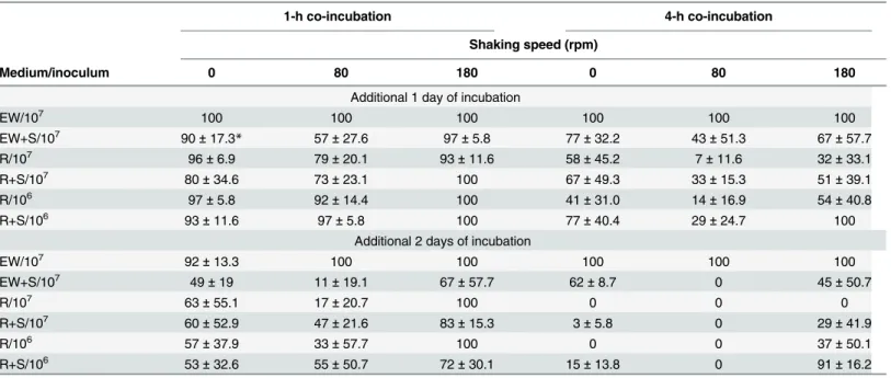
co-Table 1. Conditions for the zebrafish egg bath infection model (mean of survival rate±standard deviation).
1-h co-incubation 4-h co-incubation
Shaking speed (rpm)
Medium/inoculum 0 80 180 0 80 180
Additional 1 day of incubation
EW/107 100 100 100 100 100 100
EW+S/107 90±17.3* 57±27.6 97±5.8 77±32.2 43±51.3 67±57.7
R/107 96±6.9 79±20.1 93±11.6 58±45.2 7±11.6 32±33.1
R+S/107 80±34.6 73±23.1 100 67±49.3 33±15.3 51±39.1
R/106 97±5.8 92±14.4 100 41±31.0 14±16.9 54±40.8
R+S/106 93±11.6 97±5.8 100 77±40.4 29±24.7 100
Additional 2 days of incubation
EW/107 92±13.3 100 100 100 100 100
EW+S/107 49±19 11±19.1 67±57.7 62±8.7 0 45±50.7
R/107 63±55.1 17±20.7 100 0 0 0
R+S/107 60±52.9 47±21.6 83±15.3 3±5.8 0 29±41.9
R/106 57±37.9 33±57.7 100 0 0 37±50.1
R+S/106 53±32.6 55±50.7 72±30.1 15±13.8 0 91±16.2
EW: egg water; R: RPMI; S: 10% FBS; 106:1 × 10⁶cells/mL;107:1 × 107cells/mL
This data are from 3 repeat experiments. Approximately 30 embryos were tested for each treatment.
doi:10.1371/journal.pone.0143048.t001
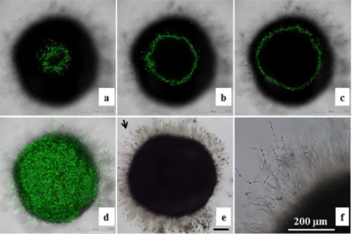
Fig 1. Zebrafish egg bath infection model in various media.Representative embryos were co-incubated with 1 × 10⁶cells/mL of SC5314 (a-d, f-i) or withoutC.albicans(control, e, j) in egg water (a, f), egg water/
serum (b, g), RPMI medium (c, h), RPMI/serum (d, i) with shaking at 80 rpm and 30°C for 4 h. The embryos were photographed immediately after non-adheredC.albicanscells were removed through washing (a-e) or after an additional 2 days of incubation (f-j). Scale bar = 200μm. This data are from 3 repeat experiments.
Approximately 30 embryos were tested for each treatment.
incubation in egg water/serum (Fig 1b), RPMI (Fig 1c) or RPMI/serum (Fig 1d), but not in egg water alone (Fig 1a). After an additional 2 days of incubation in egg water,C.albicanscells co-incubated with embryos initially in RPMI (Fig 1h), or RPMI/serum (Fig 1i) formed more hyphae than those in egg water/serum (Fig 1g). FewC.albicanscells were detected on the cho-rion of embryos when co-incubated in egg water (Fig 1f), resulting in no mortality. Further-more, we found that majority ofC.albicanscells grew on the chorion and did not reach the embryo (Fig 2).
Effects of gene deletion on
Candida albicans
virulence
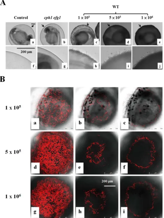
The inocula of 1 × 10⁶or 1 × 10⁷cells/mL did not appear to have significantly different effects on the killing activity (Table 1). It is likely that the amount ofC.albicanscells used has satu-rated the activity. Our preliminary data showed that abundantC.albicanscells were on the chorion and kill embryos in the inoculum equal to or greater than 5 × 10⁵cells/mL, whereas, fewerC.albicanscells were detected on the chorion after 4 h co-incubation (Fig 3A) and a high proportion of embryos survived in the inoculum of 1 × 10⁵cells/mL. Thus, the inoculum of 5 × 10⁵cells/mL was chosen to conduct subsequent experiments. Like OG1 cells, we found that majority of CAF2-dTomatoC.albicanscells (Fig 3B) grew on the chorion and did not reach the embryo.
To evaluate our infection model, we examined the virulence of severalC.albicansstrains. Embryos co-incubated with various mutant strains in RPMI/serum instead of RPMI to mimic the bloodstream of human were shaken at 80 rpm at 30°C for 4 h. The results are summarized inTable 2andFig 4. Few embryos survived after co-incubated with the wild-type cells (Fig 4A, crosses with dot line) orsap6/sap6mutant cells (Fig 4A, circles with dot line). In contrast, the deletion ofBCR1,CPH1,EFG1, andTEC1significantly reducedC.albicansvirulence. More embryos survived when co-incubated withcph1/cph1mutant cells than those with wild-type SC5314 cells after an additional 1 day and 2 days of incubation. Thus,cph1/cph1mutant cells exhibited decreased level of virulence in this model (p = 0.0006). Furthermore, all embryos sur-vived when co-incubated with the adhesion and biofilm deficient mutants,bcr1/bcr1,cph1/ cph1 efg1/efg1,efg1/efg1, ortec1/tec1(p<0.0001). Hence, the virulence levels of those strains
Fig 2. Localization of OG1Candida albicanscells in zebrafish egg bath infection model.Embryos were co-incubated with 1 × 10⁶cells/mL of OG1C.albicans. The representative slices of confocal images (a-c) are
shown. The distance between two slices was approximately 55μm. The whole merged images are presented
(d). The phase contrast photos showingC.albicanshyphae were taken by an inverted microscope (e, f). f is the enlargement of the arrow area in e. Scale bars = 200μm.
in this model are Wild-type =sap6/sap6>cph1/cph1>>bcr1/bcr1=cph1/cph1 efg1/efg1=
efg1/efg1=tec1/tec1.
Like the wild-type SC5314 cells (Fig 4Bg), there were abundantcph1/cph1(Fig 4Be), and
sap6/sap6(Fig 4Bf) hyphal cells on the chorion after an additional 2 days of incubation. Few
Fig 3. Zebrafish egg bath infection model with different inocula.(A) Embryos were co-incubated in the absence ofC.albicans(a, f) or in the presence of 1 × 10⁵(c, h), 5 × 10⁵(d-i), or 1 × 10⁶(e-j) cells/mL of
wild-type SC531cells, 1 × 10⁶(e-j) cells/mL ofcph1/cph1 efg1/efg1mutant cells (b, g) for 4 h. f-j are the
enlargement of the arrow areas in a-e. (B) Embryos were co-incubated with 1 × 10⁵(a-c), 5 × 10⁵(d-f), or 1 × 10⁶(g-i) cells/mL of CAF2-dTomatoC.albicans. The representative slices (b-c, e-f, h-i) are shown. The
distance between two slices was approximately 16μm. The whole merged images for 1 × 10⁵(a), 5 × 10⁵(d)
or 1 × 10⁶(g) cells/mL are presented. Scale bars = 200 μm.
or nobcr1/bcr1(Fig 4Ba),cph1/cph1 efg1/efg1(Fig 4Bb),efg1/efg1(Fig 4Bc), ortec1/tec1(Fig 4Bd) mutant cells were observed on the chorion. These mutant cells were not associated with mortality.
Discussion
In this study, we identified the conditions (bleached treated 1 day old embryos co-incubated with 5 × 10⁵cell/mLC.albicansat stationary phase in 4 mL RPMI or RPMI/serum at 80 rpm at 30°C for 4 h) for conducting zebrafish egg bath infection model. We also evaluated this model with various strains known to be defective in virulence in other models. To mimic the bloodstream of human, RPMI/serum was used in the present study. We observed an
Table 2. Mean of survival rates of embryos after co-incubated with various mutant strains.
Control (n = 76) bcr1(n = 71) cph1 efg1(n = 72) efg1(n = 73) cph1(n = 77) sap6(n = 81) WT (n = 82)
hours Mean Mean Mean Mean Mean SEM Mean SEM Mean SEM
0 100 100 100 100 100 100 100
1 day 100 100 100 100 58.4 5.6 29.7 5.1 32.9 5.2
2 days 100 100 100 100 10.4 3.5 2.5 1.7 2.4 1.7
doi:10.1371/journal.pone.0143048.t002
Fig 4. Virulence ofC.albicansmutant strains in the infection model.(A) Survival rates of embryos. Embryos alone (Control) or embryos with 5 × 10⁵cells/mL ofbcr1/bcr1,cph1/cph1 efg1/efg1,efg1/efg1,
tec1/tec1,cph1/cph1,sap6/sap6, or WT (SC5314) cells in RPMI/serum were incubated at 30°C for 4 h. Survival rates were determined after an additional 1 day and 2 days of incubation. (B) Representative embryos were co-incubated with (a)bcr1/bcr1, (b)cph1/cph1 efg1/efg1, (c)efg1/efg1, (d)tec1/tec1, (e)
cph1/cph1, (f)sap6/sap6, or (g) WT (SC5314) cells, and photographed after an additional 2 days of incubation. Scale bar = 200μm. The data are from 4 repeat experiments. Approximately 70 embryos were
tested for each strain.
appropriate shaking speed not only mimic the condition of blood flow but also enable embryos to mix evenly with theC.albicanscells, as suggested previously [17]. This novel zebrafish model is time-saving and cost effective.
To establish an infection, pathogenic cells adhere to the surfaces of host epidermal or endo-thelial cells, invade host cells, evade the immune system, survive and propagate in the host environment, and then spread to new tissues [39]. However, whether adhesion and hyphal for-mation are essential for the lethality ofC.albicanscells remains unclear. Our observation that
bcr1/bcr1mutant cells were not lethal to the embryos suggests that the adhesion and biofilm formation capabilities during the co-incubation period are critical. The mouse systemic infec-tion model is commonly used for investigating the funcinfec-tions of interested genes related to the virulence ofC.albicans. However, whenC.albicanscells are injected directly into the mouse tail vein, some of the genes involved in adhesion may not be detected. Our zebrafish egg bath infection model provides an alternative model to identify virulence genes, particularly those involved in adhesion. SinceC.albicanshyphal cells did not reach the embryo, the potential cause(s) for the death of the embryos, including failure of transporting toxicities, either secreted byC.albicansor generated by embryos, and/or lack of oxygen are under investigation.
The morphological transition between yeasts and hyphae byC.albicans, generally consid-ered essential for full virulence of this fungus [40], is induced by growth at 37°C and other sti-muli, including serum, in vitro. Interestingly, we found thatC.albicanscan form hyphae at lower temperature, consistent with previous report [16]. Furthermore, we observed thatC. albi-cansadhering to the chorion of embryos formed long hyphae/biofilms (Fig 4Be–4Bg) in egg water at 30°C, indicating that additional host-related factors from embryos are crucial for superseding the need for increased temperature and other stimuli, such as serum. Compared to other in vitro systems, such as polystyrene microplates, mammalian cell lines, and reconsti-tuted human epithelium [41–43], the zebrafish egg bath model can be applied to identify the receptors on the chorion of the embryos which can be beneficial for future study ofC.albicans -host interaction.
Acknowledgments
We would also like to thank Dr. Y. C. Chen for kindly providing us with thesap6/sap6strain, Dr. C. H. Lin for thebcr1/bcr1andtec1/tec1strains, and Drs. C. Y. Lan and R. T. Wheeler for the OG1 strain and the CAF2-dTomato strain, respectively. We also thank Drs. F. Fang and C. H. Lin for their helpful suggestions and comments on the manuscript. We thank Mr. M. Swof-ford for editing the manuscript. We would also thank the NHRI zebrafish core facility for its help in establishing the bath infection model.
Author Contributions
Conceived and designed the experiments: YZC YLY MSY HJL. Performed the experiments: YZC WLC. Analyzed the data: YZC YLY MSY HJL. Contributed reagents/materials/analysis tools: MSY. Wrote the paper: YZC YLY HJL.
References
1. Chen YC, Chang SC, Sun CC, Yang LS, Hsieh WC, Luh KT. Secular trends in the epidemiology of nos-ocomial fungal infections at a teaching hospital in Taiwan, 1981 to 1993. Infect Control Hosp Epidemiol. 1997; 18: 369–375. PMID:9154483
3. White TC, Holleman S, Dy F, Mirels LF, Stevens DA. Resistance mechanisms in clinical isolates of
Candida albicans. Antimicrob Agents Chemother. 2002; 46: 1704–1713. PMID:12019079
4. Yang YL, Lo HJ. Mechanisms of antifungal agent resistance. J Microbiol Immunol Infect. 2001; 34: 79– 86. PMID:11456364
5. Cheng MF, Yu KW, Tang RB, Fan YH, Yang YL, Hsieh KS, et al. Distribution and antifungal susceptibil-ity ofCandidaspecies causing candidemia from 1996 to 1999. Diagn Microbiol Infect Dis. 2004; 48: 33–37. PMID:14761719
6. Pfaller MA, Diekema DJ, Jones RN, Sader HS, Fluit AC, Hollis RJ, et al. International surveillance of bloodstream infections due toCandidaspecies: frequency of occurrence and in vitro susceptibilities to fluconazole, ravuconazole, and voriconazole of isolates collected from 1997 through 1999 in the SEN-TRY antimicrobial surveillance program. J Clin Microbiol. 2001; 39: 3254–3259. PMID:11526159
7. Yang YL, Cheng MF, Wang CW, Wang AH, Cheng WT, Lo HJ, et al. The distribution of species and susceptibility of amphotericin B and fluconazole of yeast pathogens isolated from sterile sites in Tai-wan. Med Mycol. 2010; 48: 328–334. doi:10.3109/13693780903154070PMID:20141372
8. Alarco AM, Marcil A, Chen J, Suter B, Thomas D, Whiteway M. Immune-deficientDrosophila melano-gaster: a model for the innate immune response to human fungal pathogens. J Immunol. 2004; 172: 5622–5628. PMID:15100306
9. Breger J, Fuchs BB, Aperis G, Moy TI, Ausubel FM, Mylonakis E. Antifungal chemical compounds iden-tified using aC.eleganspathogenicity assay. PLoS Pathog. 2007; 3: e18. PMID:17274686
10. Chamilos G, Lionakis MS, Lewis RE, Lopez-Ribot JL, Saville SP, Albert ND, et al.Drosophila melano-gasteras a facile model for large-scale studies of virulence mechanisms and antifungal drug efficacy in
Candidaspecies. J Infect Dis. 2006; 193: 1014–1022. PMID:16518764
11. Chamilos G, Nobile CJ, Bruno VM, Lewis RE, Mitchell AP, Kontoyiannis DP.Candida albicansCas5, a regulator of cell wall integrity, is required for virulence in murine and toll mutant fly models. J Infect Dis. 2009; 200: 152–157. doi:10.1086/599363PMID:19463063
12. Cotter G, Doyle S, Kavanagh K. Development of an insect model for the in vivo pathogenicity testing of yeasts. FEMS Immunol Med Microbiol. 2000; 27: 163–169. PMID:10640612
13. Pukkila-Worley R, Peleg AY, Tampakakis E, Mylonakis E.Candida albicanshyphal formation and viru-lence assessed using aCaenorhabditis elegansinfection model. Eukaryot Cell. 2009; 8: 1750–1758. doi:10.1128/EC.00163-09PMID:19666778
14. Mylonakis E, Casadevall A, Ausubel FM. Exploiting amoeboid and non-vertebrate animal model sys-tems to study the virulence of human pathogenic fungi. PLoS Pathog. 2007; 3: e101. PMID:17676994
15. Chao CC, Hsu PC, Jen CF, Chen IH, Wang CH, Chan HC, et al. Zebrafish as a model host forCandida albicansinfection. Infect Immun. 2010; 78: 2512–2521. doi:10.1128/IAI.01293-09PMID:20308295
16. Brothers KM, Newman ZR, Wheeler RT. Live imaging of disseminated candidiasis in zebrafish reveals role of phagocyte oxidase in limiting filamentous growth. Eukaryot Cell. 2011; 10: 932–944. doi:10. 1128/EC.05005-11PMID:21551247
17. Gratacap RL, Rawls JF, Wheeler RT. Mucosal candidiasis elicits NF-kappaB activation, proinflamma-tory gene expression and localized neutrophilia in zebrafish. Disease models & mechanisms. 2013; 6: 1260–1270.
18. Dabrowa N, Howard DH. Proline uptake inCandida albicans. J Gen Microbiol. 1981; 127: 391–397. PMID:7045279
19. Shepherd MG, Yin CY, Ram SP, Sullivan PA. Germ tube induction inCandida albicans. Can J Micro-biol. 1980; 26: 21–26. PMID:6996798
20. Chen CG, Yang YL, Cheng HH, Su CL, Huang SF, Chen CT, et al. Non-lethalCandida albicans cph1/ cph1 efg1/efg1transcription factor mutant establishing restricted zone of infection in a mouse model of systemic infection. Int J Immunopathol Pharmacol. 2006; 19: 561–565. PMID:17026841
21. Lo HJ, Kohler JR, DiDomenico B, Loebenberg D, Cacciapuoti A, Fink GR. NonfilamentousC.albicans
mutants are avirulent. Cell. 1997; 90: 939–949. PMID:9298905
22. Chamilos G, Lionakis MS, Lewis RE, Lopez-Ribot JL, Saville SP, Albert ND, et al.Drosophila melano-gaster as a facile model for large-scale studies of virulence mechanisms and antifungal drug efficacy in
Candidaspecies. J Infect Dis. 2006; 193: 1014–1022. PMID:16518764
23. Monod M, Togni G, Hube B, Sanglard D. Multiplicity of genes encoding secreted aspartic proteinases inCandidaspecies. Mol Microbiol. 1994; 13: 357–368. PMID:7984113
25. Correia A, Lermann U, Teixeira L, Cerca F, Botelho S, da Costa RM, et al. Limited role of secreted aspartyl proteinases Sap1 to Sap6 inCandida albicansvirulence and host immune response in murine hematogenously disseminated candidiasis. Infect Immun. 2010; 78: 4839–4849. doi:10.1128/IAI. 00248-10PMID:20679440
26. Schweizer A, Rupp S, Taylor BN, Rollinghoff M, Schroppel K. The TEA/ATTS transcription factor CaTec1p regulates hyphal development and virulence inCandida albicans. Mol Microbiol. 2000; 38: 435–445. PMID:11069668
27. Harriott MM, Lilly EA, Rodriguez TE, Fidel PL Jr., Noverr MC.Candida albicansforms biofilms on the vaginal mucosa. Microbiology. 2010; 156: 3635–3644. doi:10.1099/mic.0.039354-0PMID:20705667
28. Nobile CJ, Andes DR, Nett JE, Smith FJ, Yue F, Phan QT, et al. Critical role of Bcr1-dependent adhe-sins inC.albicansbiofilm formation in vitro and in vivo. PLoS Pathog. 2006; 2: e63. PMID:16839200
29. Nobile CJ, Mitchell AP. Regulation of cell-surface genes and biofilm formation by theC.albicans tran-scription factor Bcr1p. Curr Biol. 2005; 15: 1150–1155. PMID:15964282
30. Gillum AM, Tsay EY, Kirsch DR. Isolation of theCandida albicansgene for orotidine-5'-phosphate decarboxylase by complementation ofS.cerevisiae ura3andE.coli pyrFmutations. Mol Gen Genet. 1984; 198: 179–182. PMID:6394964
31. Liu H, Kohler J, Fink GR. Suppression of hyphal formation inCandida albicansby mutation of aSTE12
homolog. Science. 1994; 266: 1723–1726. PMID:7992058
32. Lin CH, Kabrawala S, Fox EP, Nobile CJ, Johnson AD, Bennett RJ. Genetic control of conventional and pheromone-stimulated biofilm formation inCandida albicans. PLoS Pathog. 2013; 9: e1003305. doi: 10.1371/journal.ppat.1003305PMID:23637598
33. Buu LM, Chen YC. Sap6, a secreted aspartyl proteinase, participates in maintenance the cell surface integrity ofCandida albicans. J Biomed Sci. 2013; 20: 101. doi:10.1186/1423-0127-20-101PMID: 24378182
34. Brothers KM, Gratacap RL, Barker SE, Newman ZR, Norum A, Wheeler RT. NADPH Oxidase-Driven Phagocyte Recruitment ControlsCandida albicansFilamentous Growth and Prevents Mortality. PLoS Pathog. 2013; 9: e1003634. doi:10.1371/journal.ppat.1003634PMID:24098114
35. Sherman F. Getting started with yeast. Methods Enzymol. 2002; 350: 3–41. PMID:12073320
36. Westerfield M. The Zebrafish Book. A Guide for the Laboratory Use of Zebrafish (Brachydanio rerio). Eugene, OR: University of Oregon Press. 1993.
37. Kimmel CB, Ballard WW, Kimmel SR, Ullmann B, Schilling TF. Stages of embryonic development of the zebrafish. Dev Dyn. 1995; 203: 253–310. PMID:8589427
38. Kais B, Schneider KE, Keiter S, Henn K, Ackermann C, Braunbeck T. DMSO modifies the permeability of the zebrafish (Danio rerio) chorion-implications for the fish embryo test (FET). Aquatic toxicology (Amsterdam, Netherlands). 2013; 140–141: 229–238.
39. Yang YL. Virulence factors ofCandidaspecies. J Microbiol Immunol Infect. 2003; 36: 223–228. PMID: 14723249
40. Jacobsen ID, Wilson D, Wachtler B, Brunke S, Naglik JR, Hube B.Candida albicansdimorphism as a therapeutic target. Expert Rev Anti Infect Ther. 2012; 10: 85–93. doi:10.1586/eri.11.152PMID: 22149617
41. Lewis RE, Lo HJ, Raad II, Kontoyiannis DP. Lack of Catheter Infection by theefg1/efg1 cph1/cph1
Double-Null Mutant, aCandida albicansStrain That Is Defective in Filamentous Growth. Antimicrob Agents Chemother. 2002; 46: 1153–1155. PMID:11897612
42. Chandra J, Mukherjee PK, Ghannoum MA. In vitro growth and analysis of Candida biofilms. Nat Protoc. 2008; 3: 1909–1924. doi:10.1038/nprot.2008.192PMID:19180075