Colon Cancer Cells
Yu-Lin Lin1,9, Jau-Yu Liau2., Shan-Chi Yu2., Da-Liang Ou6,8
, Liang-In Lin4,7, Li-Hui Tseng5, Yih-Leong Chang2,10, Kun-Huei Yeh1,8,9*, Ann-Lii Cheng1,3,8
1Department of Oncology, National Taiwan University Hospital, Taipei, Taiwan, 2Department of Pathology, National Taiwan University Hospital, Taipei, Taiwan,
3Department of Internal Medicine, National Taiwan University Hospital, Taipei, Taiwan,4Department of Laboratory Medicine, National Taiwan University Hospital, Taipei, Taiwan,5Department of Medical Genetics, National Taiwan University Hospital, Taipei, Taiwan,6National Center of Excellence for Clinical Trial and Research, National Taiwan University Hospital, Taipei, Taiwan,7Department of Clinical Laboratory Sciences and Medical Biotechnology, College of Medicine, National Taiwan University, Taipei, Taiwan,8Graduate Institute of Oncology, College of Medicine, National Taiwan University, Taipei, Taiwan,9Graduate Institute of Clinical Medicine, College of Medicine, National Taiwan University, Taipei, Taiwan,10Department and Graduate Institute of Pathology, College of Medicine, National Taiwan University, Taipei, Taiwan
Abstract
Molecular biomarkers to determine the effectiveness of targeted therapies in cancer treatment have been widely adopted in colorectal cancer (CRC), but those to predict chemotherapy sensitivity remain poorly defined. We tested our hypothesis that KRAS mutation may be a predictor of oxaliplatin sensitivity in CRC. KRAS was knocked-down in KRAS-mutant CRC cells (DLD-1G13Dand SW480G12V) by small interfering RNAs (siRNA) and overexpressed in KRAS-wild-type CRC cells (COLO320DM) by
KRAS-mutant vectors to generate paired CRC cells. These paired CRC cells were tested by oxaliplatin, irinotecan and 5FU to determine the change in drug sensitivity by MTT assay and flow cytometry. Reasons for sensitivity alteration were further determined by western blot and real-time quantitative reverse transcriptase polymerase chain reaction (qRT -PCR). In KRAS-wild-type CRC cells (COLO320DM), KRAS overexpression by mutant vectors caused excision repair cross-complementation group 1 (ERCC1) downregulation in protein and mRNA levels, and enhanced oxaliplatin sensitivity. In contrast, in KRAS-mutant CRC cells (DLD-1G13Dand SW480G12V), KRAS knocked-down by KRAS-siRNA led to ERCC1 upregulation and increased oxaliplatin resistance. The sensitivity of irinotecan and 5FU had not changed in the paired CRC cells. To validate ERCC1 as a predictor of sensitivity for oxaliplatin, ERCC1 was knocked-down by siRNA in KRAS-wild-type CRC cells, which restored oxaliplatin sensitivity. In contrast, ERCC1 was overexpressed by ERCC1-expressing vectors in KRAS-mutant CRC cells, and caused oxaliplatin resistance. Overall, our findings suggest that KRAS mutation is a predictor of oxaliplatin sensitivity in colon cancer cells by the mechanism of ERCC1 downregulation.
Citation:Lin Y-L, Liau J-Y, Yu S-C, Ou D-L, Lin L-I, et al. (2012) KRAS Mutation Is a Predictor of Oxaliplatin Sensitivity in Colon Cancer Cells. PLoS ONE 7(11): e50701. doi:10.1371/journal.pone.0050701
Editor:Ajay Goel, Baylor University Medical Center, United States of America
ReceivedMay 8, 2012;AcceptedOctober 25, 2012;PublishedNovember 28, 2012
Copyright:ß2012 Lin et al. This is an open-access article distributed under the terms of the Creative Commons Attribution License, which permits unrestricted use, distribution, and reproduction in any medium, provided the original author and source are credited.
Funding:This study was supported by a grant from the Department of Health, Executive Yuan (DOH100-TD-C-111-001), Taipei, Taiwan. The funders had no role in study design, data collection and analysis, decision to publish, or preparation of the manuscript.
Competing Interests:The authors have declared that no competing interests exist.
* E-mail: [email protected]
.These authors contributed equally to this work.
Introduction
Biomarkers to determine treatment efficacy have been investi-gated in the traditional chemotherapy era, but only a limited number of biomarkers has been found thus far. Examples are excision repair cross-complementation group 1 (ERCC1) expres-sion to predict the resistance of oxaliplatin [1], and thymidylate synthase (TS) expression to determine 5FU sensitivity [2]. This concept has evolved and has become more relevant while treatment has advanced to molecular-targeted era. Most molec-ular-targeted agents have predefined targets, which facilitate predicting the efficacy of the treatment or prognosis of diseases. Good examples are epidermal growth factor receptor (EGFR) mutation for predicting the effectiveness of EGFR tyrosine kinase inhibitors (TKIs) in lung adenocarcinoma [3], as well as KRAS mutation for predicting the unresponsiveness of EGFR monoclo-nal antibody in colorectal cancer (CRC) [4]. Although extensive studies have been undertaken to identify new predictors from
known signaling pathways or microarray-based studies [5,6], biomarkers to predict chemotherapy sensitivity remain poorly defined.
first-Figure 1. Knocking down KRAS expression in KRAS-mutant (G13D) CRC cells confers oxaliplatin resistance and ERCC1 upregulation.(A) KRAS-knocked-down DLD-1G13Dcells were more resistant to oxaliplatin, but have the same sensitivity to irinotecan, 5FU, and doxorubicin than parental DLD-1G13Dcells, as demonstrated by MTT assay. (B) The protein level of ERCC1, but not those of TOPO1 or TS, was upregulated after DLD-1G13Dcells were knocked-down by KRAS siRNA. (C) The mRNA level of ERCC1, but not those of TOPO1 or TS, was upregulated after DLD-1G13Dcells were knocked-down by KRAS siRNA. ***:p,0.001.
doi:10.1371/journal.pone.0050701.g001
line PFS in KRAS-mutant and wild-type patients was 7.7 and 8.4 months, respectively.
According to these observations, we hypothesized that KRAS mutation may be a predictor of oxaliplatin sensitivity in CRC. First, KRAS was knocked-down in KRAS-mutant CRC cells and overexpressed in KRAS-wild-type CRC cells. These paired CRC cells were tested by oxaliplatin, irinotecan and 5FU to evaluate the change in drug sensitivity. Reasons for sensitivity alteration were further determined by western blot and real-time quantitative reverse transcriptase polymerase chain reaction (qRT -PCR). Finally, the target responsible for sensitivity alteration was validated by knocking-down and overexpressing the target.
Materials and Methods
Cell Culture and Reagents
Human CRC cell lines COLO320DM (KRAS-wild-type), DLD-1G13D (KRAS G13D mutation), and SW480G12V (KRAS G12V mutation) were all obtained from American Type Culture Collection. Cells were all maintained in RPMI-1640 containing 10% fetal bovine serum, 2 mmol/L of L-glutamine (Life Technologies, Carlsbad, CA, USA), and PSA (10,000 units/ml of penicillin, 10 mg/ml of streptomycin, and 25mg/ml ampho-tericin B; Biological Industries, Kibbutz Beit Haemek, Israel) and cultured at 37uC in a humidified incubator containing 5% CO2. Oxaliplatin (EloxatinH injection 5 mg/ml) was obtained from Sanofi-Aventis Co., Ltd. (Taipei, Taiwan). Irinotecan, 5FU, and doxorubicin were all purchased from Sigma-Aldrich (St. Louis, MO, USA). Rabbit antibodies for western blot against ERCC1 and KRAS were purchased from Cell Signaling Technology, Inc. (Beverly, MA, USA). Mouse antibodies against TS, topoisomerase I (TOPO I), andb-actin were obtained from Millipore (Bedford, MA, USA), BD Biosciences (San Jose, CA, USA) and Cell Biolabs, Inc. (San Diego, CA, USA), respectively.
Knocking-down of KRAS and ERCC1
Two types of both KRAS and ERCC1 small interfering RNAs (siRNA) and scrambled nonspecific (negative control) siRNA were purchased from Applied Biosystems, Inc. (Foster City, CA, USA). For KRAS gene knockdown, DLD-1G13Dand SW480G12V cells were first transfected with KRAS- or scrambled siRNAs for 1 day using the Lipofectamine2000 transfection reagent (Invitrogen, Carlsbad, CA, USA) according to the manufacturer’s instructions. The transfected cells were then treated with oxaliplatin, irinotecan, 5FU and doxorubicin with various concentrations for the following 72 hours. The protein lysate and mRNA of parental and KRAS knockdown DLD-1G13Dand SW480G12V cells were collected in 24, 48, 72 and 96 hours after transfection for evaluation of KRAS knockdown magnitude by western blot. For ERCC1 gene knockdown, COLO320DM cells transfected with two different ERCC1- or scrambled SiRNAs were treated with oxaliplatin for 72 hours. The protein lysate and mRNA of parental and ERCC-knocked-down COLO320DM cells were collected in 24, 48, 72 and 96 hours post-transfection for evaluating the ERCC1 knockdown effect by western blot and qRT-PCR.
Overexpression of KRAS and ERCC1
The pCMV6-Myc-DDK-tagged-KRAS vector was purchased from OriGene Technologies (Rockville, MD, USA). DNA-sequence-encoding KRAS G12V and G13D mutation were generated by site-directed mutagenesis and cloned into the pCMV6-Myc-DDK-tagged-KRAS vector. The sequences of KRAS G12V and G13D mutation were as follows: 59
-GTTGTGGTAGTTGGAGCTGTT
GGCGTAGGCAA-GAATGCC-39; reverse: 59
-GGCACTCTTGCCTACGCCAA-CAGCTCCAACTACCACAAG-39 and forward: 59
-GGTAGTTGGAGCTGGTGACGTAGGCAAGAGTGCC-39; reverse: 59 -GGCACTCTTGCCTACGTCACCAGCTCCAAC-TACC-39, respectively. For KRAS overexpression, CO-LO320DM cells were transiently transfected with the pCMV6-Myc-DDK-tagged-KRAS, -KRASG12V, and -KRASG13Dvectors. After 24-hour of transfection, cells were treated with oxaliplatin, irinotecan, 5FU, and doxorubicin with various concentrations for the following 72 hours. The protein lysate and mRNA of COLO320DM cells transfected by the pCMV6-Myc-DDK-tagged-KRAS, -KRASG12V, and -KRASG13D vectors were collected at 24, 48, 72, and 96 hours for evaluation of KRAS overexpression magnitude by western blot. For ERCC1 overex-pression, SW480G12V cells were transfected by the pCMV6-ERCC1-GFP vector (OriGene Technologies, Rockville, MD, USA) for 24 hours, and treated with oxaliplatin for 72 hours. The protein lysate of SW480G12Vcells transfected by the ERCC1-GFP vector was collected at 24, 48, 72, and 96 hours for evaluation of ERCC1 overexpression magnitude by western blot.
Cell Viability and Apoptotic Analyses
Cell viability was assessed by using the 3-(4,5-dimethylthiazol-2-yl)-2,5-diphenyltetrazolium bromide (MTT; Tokyo Chemical Industry Inc., Tokyo, Japan) assay in 6 replicates. Initially, COLO320DM, SW480G12V, and DLD-1G13D cells were seeded at 3500, 4500, and 3000 cells per well in 96-well flat-bottomed plates, respectively. After 24-hour incubation, SW480G12V and DLD-1G13D cells were transfected by KRAS- and scrambled siRNAs, and COLO320DM cells were transfected by the pCMV6-Myc-DDK-tagged KRAS, -KRASG12V, and -KRASG13D vectors, as described. After KRAS-siRNAs were transfected to DLD-1G13D/SW480G12V cells and KRAS-mutant vectors to COLO320DM cells for 24 hours, cells were treated with oxaliplatin, irinotecan, 5FU, and doxorubicin at various concen-trations in 10% FBS-supplemented RPMI-1640 for 72 hours. The control cells were mixed with DMSO at a concentration equal to that in drug-treated cells. Cell viability of these treated cells was measured by adding 200ml of 0.5 mg/ml MTT solubilized in DMSO to each well, and cells were incubated in the CO2 incubator at 37uC for 2 hours after removal of the medium. Absorbance was determined at 570 nm. Concentrations of compounds that inhibited viability by 50% (IC50) were determined using the median effect method by employing CalcuSyn software (Biosoft, Ferguson, MO, USA).
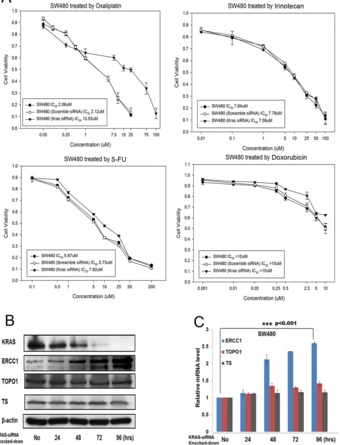
Figure 2. Knocking down KRAS expression in other KRAS-mutant subtype (G12V) CRC cells results in oxaliplatin resistance and ERCC1 upregulation.(A) KRAS-knocked-down SW480G12Vcells were more resistant to oxaliplatin, but have the same sensitivity to irinotecan, 5FU, and doxorubicin than parental SW480G12Vcells, as demonstrated by MTT assay. (B) The protein level of ERCC1, but not those of TOPO1 or TS, was upregulated after SW480G12Vcells were knocked-down by KRAS siRNA. (C) The mRNA level of ERCC1, but not those of TOPO1 or TS, was upregulated after SW480G12Vcells were knocked-down by KRAS siRNA. ***:
p,0.001. doi:10.1371/journal.pone.0050701.g002
Figure 3. Overexpressing KRAS in KRAS wild-type CRC cells leads to oxaliplatin sensitivity and ERCC1 downregulation.(A) KRASG13D -DDK-myc-COLO320DM cells were more sensitive to oxaliplatin, but have the same sensitivity to irinotecan, 5FU, and doxorubicin than parental COLO320DM cells, as demonstrated by MTT assay. (B) The protein level of ERCC1, but not those of TOPO1 or TS, was downregulated after COLO320DM cells were transfected by the KRASG13Dmutant vector. (C) The mRNA level of ERCC1, but not those of TOPO1 or TS, was downregulated after COLO320DM cells were transfected by the KRASG13Dmutant vector. **:p,0.01.
Western Blot Analysis
KRAS-overexpressed COLO320DM and KRAS-knocked-down SW480G12V and DLD-1G13D cells treated with various concentrations of oxaliplatin, irinotecan, and 5FU for 72 hours in 6-cm dishes (16105cells per dish) were collected and lysed with a RIPA lysis buffer [50 mmol/L Tris-HCl (pH 8.0), 150 mmol/L NaCl, 1% NP40, 0.5% sodium deoxycholate, 0.1% SDS] (Sigma
Cat. No. R0278). Protein concentrations of the lysate were determined using a Pierce BCA protein assay kit (Thermal Scientific, Odessa, Texas, USA). Equivalent amounts of protein from each lysate were subjected to SDS-PAGE and then transferred to nitrocellulose membranes for immunoblotting. The transblotted membranes were washed twice with Tris-buffered saline (TBS) containing 0.1% Tween 20 (TBST). After
Figure 4. Overexpressing KRAS by another KRAS overexpression vector (G12V) in KRAS wild-type CRC cells leads to oxaliplatin sensitivity and ERCC1 downregulation.(A) KRASG12V-DDK-myc-COLO320DM cells were more sensitive to oxaliplatin, but have the same sensitivity to irinotecan, 5FU, and doxorubicin than parental COLO320DM cells, as demonstrated by MTT assay. (B) The protein level of ERCC1, but not those of TOPO1 or TS, was downregulated after COLO320DM cells were transfected by the KRASG12Vmutant vector. (C) The mRNA level of ERCC1, but not those of TOPO1 or TS, was downregulated after COLO320DM cells were transfected by the KRASG12Vmutant vector. **:
p,0.01. (D) Increased percentage of apoptosis, from 22.5%60.2% to 39.1%60.2% of apoptosis (P,0.001), has been demonstrated when KRASwt-DDK-myc-COLO320DM cells, were compared to KRASG12V-DDK-myc-COLO320DM cells, in which, both were treated by the same concentration of oxaliplatin in 5
mM.
*:p,0.001.
doi:10.1371/journal.pone.0050701.g004
blocking with TBST containing 5% nonfat milk for 40 minutes, the membranes were incubated with the appropriate primary antibody in TBST containing 1% nonfat milk at 4uC overnight. All of the primary antibodies were diluted in an appropriate concentration of 1% nonfat milk-containing TBST. After treat-ment with the primary antibody, the membranes were washed twice with TBST for 20 minutes, followed by goat anti-rabbit or anti-mouse IgG-horseradish peroxidase–conjugated secondary antibody (diluted 1:3000) for 1 hour at room temperature and washed 3 times with TBST for 1 hour. The membranes were developed using an enhanced chemiluminescence horseradish peroxidase substrate (Millipore, Bedford, MA, USA) according to the manufacturer’s instructions.
Real-time Quantitative Reverse Transcriptase Polymerase Chain Reaction (qRT-PCR)
KRAS-overexpressed COLO320DM, KRAS-knocked-down SW480G12V and DLD-1G13Dcells treated with various concen-trations of oxaliplatin, irinotecan, and 5FU for 24, 48, and 72 hours, respectively, were collected and lysed in a Trizol reagent (Invitrogen, Carlsbad, CA, USA) and stored at220uC. The RNA of these cells was extracted according to the manufacturer’s
instructions. cDNAs were synthesized from total RNA (1mg) using the Applied Biosystems High-Capacity cDNA Archive kit accord-ing to the manufacturer’s instructions. The cDNAs from 50-ng total RNA were quantified using the Taqman Universal or SYBR Green PCR Master Mix (Applied Biosystems, Foster City, CA, USA) on an ABI PRISM 7900 Sequence Detection System (Perkin-Elmer/Applied Biosystems). The primer sequences of ERCC1 (ABI Taqman assay ID: Hs01012158_ml), TOPO I (ABI Taqman assay ID: Hs00243257_ml), TS (ABI Taqman assay ID: Hs00426586_ml), andb-actin gene (ABI Taqman assay ID: Hs99999903_ml) as an endogenous control were all purchased from Applied Biosystems (Foster City, CA, USA). Conditions for PCR were 50uC for 2 minutes, 95uC for 10 minutes, and 40 cycles of 95uC for 15 seconds (denaturation) and 60uC for 1 minute (annealing/extension). The relative mRNA amount of the target gene/endogenous control gene (b-actin) was calculated using the DCt (threshold cycle) method, as follows: relative expression = 2-DCt, whereDCt = Ct (target gene) - Ct (b-actin).
Statistical Analysis
For cell line studies, all data were repeated for at least 3 independent experiments. Quantitative data are represented as mean 6 SD. Comparisons between data within the same
Figure 6. Downregulation of ERCC1 expression in KRAS-mutant CRC cells might be related to hypermethylation of ERCC1gene, which possibly induced by up-regulation of DNMT3B (DNA methyltransferase 3B).(A) Protein expression of ERCC1 in DLD-1(KRASG13D mutation) cells is up-regulated after 59-azacitidine (de-methylating agent) treatment for 96 hours, which implied that the downregulation of ERCC1 in KRAS-mutant CRC cells might be partly through ERCC1 hypermethylation. (B) Downregulation of ERCC1 in COLO320DM (KRAS wild-type) cells transfected by KRASG13D-mutant-vector for 24 and 96 hours may not only be restored by 5
9-azacitidine in 10mM, but also caused up-regulation of
DNMT3B.
doi:10.1371/journal.pone.0050701.g006
experiments were analyzed using the Student’sttest. Ap-value of
,0.05 was considered statistically significant.
Results
Knocking-down KRAS in KRAS-mutant CRC Cells Increases Oxaliplatin Resistance and Causes ERCC1 Overexpression
Knocking-down KRAS in DLD-1G13D cells resulted in cells more resistant to oxaliplatin, but not to irinotecan, and 5FU, standard chemotherapeutic agents for CRC, and not to doxoru-bicin, broad spectrum chemotherapeutic agent for other major cancers. Two different KRAS-siRNAs were transfected to DLD-1G13D cells. The IC
50 of the first-paired parental DLD-1G13D/ KRAS-siRNA(1)-DLD-1G13Dcells treated with oxaliplatin for 72 hours was 3.97/33.07mM. The second-paired parental DLD-1G13D/KRAS-siRNA(2)-DLD-1G13D cells was 3.97/13.49mM. The IC50 of paired parental DLD-1G13D /KRAS-siRNA-DLD-1G13D cells treated with irinotecan, 5FU and doxorubicin remained unchanged (Figure 1A). ERCC1, TOPO I and TS, which were thought to be biomarkers for predicting the sensitivity of oxaliplatin [1], irinotecan [10] and 5FU [2], respectively, were further checked by western blot and qRT-PCR (Figures 1B, and1C) to explore mechanisms behind our findings. Only ERCC1 expression was upregulated after KRAS knockdown; in contrast, TOPO I and TS remained constant both in protein and mRNA levels. KRAS knockdown efficiency was evaluated by western blot, which showed diminished of KRAS expression after 72-hour of knocking-down the KRAS gene (Figure 1B).
To further consolidate our observation, we used another CRC cell, SW480, which harbored another KRAS mutant subtype, G12V, to repeat the same experimental procedures. In summary, parental SW480G12V cells was, as expected, more sensitive to oxaliplatin than KRAS knocked-down SW480G12Vcells. The IC50 of parental SW480G12V/KRAS-siRNA-SW480G12V cells treated by oxaliplatin for 72 hours was 2.08/13.53mM, but that of parental SW480G12V/KRAS-siRNA-SW480G12V cells treated with irinotecan, 5FU, and doxorubicin remained unchanged (Figure 2A). ERCC1, TOPO I, and TS were simultaneously checked by western blot and qRT-PCR (Figures 2B and 2C), and again only ERCC1 was upregulated after KRAS knockdown, with TOPO I and TS levels remaining unchanged in protein and mRNA levels. Similarly, KRAS knockdown efficiency was measured by western blot, which showed a decline in KRAS expression after 72-hour of knocking-down the KRAS gene (Figure 2B).
Overexpressing KRAS in KRAS Wild-type CRC Cells Leads to Oxaliplatin Sensitivity and ERCC1 Downregulation
Overexpression of KRAS in COLO320DM cells by KRAS-mutant vectors resulted in cells more sensitive to oxaliplatin. The IC50 of parental COLO320DM/KRASG13D -DDK-myc-CO-LO320DM cells treated by oxaliplatin for 72 hours was 2.86/ 0.26mM, but that of parental COLO320DM/KRASG13D -DDK-myc-COLO320DM cells treated with irinotecan, 5FU, and doxorubicin remained unchanged (Figure 3A). ERCC1, TOPO I, and TS were checked by western blot and qRT-PCR (Figures 3B and 3C), which showed that only ERCC1 was downregulated without any change in protein and mRNA levels in TOPO I and TS after KRAS overexpression. The expression of ectopic KRAS and endogenous KRAS was measured by western blot, which showed a robust expression of ectopic KRAS, with a constant expression of endogenous KRAS after 24-hour overexpression of the KRAS gene (Figure 3B).
The same results were also found in COLO320DM cells transfected with the KRASG12V-mutant vector. The IC50 of parental COLO320DM/KRASG12V-DDK-myc-COLO320DM cells treated with oxaliplatin for 72 hours was 2.55/0.25mM, but that of parental COLO320DM/KRASG12V -DDK-myc-CO-LO320DM cells treated with irinotecan, 5FU, and doxorubicin remained unchanged (Figure 4A). Again, only ERCC1 was downregulated without any change of TOPO I and TS in protein (Figure 4B) and mRNA levels (Figure 4C) after KRAS overex-pression. The expression of ectopic KRAS and endogenous KRAS after 24-hour overexpression of the KRAS gene is shown in Figure 4B. To further strengthen the finding that KRASG12V -DDK-myc-COLO320DM cells were more sensitive to oxaliplatin than parental COLO320DM cells, flow cytometry with annexin V-FITC was performed. Consequently, increased percentage of apoptosis, from 22.5%60.2% to 39.1%60.2% of apoptosis (P,0.001), has been found when parental COLO320DM cells, transfected by KRASwt-DDK-myc-vector, were compared to COLO320DM cells, transfected by KRASG12V-DDK-myc-vector, in which, both were treated by the same concentration of oxaliplatin in 5mM (Figure 4D).
Validating ERCC1 Expression as the Predictor of Oxaliplatin Sensitivity
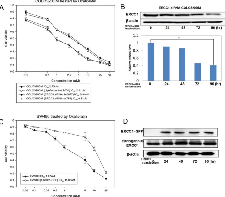
Knocking-down ERCC1 in KRAS wild-type CRC cells restores oxaliplatin sensitivity. To further confirm the relationship between ERCC1 expression and oxaliplatin sensitiv-ity, we knocked-down the ERCC1 gene using 2 different ERCC1-siRNAs in KRAS-wild-type cells (COLO320DM). We found that the IC50 of the first-paired parental COLO320DM/ERCC1-siRNA(1)-COLO320DM cells treated with oxaliplatin for 72 hours was 2.75/0.91mM (Figure 5A). The second-paired parental COLO320DM/ERCC1-siRNA(2)-COLO320DM cells was 2.75/ 0.83mM (Figure 5A). The protein and mRNA expression levels of ERCC1 were downregulated after ERCC1 was knocked-down by ERCC1-siRNA in COLO320DM cells (Figure 5B).
Overexpressing ERCC1 in KRAS-mutant CRC cells causes oxaliplatin resistance. Overexpression of ERCC1 in SW480G12V cells by the ERCC1-overexpressing vector caused SW480G12V cells to become more resistant to oxaliplatin. The IC50 of parental SW480G12V/ERCC1-GFP-SW480G12V cells treated with oxaliplatin for 72 hours was 1.87/11.03mM (Figure 5C). Western blot was used to determine the ERCC1-GFP overexpression level after transfection (Figure 5D).
Discussion
Our study shows that KRAS mutation is a predictor of oxaliplatin sensitivity in colon cancer cells by ERCC1 downreg-ulation. This may provide an important step to personalized chemotherapy in colon cancer.
KRAS-mutant CRC patients is warranted. Our study was initiated to find better predictors in current chemotherapy, for which the hypothesis was generated from subgroup analyses of randomized prospective clinical trials, PRIME [8] and OPUS [7], versus CRYSTAL [9]. According to our findings, KRAS-mutant CRC patients might benefit more from receiving first-line oxaliplatin-based regimens than KRAS-wild-type patients. This phenomenon warrants further confirmation by large prospective clinical trials.
Our data demonstrated that KRAS mutation in CRC cells caused ERCC1 downregulation. This significant finding might imply that some other unknown druggable targets may still be responsible for KRAS-mutant CRC treatment in addition to the traditional RAS/RAF/MEK/ERK pathway. To explore these unknown targets, studies designed from epigenetic and/or genetic point of views may be helpful. From epigenetic point of view, hypermethylation causes gene silencing is well-known [12,13]. In our study, we have found that the protein expression of ERCC1 in DLD-1(KRASG13D mutation) cells is up-regulated after 59 -azacitidine (de-methylating agent) treatment for 96 hours (Figure 6A), which indicated that the downregulation of ERCC1 in KRAS-mutant CRC cells might be partly through ERCC1 hypermethylation. We also found that the downregulation of protein expression of ERCC1 in COLO320DM (KRAS wild-type) cells transfected by KRAS-mutant-vector for 24 and 96 hours may be restored by 59-azacitidine (Figure 6B). This further implied that the downregulation of ERCC1 expression in CRC cells is not only partly through hypermethylation, but also determined by the changes of KRAS expression in CRC cells. Because downregu-lation of ERCC1 in COLO320DM cells, transfected by KRASG13D-mutant-vector, might be caused by hypermethylation of ERCC1, we further checked DNMT3B (DNA methyltransfer-ase 3B), whose major role is to proceed the process of methylation. We found that DNMT3B was upregulated when ERCC1 was dowenregulated in COLO320DM cells, transfected by KRASG13D-mutant-vector (Figure 6B). DNMT3B may again suppress by 59-azacitidine, which depicted that DNMT3B is probably responsible for the methylation process. Therefore, our data showed that ERCC1 downregulation in KRAS-mutant CRC cells might be through ERCC1 hypermethylation. We proposed that KRAS-mutant CRC cells might have higher methylation rate on CpG islands of ERCC1 promoter region than KRAS-mutant cells transfected by KRAS-siRNA. This hypothesis may be validated by comparing the possible differences of hypermethyla-tion on ERCC1 promoter region between KRAS-mutant cells and KRAS-mutant cells transfected by KRAS-siRNA by methylation specific PCR and/or sodium bisulfite sequencing analysis [14]. The whole concept would be that KRAS activating mutation might cause DNMT3B upregulation. Subsequently, DNMT3B might bind to the promoter region of ERCC1 to result in hypermethylation of ERCC1 gene. Finally, hypermethylation of ERCC1 gene results in downregulation of ERCC1 expression. Alternatively, from genetic point of view, as KRAS mutation is an activating mutation, which has been widely accepted [15,16], we
proposed that there might be an existed unknown factor, which may be inhibited by the activation of KRAS gene or its downstream signals. This factor needs also to be an activating factor to activate ERCC1 gene. Then, once KRAS gene is mutated (activated), this factor might be inhibited, and the amount of this factor might be declined. Subsequently, the expression of ERCC1 might be suppressed due to the lack of this factor. To conduct this kind of studies, reporter gene constructs using ERCC1 promoter, luciferase activity assay and chromatin immunoprecipitation may be thus needed [17].
Although the detailed mechanisms behind these findings remain elusive, crosstalks between the mutated KRAS gene and DNA repair machinery pathways, which might also be responsible for the effect of oxaliplatin-based treatment, have been investigated [18,19]. Additionally, various new generations of microarray-based technologies, comparing same given cells with/without KRAS mutation by knocking-down and overexpressing the KRAS gene accordingly, may be another helpful way in defining new targets for KRAS-mutant CRC treatment [5,6].
Overexpression of ERCC1 is associated to the resistance to platinum-based chemotherapy [1,20,21,22,23,24], which has been demonstrated in various kinds of cancers, including esophageal cancers [25], non-small cell lung cancers [26], and bladder cancers [27]. These findings are also compatible to the current study. In our study, we demonstrated that KRAS wild-type (COLO320DM) CRC cells were more resistant to oxaliplatin than the same given cells (COLO320DM) transfected by KRAS-mutant-vectors. We also demonstrated one of the reasons for the resistance may be related to higher ERCC1 expression in parental COLO320DM cells compared to COLO320DM cells transfected by KRAS-mutant-vectors.
Our in vitro experiments had limitations. First, 2 of 7 KRAS-mutant subtypes, which represented 40% of total KRAS mutation [28], were chosen as models in our study; this might not represent the biological behaviors of all KRAS-mutant subtypes in CRC cells. Further comprehensive studies with all KRAS mutant subtypes may be warranted. Second, mechanisms behind ERCC1 downregulation caused by KRAS mutation remain elusive. Although we proposed two possible ways to approach this issue, deeper understanding the biology of KRAS gene and the crosstalk between KRAS gene and DNA repair machinery may facilitate the advances on this issue.
In conclusion, our data suggested that KRAS mutation is a predictor of oxaliplatin sensitivity in colon cancer cells by ERCC1 downregulation.
Author Contributions
Conceived and designed the experiments: YL KY AC. Performed the experiments: YL JL SY DO LL LT. Analyzed the data: YL DO LL LT YC KY AC. Contributed reagents/materials/analysis tools: YL JL SY DO LL LT YC KY AC. Wrote the paper: YL JL SY DO LL LT YC KY AC.
References
1. Dabholkar M, Vionnet J, Bostick-Bruton F, Yu JJ, Reed E (1994) Messenger RNA levels of XPAC and ERCC1 in ovarian cancer tissue correlate with response to platinum-based chemotherapy. J Clin Invest 94: 703–708. 2. Van Triest B, Pinedo HM, van Hensbergen Y, Smid K, Telleman F, et al. (1999)
Thymidylate synthase level as the main predictive parameter for sensitivity to 5-fluorouracil, but not for folate-based thymidylate synthase inhibitors, in 13 nonselected colon cancer cell lines. Clin cancer Res 5: 643–654.
3. Paez JG, Janne PA, Lee JC, Tracy S, Greulich H, et al. (2004) EGFR mutations in lung cancer: correlation with clinical response to gefitinib therapy. Science 304: 1497–1500.
4. Lievre A, Bachet JB, Le Corre D, Boige V, Landi B, et al. (2006) KRAS mutation status is predictive of response to cetuximab therapy in colorectal cancer. Cancer Res 66: 3992–3995.
5. Scholl C, Frohling S, Dunn IF, Schinzel AC, Barbie DA, et al. (2009) Synthetic lethal interaction between oncogenic KRAS dependency and STK33 suppres-sion in human cancer cells. Cell 137: 821–834.
6. Luo J, Emanuele MJ, Li D, Creighton CJ, Schlabach MR, et al. (2009) A genome-wide RNAi screen identifies multiple synthetic lethal interactions with the Ras oncogene. Cell 137: 835–848.
7. Bokemeyer C, Bondarenko I, Makhson A, Hartmann JT, Aparicio J, et al. (2009) Fluorouracil, leucovorin, and oxaliplatin with and without cetuximab in
the first-line treatment of metastatic colorectal cancer. J Clin Oncol 27: 663– 671.
8. Douillard JY, Siena S, Cassidy J, Tabernero J, Burkes R, et al. (2010) Randomized, phase III trial of panitumumab with infusional fluorouracil, leucovorin, and oxaliplatin (FOLFOX4) versus FOLFOX4 alone as first-line treatment in patients with previously untreated metastatic colorectal cancer: the PRIME study. J Clin Oncol 28: 4697–4705.
9. Van Cutsem E, Kohne CH, Hitre E, Zaluski J, Chang Chien CR, et al. (2009) Cetuximab and chemotherapy as initial treatment for metastatic colorectal cancer. N Engl J Med 360: 1408–1417.
10. Sugimoto Y, Tsukahara S, Oh-hara T, Isoe T, Tsuruo T (1990) Decreased expression of DNA topoisomerase I in camptothecin-resistant tumor cell lines as determined by a monoclonal antibody. Cancer Res 50: 6925–6930. 11. Tournigand C, Andre T, Achille E, Lledo G, Flesh M, et al. (2004) FOLFIRI
followed by FOLFOX6 or the reverse sequence in advanced colorectal cancer: a randomized GERCOR study. J Clin Oncol 22: 229–237.
12. Esteller M, Toyota M, Sanchez-Cespedes M, Capella G, Peinado MA, et al. (2000) Inactivation of the DNA repair gene O6-methylguanine-DNA methyl-transferase by promoter hypermethylation is associated with G to A mutations in K-ras in colorectal tumorigenesis. Cancer Res 60: 2368–2371.
13. Eden S, Cedar H (1994) Role of DNA methylation in the regulation of transcription. Curr Opin Genet Dev 4: 255–259.
14. Chen HY, Shao CJ, Chen FR, Kwan AL, Chen ZP (2010) Role of ERCC1 promoter hypermethylation in drug resistance to cisplatin in human gliomas. Int J Cancer 126: 1944–1954.
15. Trahey M, McCormick F (1987) A cytoplasmic protein stimulates normal N-ras p21 GTPase, but does not affect oncogenic mutants. Science 238: 542–545. 16. Bos JL (1989) ras oncogenes in human cancer: a review. Cancer Res 49: 4682–
4689.
17. Ou DL, Shen YC, Yu SL, Chen KF, Yeh PY, et al. (2010) Induction of DNA damage-inducible gene GADD45beta contributes to sorafenib-induced apopto-sis in hepatocellular carcinoma cells. Cancer Res 70: 9309–9318.
18. Golding SE, Rosenberg E, Neill S, Dent P, Povirk LF, et al. (2007) Extracellular signal-related kinase positively regulates ataxia telangiectasia mutated, homol-ogous recombination repair, and the DNA damage response. Cancer Res 67: 1046–1053.
19. Squatrito M, Holland EC (2011) DNA damage response and growth factor signaling pathways in gliomagenesis and therapeutic resistance. Cancer Res 71: 5945–5949.
20. Lee KB, Parker RJ, Bohr V, Cornelison T, Reed E (1993) Cisplatin sensitivity/ resistance in UV repair-deficient Chinese hamster ovary cells of complemen-tation groups 1 and 3. Carcinogenesis 14: 2177–2180.
21. Sancar A (1994) Mechanisms of DNA excision repair. Science 266: 1954–1956. 22. Zhen W, Link CJ Jr, OConnor PM, Reed E, Parker R, et al. (1992) Increased gene-specific repair of cisplatin interstrand cross-links in cisplatin-resistant human ovarian cancer cell lines. Mol Cell Biol 12: 3689–3698.
23. Jones JC, Zhen WP, Reed E, Parker RJ, Sancar A, et al. (1991) Gene-specific formation and repair of cisplatin intrastrand adducts and interstrand cross-links in Chinese hamster ovary cells. J Biol Chem 266: 7101–7107.
24. Dabholkar M, Bostick-Bruton F, Weber C, Bohr VA, Egwuagu C, et al. (1992) ERCC1 and ERCC2 expression in malignant tissues from ovarian cancer patients. J Natl Cancer Inst 84: 1512–1517.
25. Joshi MB, Shirota Y, Danenberg KD, Conlon DH, Salonga DS, et al. (2005) High gene expression of TS1, GSTP1, and ERCC1 are risk factors for survival in patients treated with trimodality therapy for esophageal cancer. Clin Cancer Res 11: 2215–2221.
26. Takenaka T, Yoshino I, Kouso H, Ohba T, Yohena T, et al. (2007) Combined evaluation of Rad51 and ERCC1 expressions for sensitivity to platinum agents in non-small cell lung cancer. Int J Cancer 121: 895–900.
27. Bellmunt J, Paz-Ares L, Cuello M, Cecere FL, Albiol S, et al. (2007) Gene expression of ERCC1 as a novel prognostic marker in advanced bladder cancer patients receiving cisplatin-based chemotherapy. Ann Oncol 18: 522–528. 28. Winder T, Mundlein A, Rhomberg S, Dirschmid K, Hartmann BL, et al. (2009)