RBCCV 44205-1500 DOI: 10.5935/1678-9741.20130078
Fetal heart assessment in the irst trimester of
pregnancy: inluence of crown-rump length and
maternal body mass index
Avaliação do coração fetal no primeiro trimestre de gestação: inluência do comprimento cabeça-nádega
e índice de massa corporal materna
David Baptista Silva Pares
1, MD, MSc, PhD; Angélia Iara Felipe Lima
1, MD, MSc; Edward Araujo
Júnior
1, MD, MSC, PhD; Luciano Marcondes Machado Nardozza
1, MD, MSC, PhD; Wellington
P. Martins
2, MD, PhD; Antonio Fernandes Moron
1, MD, MSC, PhD
1 Federal University of São Paulo (UNIFESP). São Paulo, SP, Brazil. 2 Faculty of Medicine of Ribeirão Preto of the University of São Paulo (FMRP USP), Ribeirão Preto, SP, Brazil.
This study was carried out at Fetal Cardiology Unit of the Obstetric De
-partment of the Federal University of São Paulo (UNIFESP), São Paulo, SP, Brazil. Gynecology and Obstetric Unit at the Ribeirão Preto Medical School, Universitu of São Paulo (FMRP- USP), Ribeirão Preto, SP, Brazil.
Ultrasonography and Medical Recycling School of Ribeirão Preto (EURP), Ribeirão Preto, SP, Brazil.
No inancial support.
Correspondence address:
Edward Araújo Júnior
Obstetric Department of the Federal University of São Paulo (UNIFESP) Rua Napoleão de Barros, 875 – Vila Clementino, São Paulo, SP
Brazil – Zip Code: 04024-002 E-mail: araujojred@terra.com.br
Article received on April 28th, 2013
Article accepted on June 17th, 2013
Abstract
Objective: To evaluate the inluence of the crown-rump length and body mass index on sonographic evaluation of the fetal heart using abdominal and vaginal routes in the irst trimester of pregnancy.
Methods: We conducted a cross-sectional study with 57 pregnant women between 12-14 weeks (CRL≤ 84 mm). We evaluated the following fetal cardiac plans using the abdominal and vaginal routes: four-chamber view, right ventricle outlow tract, left ventricle outlow tract and aortic arch. We used the B-mode, color Doppler and four-dimensional ultrasonography (spatio-temporal image correlation). To evaluate the inluence of crown-rump length and body mass index in the assessment of fetal cardiac planes, we used the t test unpaired.
Results: There were no statistically signiicant differences in the rates of success and failure between abdominal and vaginal routes in relation to body mass index, however, there was a higher failure rate in vaginal assessment using B mode associated with color Doppler (P<0.01).
Conclusion: The crown-rump length and body mass index had no interference in fetal cardiac assessment in the irst trimester of pregnancy.
Descriptors: Fetal heart. Color Doppler. Ultrasonography, Doppler, Color. Crown-rump length. Body mass index.
Resumo
Objetivo: Avaliar a inluência do comprimento cabeça-nádega e do índice de massa corporal na avaliação ultrassonográica do coração fetal, pelas vias abdominal e vaginal, no primeiro trimestre de gestação.
Métodos: Realizou-se um estudo de corte transversal com 57
Utilizou-vaginal and abdominal approaches, using B mode, color Doppler and STIC.
METHODS
We performed a prospective cross-sectional study with healthy pregnant women between 12 and 14 weeks of gestation, from July 2011 to July 2012. This study was approved by the Research Ethics Committee of the Federal University of São Paulo (UNIFESP), whereas women who agreed to participate signed a voluntary written informed consent. Inclusion criteria were singleton pregnancies, with measurement of CRL from 45 mm to 84 mm. We performed a single measure of CRL, which also served to the correct dating of gestational age.
Patients were randomly selected, with the examinations in two Voluson 730 Expert and E8 devices (General Electric Healthcare, Zipf, Austria) equipped with volumetric convex (RAB4-8L) and endocardial (RIC5-9W) transducers. Cardiac assessment was performed by a single examiner (AIFL) immediately after the screening of the irst trimester. Initially, we used the abdominal approach, associating the B mode to color Doppler, followed by vaginal approach. We aimed to obtain the plans of four heart chambers, left ventricular outlow tract and right ventricular outlow tract and aortic arch. In the plane of the four heart chambers, size, axis and symmetry of the chambers were assessed.
In the plans of the right and left ventricular outlow tract, there was the crossing of the great vessels, in addition to the similarity of their diameters. The plane of the aortic arch allowed the identiication of the descending aorta. We
INTRODUCTION
Congenital heart disease (CHD) are the most common birth defects, affecting 5-8 per 1,000 live births [1]. Early diagnosis of CHD allows better prenatal care and referral of pregnant women to tertiary center of cardiology and neonatal
cardiac surgery.
Fetal cardiac assessment through abdominal approach
during the examination of screening for chromosomal defects in irst trimester of pregnancy allows the evaluation of the four-chamber plane, and enable the diagnosis of 44.8% of CHD [2]. The vaginal ultrasound has been used for almost 20 years for fetal cardiac evaluation at the end of the irst trimester of pregnancy, allowing the assessment of the four-chamber plane in addition to the extended examination [3].
The three-dimensional ultrasound using spatio-temporal
image correlation (STIC) software has been used in the irst
trimester through the abdominal approach, demonstrating
that cardiac volumes sent via an internet link allowed us to obtain standard cardiac planes [4].
The irst trimester screening is important, not only for the calculation of risk for chromosomal defects, but also to correct dating of gestational age, assessment of some fetal malformations and determination of chorionicity in cases of twin pregnancies [5,6].
The body mass index (BMI) and crown-rump length (CRL) can be factors that inluence fetal cardiac assessment in the irst trimester through abdominal approach [7], however, there are no descriptions of the inluence of these
parameters in vaginal or STIC approaches.
The aim of this study is to assess the inluence of BMI Abbreviations, acronyms & symbols
CRL crown-rump length CHD Congenital Heart Disease BMI Body Mass Index
STIC Spatio-temporal image correlation
se o modo B, Doppler colorido e ultrassonograia de quarta dimensão (spatio-temporal image correlation). Para avaliar a inluência do comprimento cabeça-nádega e índice de massa corporal na avaliação dos planos cardíacos fetal, utilizou-se o teste t não-pareado.
Resultados: Não se observou diferenças estaticamente signiicativas nas taxas de sucesso e insucesso entre as vias abdominal e vaginal em relação ao índice de massa corporal, contudo, observou-se maior taxa de insucesso na avaliação vaginal utilizando o modo B associado ao Doppler colorido (P<0,01).
Conclusão: O índice de massa corporal e o comprimento cabeça-nádega não tiveram interferência na avaliação cardíaca fetal no primeiro trimestre de gestação.
Descritores: Coração fetal. Doppler colorido.
by abdominal and/or vaginal and “unsuccessful” when obtaining three or fewer plans.
After the two-dimensional evaluation, the assessment was performed by STIC associated with color Doppler. A four-dimensional assessment was performed immediately after the two-dimensional, starting with an abdominal approach, followed by vaginal. The acquisition of the STIC volumes was performed in the plane of the four heart chambers, wherever possible with the fetal back at 6h, with a scan time of 10 seconds and aperture angle of 20°.
The maximum scan time for each method was 30 minutes. All women returned at the age between 20 and 24 weeks for completion of two-dimensional echocardiography, in order to conirm normal fetal heart anatomy. The women were not followed-up, not being obtained their neonatal outcomes.
Statistical analysis was performed using SPSS software version 18.0 (SPSS Inc. Chicago, IL, USA). To assess the inluence of BMI and CRL in fetal cardiac evaluation by abdominal and vaginal approaches, we used the unpaired t test. We used a signiicance level of P<0.05.
RESULTS
We assessed 57 pregnant women between 12 and 14 weeks (19 patients in each gestational age) who agreed to participate in the study, and 4 were not included because they had not the CRL > 84 mm. Therefore, for inal statistical analysis were considered 53 pregnant women. The average maternal age was 27.8 ± 5.5 years (range of 14-39 years). The average CRL was 71.5 ± 8.6 mm (range of 55.9 to 84 mm). The median maternal BMI was 23.8 ± 2.6 kg/m2 (range of 17.5 to 29.9 kg/m2).
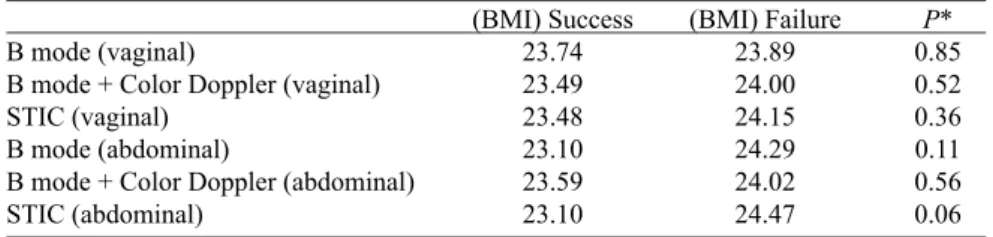
BMI did not influence the performance of the methods (B mode, color Doppler and STIC) when using
the vaginal approach, but in the abdominal approach,
although with no statistically significant differences, the findings suggest that it is more difficult for a satisfactory examination as BMI increases (Table 1). Regarding the CRL, there was greater failure rate in assessing through vaginal approach when using color Doppler and B modes (P<0.01) (Table 2).
Table 2. Review of signiicant difference between the average crown-rump length and the success or not of the method by vaginal/abdominal approaches
B mode (vaginal)
B mode + Color Doppler (vaginal) STIC (vaginal)
B mode (abdominal)
B mode + Color Doppler (abdominal) STIC (abdominal)
(CRL) Success
69.78 66.78 69.32 73.51 73.95 71.73
(CRL) Failure
72.24 73.54 73.30 70.28 68.89 71.31
P*
0.34 <0.01
0.13 0.19 0.09 0.86
* unpaired t test; STIC: spatio-temporal image correlation; CRL: crown-rump length Table 1. Review of signiicant difference between the mean body mass index and the success
or failure of the method by vaginal/abdominal approaches
B mode (vaginal)
B mode + Color Doppler (vaginal) STIC (vaginal)
B mode (abdominal)
B mode + Color Doppler (abdominal) STIC (abdominal)
(BMI) Success
23.74 23.49 23.48 23.10 23.59 23.10
(BMI) Failure
23.89 24.00 24.15 24.29 24.02 24.47
P*
0.85 0.52 0.36 0.11 0.56 0.06
DISCUSSION
In this study we assessed the inluence of BMI and CRL in evaluating standardized cardiac planes between 12-14 weeks of gestation, through abdominal and vaginal approaches,
through B mode, Color Doppler and spatio-temporal image
correlation. To our knowledge, there are no studies in the literature with similar methodology.
Obstetric oltrasonography of obese women is dificult and in some situations can become a real challenge for physicians. In a study comparing obese women (BMI ≥ 30kg/m2) versus non-obese pregnant women (BMI < 30kg/
m2) who underwent secondtrimester ultrasound, it was found
that suboptimal visualization rates increased signiicantly in obese group, both for the heart (37% versus 19%) and fetal column (43% vs. 29%) [ 8].
In the irst trimester, only one study assessede the inluence of BMI on fetal cardiac evaluation [7]. In this study, 103 pregnant women between 11 and 13 weeks and 6 days were examined, through abdominal approach using B mode and, in some cases, associated with color Doppler. They did not found no inluence of BMI in cardiac assessment (P=0.752) [7]. Similarly, in our study, assessing 54 pregnant
women between 12 and 14 weeks, there was also no inluence of BMI. One possible explanation would be the fact that at the end of the irst trimester, most of the women maintains prepregnancy BMI, in addition, the mean BMI of our group was 23.8 kg/m2, and no pregnant woman had a BMI≥ 30kg/
m2. Other studies have reported the inluence of maternal
BMI as a visualization of the fetal heart in the irst trimester of pregnancy [9-11]. Even the guideline of the American Institute of Ultrasound in Medicine (AIUM) for fetal cardiac evaluation refers to a technical limitation in the case of obese patients, due to acoustic shadows in the third trimester. They reported the need for assessments at different times, in addition to optimization of the device and focus adjustment, frequency, gain, magniication, temporal and harmonic [12] resolution. In relation to the inluence of CRL, we observed failure rate statistically signiicant only for the group assessed by vaginal approach using color Doppler and B mode (P<0.01). In a
previous study of the screening in the irst trimester, the CRL showed a factor of inluence on quality of nuchal translucency measurement [13]. Regarding fetal cardiac evaluation, there is only one study that assessed the inluence of the CRL [7].
In this study, the authors observed no inluence of CRL in fetal cardiac assessment (P=0.899), with a mean of CRL of
72.1 mm [7], however, it was performed only the assessment by laparotomy. In our study, the mean CRL was 71.5 mm and we used the abdominal and vaginal approaches in all cases. The higher failure rate in vaginal approach is due to the need for prior learning curve, plus the need for experienced
examiners to obtain the standardized cardiac plans. In a study
by Vimpeli et al. [ 14 ], who assessed 584 fetuses with CRL
between 41 and 78 mm, the rate of success in getting all cardiac plans was only 58%.
As limitation of the study, we noted that all pregnant women were randomly selected, so that the results were adversely affected due to the absence of women with BMI ≥ 30kg/ m2. Maybe if we had selected patients knowingly
obese or with certain diseases prior to pregnancy such as diabetes mellitus we could infer the real impact of maternal BMI in assessing the quality of fetal cardiac examination in the irst trimester of pregnancy.
CONCLUSION
Concluding, we found no inluence of the index of body mass and crown-rump length in fetal cardiac assessment between 12-14 weeks gestation. The assessment through
vaginal approach needs more prior training, and experienced
examiners in this pathway. Subsequent studies using populations of previously obese women are needed to prove the real inluence of body mass index in fetal cardiac assessment in the irst trimester of pregnancy.
Authors' roles & responsibilities
DBSP Main coordinatination AIFL Data collection
EAJ Preparation of the article for publication LMMN Adjunct coordenation
WPM Statistical analysis
AFM Final review
REFERENCES
1. Sharland G. Routine fetal cardiac screening: what are we doing and
what should we do? Prenat Diagn. 2004;24(13):1123-9.
2. Eleftheriades M, Tsapakis E, Sotiriadis A, Manolakos E, Hassiakos D, Botsis D. Detection of congenital heart defects throughout pregnancy: impact of first trimester ultrasound screening for cardiac abnormalities. J Matern Fetal Neonatal Med. 2012;25(12):2546-50.
4. Viñals F, Ascenzo R, Naveas R, Huggon I, Giuliano A. Fetal
echocardiography at 11 + 0 to 13 + 6 weeks using
four-dimensional spatiotemporal image correlation telemedicine
via an Internet link: a pilot study. Ultrasound Obstet Gynecol. 2008;31(6):633-8.
5. Novotná M, Hašlík L, Svabík K, Zizka Z, Belošovičová H, Břešťák M, et al. Detection of fetal major structural anomalies at the 11-14 ultrasound scan in an unselected population. Ceska Gynekol. 2012;77(4):330-5.
6. Dias T, Arcangeli T, Bhide A, Napolitano R, Mahsud-Dornan S,
Thilaganathan B. First-trimester ultrasound determination of chorionicity in twin pregnancy. Ultrasound Obstet Gynecol. 2011;38(5):530-2.
7. Abu-Rustum RS, Ziade MF, Abu-Rustum SE. Learning curve and factors influencing the feasibility of performing fetal echocardiography at the time of the first-trimester scan. J Ultrasound Med. 2011;30(5):695-700.
8. Hendler I, Blackwelll SC, Bujold E, Treadwelll MC,Wolfe HM, Sokoll RJ, Sorokin J. The impact of maternal obesity on midtrimester sonographic visualization of fetal cardiac
and craniospinal structures. Int J Obes Relat Metab Disord.
2004;28(12):1607-11.
9. Cook AC, Yates RW, Anderson RH. Normal and abnormal fetal cardiac anatomy. Prenat Diagn. 2004;24(13):1032-48.
10. Simpson J. Echocardiographic evaluation of cardiac function in the fetus. Prenat Diagn. 2004;24(13):1081-91.
11. Lohr PA, Reeves MF, Creinin MD. A comparison of transabdominal and transvaginal ultrasonography for determination of gestational age and clinical outcomes in women undergoing early medical abortion. Contraception. 2010;81(3):240-4.
12. Fetal Echocardiography Task Force; American Institute
of Ultrasound in Medicine Clinical Standards Committee;
American College of Obstetricians and Gynecologists; Society
for Maternal-Fetal Medicine. AIUM practice guideline for the performance of fetal echocardiography. J Ultrasound Med. 2011;30(1):127-36.
13. Zohav E, Dunsky A, Segal O, Peled R, Herman A, Segal S.
The effects of maternal and fetal parameters on the quality of
nuchal translucency measurement. Ultrasound Obstet Gynecol.
2001;18(6):638-40.
14. Vimpelli T, Huhtala H, Acharya G. Fetal echocardiography