FXN
Promoter Silencing in the Humanized
Mouse Model of Friedreich Ataxia
Yogesh K. Chutake1, Whitney N. Costello1, Christina C. Lam1, Aniruddha C. Parikh1, Tamara T. Hughes1, Michael G. Michalopulos1, Mark A. Pook3, Sanjay I. Bidichandani1,2*
1Department of Pediatrics, University of Oklahoma College of Medicine, Oklahoma City, OK 73104, United States of America,2Department of Biochemistry & Molecular Biology, University of Oklahoma College of Medicine, Oklahoma City, OK 73104, United States of America,3Ataxia Research Group, Division of Biosciences, Department of Life Sciences, College of Health & Life Sciences, Brunel University London, Uxbridge, UB8 3PH, United Kingdom
*Sanjay-Bidichandani@ouhsc.edu
Abstract
Background
Friedreich ataxia is caused by an expanded GAA triplet-repeat sequence in intron 1 of the
FXNgene that results in epigenetic silencing of theFXNpromoter. This silencing mecha-nism is seen in patient-derived lymphoblastoid cells but it remains unknown if it is a wide-spread phenomenon affecting multiple cell types and tissues.
Methodology / Principal Findings
The humanized mouse model of Friedreich ataxia (YG8sR), which carries a single trans-genic insert of the humanFXNgene with an expanded GAA triplet-repeat in intron 1, is deficient forFXNtranscript when compared to an isogenic transgenic mouse lacking the expanded repeat (Y47R). We found that in YG8sR the deficiency ofFXNtranscript extended both upstream and downstream of the expanded GAA triplet-repeat, suggestive of deficient transcriptional initiation. This pattern of deficiency was seen in all tissues tested, irrespective of whether they are known to be affected or spared in disease pathogenesis, in both neuronal and non-neuronal tissues, and in cultured primary fibroblasts.FXNpromoter function was directly measured via metabolic labeling of newly synthesized transcripts in fibroblasts, which revealed that the YG8sR mouse was significantly deficient in transcrip-tional initiation compared to the Y47R mouse.
Conclusions / Significance
Deficient transcriptional initiation accounts forFXNtranscriptional deficiency in the human-ized mouse model of Friedreich ataxia, similar to patient-derived cells, and the mechanism underlying promoter silencing in Friedreich ataxia is widespread across multiple cell types and tissues.
OPEN ACCESS
Citation:Chutake YK, Costello WN, Lam CC, Parikh AC, Hughes TT, Michalopulos MG, et al. (2015)FXN Promoter Silencing in the Humanized Mouse Model of Friedreich Ataxia. PLoS ONE 10(9): e0138437. doi:10.1371/journal.pone.0138437
Editor:Annalisa Pastore, National Institute for Medical Research, Medical Research Council, London, UNITED KINGDOM
Received:June 30, 2015
Accepted:August 31, 2015
Published:September 22, 2015
Copyright:© 2015 Chutake et al. This is an open access article distributed under the terms of the Creative Commons Attribution License, which permits unrestricted use, distribution, and reproduction in any medium, provided the original author and source are credited.
Data Availability Statement:All relevant data are within the paper and its Supporting Information files.
Introduction
Friedreich ataxia (FRDA) is the most common inherited ataxia and it is characterized clinically by sensory ataxia, cardiomyopathy, and a predisposition to diabetes [1]. The disease is progres-sive, and there is currently no effective therapy to slow the deterioration. FRDA is inherited as an autosomal recessive condition, and the vast majority of patients are homozygous for an
abnormally expanded GAA triplet-repeat (GAA-TR) sequence in intron 1 of theFXNgene [2].
Non-FRDA alleles contain<30 triplets, while disease-causing expanded alleles typically
con-tain 100–1300 triplets. Cells and tissues from patients who are homozygous for the expanded GAA-TR sequence have a severe deficiency ofFXNtranscript [3]. This produces a deficiency of frataxin, a mitochondrial protein that plays an important role in Fe-S cluster biogenesis [4,5], ultimately leading to pathological changes in susceptible tissues such as dorsal root gan-glia, myocardium, and the cerebellar dentate nucleus [6]. A precise delineation of the mecha-nism(s) by which the expanded GAA-TR sequence results in transcriptional deficiency will be crucial for the development of rationally designed therapies for FRDA.
The expanded GAA-TR sequence is thought to lead to deficiency ofFXNtranscript by more
than one molecular mechanism. Abnormal secondary DNA structures and repeat-proximal heterochromatin, both mediated by the expanded GAA-TR sequence, result in impedance of transcriptional elongation through intron 1 of theFXNgene [3,7–9]. However, the predomi-nant mechanism of transcriptional deficiency in FRDA seems to be via epigenetic silencing of theFXNgene promoter [9,10]. This mechanism of silencing is reliant on the spread of repres-sive chromatin from the expanded GAA-TR sequence in intron 1 [9,10,11–13], which is a
known source of heterochromatin [14]. This spread encompasses theFXNpromoter, renders it
transcriptionally non-permissive, and thereby causes a severe deficiency of transcriptional ini-tiation [10]. Indeed, the length of the expanded GAA-TR sequence correlates well with the severity ofFXNpromoter silencing [15], further substantiating the etiological relationship
between epigenetic silencing of theFXNpromoter and the expanded GAA-TR mutation in
intron 1. This mechanism of gene silencing, while compelling, has so far only been demon-strated in patient-derived lymphoblastoid cells [10,15]. It remains unknown if repeat-mediated
epigenetic promoter silencing is an important underlying mechanism forFXNtranscriptional
deficiency in multiple cell types and tissues, and thus its pathophysiological significance in FRDA remains unclear.
The YG8sR humanized mouse model of FRDA, which contains the entire humanFXNgene
with an expanded GAA-TR mutation in a murineFxn-/-background, is known to mimic the
transcriptional deficiency seen in FRDA patients [16]. Admittedly, the YG8sR mouse has a mild, variable and very late-onset phenotype. However, the deficiency ofFXNtranscript is clearly seen across multiple tissues, making it an adequate model to study the mechanism of
FXNtranscriptional deficiency in the context of the humanFXNgene. Indeed, the mild and
late-onset phenotype in the YG8sR mouse has the advantage that tissues isolated from young YG8sR mice would allow analysis prior to the onset of disease-associated pathology and thus “affected”cell types would likely be well represented in the tissue samples. The Y47R mouse
[16], which has the same genomic makeup as YG8sR except that it lacks the expanded
GAA-TR mutation and does not exhibit any FRDA-related phenotype, serves as a useful
“non-FRDA”control.
We provide evidence that deficient transcriptional initiation accounts for the transcriptional deficiency in the YG8sR mouse model of FRDA, similar to patient-derived cells. Thus, the mechanism underlying promoter silencing in Friedreich ataxia, originally discovered in patient-derived lymphoblastoid cells, is likely widespread across multiple cell types and tissues. respectively. The funders had no role in study design,
data collection and analysis, decision to publish, or preparation of the manuscript.
Materials and Methods
Mouse tissues and cell lines
YG8sR and Y47R mice were bred, sacrificed and autopsied at 1 and 12 months of age at Brunel University London (U.K.) under humane conditions in accordance with the U.K. Home Office “Animals (Scientific Procedures) Act 1986”and with approval from the Brunel University Lon-don Animals Welfare and Ethical Review Board. Frozen tissue samples were sent on dry ice by overnight courier service to the University of Oklahoma Health Sciences Center for further analysis. DNA and RNA from tissues and cell lines were extracted using the DNeasy Blood and Tissue kit (Qiagen) and the RNeasy kit (Qiagen), respectively. The length of the GAA-TR sequence was analyzed by PCR amplification using previously described primers [17], and by direct sequencing of purified PCR products in both directions. Fibroblast cell lines from YG8sR and Y47R mice, established and characterized as described previously [16], were main-tained in culture in DMEM supplemented with 10% FBS and penicillin and streptomycin.
Quantitative measurement of
FXN
transcript
This was done by reverse transcription followed by quantitative polymerase chain reaction (quantitative RT-PCR), using protocols and primer sequences as previously described [10]. Briefly, reverse transcription was performed either with a mixture of random hexamers and
oligo dT primers (for the Exon 3–4 amplicon;“Ex3-Ex4”) or with a strand-specific RT primer
(for the Exon 1 amplicon;“Ex1”) using the QuantiTect1reverse transcription kit (Qiagen). Transcript levels were quantified by real-time PCR and normalized relative to expression of the
controlTbpgene (forward primer: 5’-CCTTGTACCCTTCACCAATGAC-3’, reverse primer:
5’-ACAGCCAAGATTCACGGTAGA-3’) for experiments using fibroblasts, and normalized
relative to the geometric mean of the control genesPsmd4(forward primer: 5’-TGGAGAGCA
CTATGGTTTGTGT-3’; reverse primer: 5’-ACGTTATTCTCAGGGTTGCTTC-3’) andEef2
(forward primer: 5’-TGTCAGTCATCGCCCATGTG-3’; reverse primer: 5’-CATCCTTGCGA
GTGTCAGTGA-3’) [18] for mouse tissues, using theΔΔCt method on the LightCycler196
Real-Time PCR system (Roche Diagnostics Corp) with SsoAdvanced™Universal SYBR1Green
Supermix (BioRad) orPowerSYBR1Green PCR Master Mix (Life Technologies).
Methylation sensitive
—
high resolution melting (MS-HRM) assay to test
for CpG DNA methylation
The MS-HRM assay [19] is based on differential melting of PCR products containing different amounts of C’s and T’s following bisulfite treatment of DNA that contains differentially meth-ylated CpG sites. DNA is amplified in the presence of a DNA binding saturating fluorescent dye, e.g. ResoLight. As the temperature of the PCR products is raised the DNA begins to melt. In bisulfite treated DNA with high endogenous CpG methylation, the C’s that remain uncon-verted requires a longer time & higher temperature to melt compared with those with lower CpG methylation. Primers for the MS-HRM assay were designed to amplify the bisulfite con-verted DNA template. Accordingly, primers were designed to amplify a 90 bp region of interest that includes CpG-393, CpG-381 and CpG-358 (forward primer: 5’-CCAAAAAAATAATAA
AAAATATTTC-3’; reverse primer: 5’-ATTAGTTATTTTGAAGGGATTTTTTT-3’) [seeFig
1A]. Since depurination during bisulfite treatment damages the DNA template, longer than
repeats, and genomic DNA from mouse-derived fibroblasts and multiple tissues from
1-month-old and 12-month old Y47R and YG8sR mice were analyzed. Purified DNA was soni-cated using the EpiShear™sonicator (Active Motif) to an average size of ~1000 bp and treated with bisulfite at 50°C for at least 5 hours using the Bisulfite Converstion Kit (Active Motif) according to the manufacturer’s protocol. MS-HRM PCR was performed using 10 ng of
bisul-fite treated DNA with AmpliTaq Gold1DNA polymerase (Life Technologies) supplemented
with 1x LightCycler1480 ResoLight dye (Roche Diagnostics Corp). Reference templates, to
simulate 100% and 0% CpG methylation at the three sites, were generated by PCR using the same primers but starting with single-stranded long synthetic oligonucleotides either contain-ing all three CpG sites intact (100% methylated) or all three CpG sites converted to TpG (0% methylated). Differential DNA melting was monitored by generating normalized melting
curves on the LightCycler196 Real-Time PCR system (Roche Diagnostics Corp) by measuring
fluorescence 15 times per°C change between 65°C and 97°C, using algorithms supplied by the manufacturer.
Quantitative measurement of
FXN
promoter activity
This was done by metabolic labeling of nascent RNA using the Click-iT1Nascent RNA
Cap-ture Kit (Life Technologies) as previously described [10]. Briefly, fibroblast cell lines were incu-bated with 5-ethynyl uridine. Following 1, 2 and 4 hours of incubation, RNA was extracted and used for biotinylation by Click reaction. Biotinylated RNA bound to streptavidin beads was reverse transcribed using the SuperScript1VILO™cDNA synthesis kit (Life Technologies). Nascent transcript levels were quantified by real-time PCR relative to expression of the control
Tbpgene from total biotinylated RNA using theΔΔCt method on the LightCycler196
Real-Time PCR system (Roche Diagnostics Corp) with SsoAdvanced™Universal SYBR1Green
Supermix (BioRad) orPowerSYBR1Green PCR Master Mix (Life Technologies).
Results
FXN
transcriptional deficiency in the YG8sR mouse extends upstream
and downstream of the expanded GAA-TR mutation
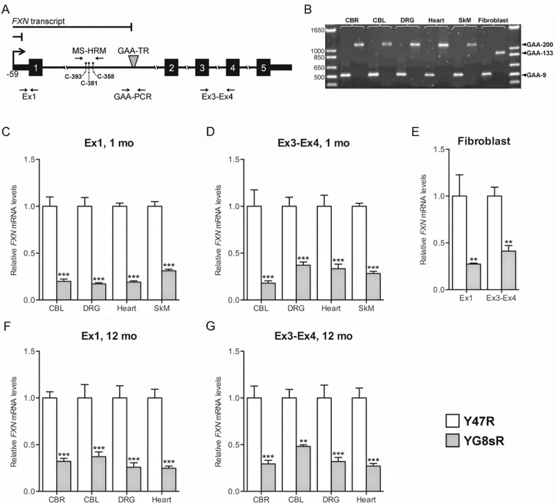
Multiple tissues (cerebrum, cerebellum, dorsal root ganglia [DRG], heart, and skeletal muscle)
and fibroblast cell lines derived from YG8sR and Y47R mice were selected for analysis ofFXN
transcript levels both upstream and downstream of the expanded GAA-TR mutation in order to differentiate between defects in transcriptional initiation and transcriptional elongation through the expanded repeat. Deficiency of transcript at both locations would suggest a defect in transcriptional initiation, but deficiency at only the downstream location would suggest a defect in transcriptional elongation (Fig 1A). The tissues and cell lines were selected to repre-sent various characteristics, including those that are known to be affected versus unaffected in FRDA, neuronal versus non-neuronal and proliferative versus post-mitotic. All tissues and cell lines from the transgenic mouse lines contained a single GAA-TR length; Y47R tissues and fibroblasts contained 9 triplets, YG8sR tissues contained ~200 triplets, and YG8sR fibroblasts contained 133 GAA triplets (the latter was confirmed by sequencing to be a pure GAA triplet-repeat [16];Fig 1B).
sequence.(F, G)Quantitative RT-PCR showing deficiency ofFXNtranscript in 12-month-old YG8sR mouse tissues compared to Y47R, both upstream (Ex1) and downstream (Ex3-Ex4) of the expanded GAA-TR sequence. CBR = cerebrum; CBL = cerebellum; DRG = dorsal root ganglia; SkM = skeletal muscle. Data shown in panels C through G represent three complete experiments using tissues isolated from two YG8sR and two Y47R individuals. Error bars represent +/-SEM.**=p<0.01,***=p<0.001.
For the upstream location, quantitative RT-PCR was performed in the immediate vicinity of the transcription start site of theFXNgene (“Ex1”), and the spliced product of exons 3 & 4 (“Ex3-Ex4”) was selected as the downstream location (Fig 1A). We found significant deficiency
ofFXNtranscript in YG8sR compared with Y47R at both the upstream and downstream
loca-tions in all tissues and cell lines tested (Fig 1C–1G), supporting the existence of a defect in tran-scriptional initiation, as was previously noted in FRDA patient-derived lymphoblastoid cell lines [10,15]. We initially focused our analysis on 1-month-old mouse tissues (as shown inFig 1C and 1D) in order to preempt any phenotypic manifestations in the YG8sR mouse and thus potentially representing all cell types including those that would be lost due to late-onset FRDA-associated pathology. However, we subsequently observed a similar deficiency in steady-stateFXNtranscript levels in 12-month-old YG8sR mouse tissues (Fig 1F and 1G) as we had seen in 1-month-old mouse tissues, suggesting that the mechanism of transcriptional deficiency persists throughout life. Overall, these data suggest that transcriptional deficiency in the YG8sR mouse model likely stems from deficient transcriptional initiation.
Increased DNA methylation at the
FXN
locus in the YG8sR mouse
Among the FRDA-specific epigenetic changes at theFXNlocus that are associated with the
expanded GAA-TR mutation is increased DNA methylation at CpG sites in the vicinity of the repeat in intron 1. The level of DNA methylation at CpG-393, CpG-381 and CpG-358 (num-bering refers to the nucleotide position of the“C”with respect to the first“G”in the GAA-TR
sequence; seeFig 1A) is known to be increased in FRDA patients who are homozygous for the
expanded GAA-TR mutation [20–22]. Indeed, methylation at CpG-381 is known to correlate
with repeat length and age of disease onset [21], and methylation at CpG-358 correlates with
FXNtranscript levels, age of onset and the FARS clinical rating scale [22]. Given the correlation of CpG methylation at these sites with phenotypically-relevant features in FRDA, we investi-gated if YG8sR tissues and fibroblasts also showed increased CpG methylation at these sites, indicative of relevant expression-related epigenetic changes at theFXNlocus despite the rela-tively short expanded GAA-TR mutation.
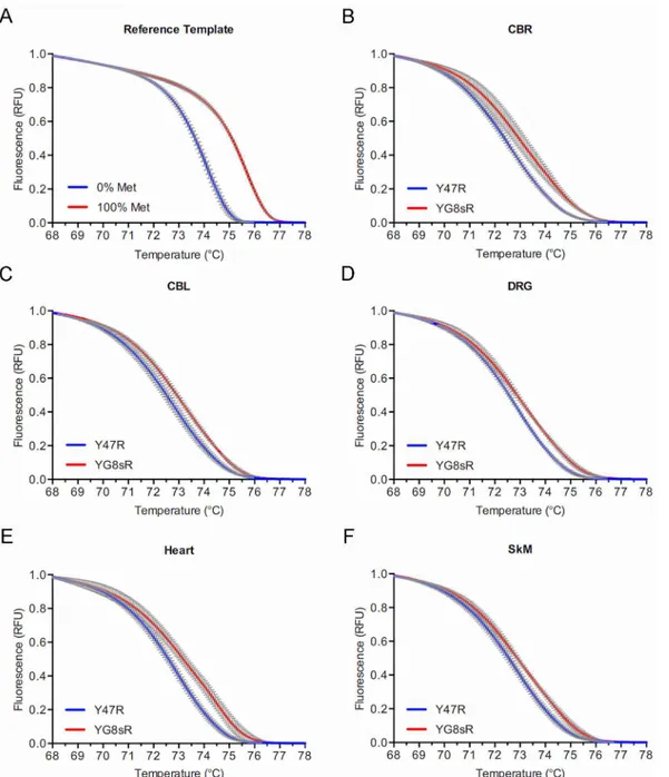
A suitable amplicon was designed to span CpG–393, CpG-381, and CpG–358 (Fig 1A). A
semi-quantitative assay involving bisulfite conversion followed by high resolution melting (methylation sensitive-high resolution melting [MS-HRM] assay [19]) was developed to assess
for increased CpG methylation in YG8sR compared to Y47R (seeMaterials and Methods).
Two positive controls were used to determine the discriminatory capacity of the MS-HRM assay. Firstly, two separate double-stranded reference templates simulating 100% methylation (i.e., C’s at the three CpG sites) and 0% methylation (i.e., T’s at the three CpG sites) were directly used for HRM analysis (i.e., without bisulfite conversion). This showed a clear separa-tion of melting curves (Fig 2A), indicating that the HRM part of the assay was capable of detecting complete methylation at the three CpG sites. Next, we tested DNA from lymphoblas-toid cell lines of FRDA patients homozygous for long GAA-TR mutations (i.e., with both expanded alleles containing>400 triplets), which are known to have increased CpG
methyla-tion compared to non-FRDA controls. This also showed a clear separamethyla-tion of the high resolu-tion melting curves due to increased DNA methylaresolu-tion in FRDA versus non-FRDA cell lines, indicating that the MS-HRM assay was capable of detecting even relative increases in
methyla-tion at the three CpG sites contained within the amplicon (Fig 2B). The MS-HRM assay was
individuals (S1andS2Figs), and two 12-month-old YG8sR and Y47R individuals (Fig 3and
S3 Fig), suggesting that this epigenetic change persists throughout life. It should be noted that while this assay simultaneously measures DNA methylation at the three CpG sites in a large number of cells (as opposed to typical assays that involve sequencing of a limited number of cloned templates), it lacks the ability to detect relative methylation levels at the three individual CpG sites contained within the amplicon. These data indicate there are repressive epigenetic changes in the form of increased DNA methylation at phenotypically-relevant CpG sites located upstream of the expanded GAA-TR in intron 1 in multiple tissues and fibroblasts from YG8sR versus Y47R.
Deficiency of
FXN
transcriptional initiation in the YG8sR mouse
The combination of repressive epigenetic changes and transcriptional deficiency extending upstream of the expanded GAA-TR mutation in tissues and fibroblasts from the YG8sR mouse model suggested that there was a deficiency of transcriptional initiation similar to the epige-netic promoter silencing in lymphoblastoid cells from FRDA patients [10,15]. To directly test iftheFXNpromoter is rendered less active in YG8sR,FXNtranscriptional initiation was
mea-sured via metabolic labeling of nascent transcripts in fibroblast cell lines derived from YG8sR and Y47R mice. Nascent transcripts were labeled with ethynyl uridine, to which biotin was sub-sequently added via click chemistry, thus permitting a quantitative, dynamic,in vivoanalysis of
newly synthesizedFXNtranscript. Quantitative RT-PCR was performed to measureFXN
tran-script levels both upstream (“Ex1”which maps immediately downstream of theFXN
transcrip-tion start site [FXN-TSS;Fig 1A]) and downstream (“Ex3-Ex4”[Fig 1A]) of the expanded GAA-TR mutation following 1, 2, and 4 hours of labeling. This revealed a significant, 2.0 to 3.4-fold deficiency of newly synthesizedFXNtranscript in YG8sR at both locations (Fig 4A &
4B). The deficiency of dynamic accumulation ofFXNtranscript levels observed immediately
downstream ofFXN-TSS (and upstream of the expanded GAA-TR mutation) indicates that
the YG8sR mouse is deficient in transcriptional initiation. Moreover, the fold-difference in
accumulation ofFXNtranscript at the various time points in YG8sR versus Y47R were similar
at both the upstream and downstream locations (Fig 4A & 4B), suggesting that deficient tran-scriptional initiation likely accounts for most of the trantran-scriptional deficiency in the YG8sR
mouse. Thus transcriptional deficiency in the YG8sR mouse model is largely due toFXN
pro-moter silencing, which leads to deficiency of transcriptional initiation, similar to lymphoblas-toid cell lines from FRDA patients.
Discussion
Our results indicate thatFXNtranscriptional deficiency in the YG8sR humanized mouse
model of FRDA is caused by deficient transcriptional initiation as a result of promoter silenc-ing. While this mechanism has previously been noted in patient-derived lymphoblastoid cell
Fig 2. Increased DNA methylation at theFXNlocus in the 1-month-old YG8sR mouse. (A)Normalized melting curves in a high resolution melting (HRM) assay of two reference double-stranded templates simulating 100% (red curve) and 0% (blue curve) DNA methylation at three CpG sites upstream of the GAA-TR mutation (seeFig 1A) showing a clear separation of the curves indicating that the HRM assay is able to detect methylation at the three CpG sites. (B)Normalized melting curves in a methylation sensitive—high resolution melting (MS-HRM) assay to detect CpG methylation in lymphoblastoid cell lines from three FRDA (red curve) and three non-FRDA control subjects (blue curve) at the three CpG sites upstream of the GAA-TR mutation (seeFig 1A) showing a clear separation of the curves indicating that the MS-HRM assay is able to detect a relative increase in methylation at the three CpG sites.(C-H) Normalized melting curves in a MS-HRM assay to detect CpG methylation in fibroblast cell lines and multiple tissues from 1-month-old YG8sR (red curves) and Y47R (blue curves) mice at the three CpG sites upstream of the GAA-TR mutation (seeFig 1A) showing a clear separation of the curves indicating a relative increase in methylation at the three CpG sites in YG8sR tissues and fibroblasts. For all HRM curves, X-axis = melting temperature, Y-axis = relative fluorescence, and error bars represent 95% confidence intervals at each of 15 points assayed in triplicate for fluorescence per°C change.
LBCLs = lymphoblastoid cell lines; CBR = cerebrum; CBL = cerebellum; DRG = dorsal root ganglia; SkM = skeletal muscle.
lines [10,15], our present data provide supportive evidence for the existence of this mechanism of transcriptional deficiency in fibroblasts and in multiple tissues. Our data also suggest that
the mechanism underlyingFXNtranscriptional deficiency in FRDA is unlikely to be
tissue-specific.
Fig 3. Increased DNA methylation at theFXNlocus in the 12-month-old YG8sR mouse. (A)Normalized melting curves in a high resolution melting (HRM) assay of two reference double-stranded templates simulating 100% (red curve) and 0% (blue curve) DNA methylation at three CpG sites upstream of the GAA-TR mutation (seeFig 1A) showing a clear separation of the curves indicating that the HRM assay is able to detect methylation at the three CpG sites.(B-F)Normalized melting curves in a MS-HRM assay to detect CpG methylation in multiple tissues from 12-month-old YG8sR (red curves) and Y47R (blue curves) mice at the three CpG sites upstream of the GAA-TR mutation (seeFig 1A) showing a clear separation of the curves indicating a relative increase in methylation at the three CpG sites in YG8sR tissues. For all HRM curves, X-axis = melting temperature, Y-axis = relative fluorescence, and error bars represent 95% confidence intervals at each of 15 points assayed in triplicate for fluorescence per°C change. CBR = cerebrum; CBL = cerebellum; DRG = dorsal root ganglia; SkM = skeletal muscle.
It is noteworthy that tissues from the YG8sR mouse have a GAA-TR length of ~200 triplets and the fibroblast cell line contains only 133 triplets [16]. In FRDA patients, these shorter than average repeat lengths would be expected to result in a later age of onset [23] and a slowly pro-gressive clinical phenotype [24,25]. It is therefore not surprising that the YG8sR mouse has a phenotype that is mild, variable, and of late onset [16]. Indeed, the 2- to 3-fold reduction in promoter activity in the YG8sR mouse is comparable to the magnitude of deficiency of tran-scriptional initiation seen in cell lines from FRDA patients who have at least one short GAA-TR allele (containing<400 GAA triplets) [15]. Therefore, a humanized mouse model
based on YG8sR but containing>400 GAA triplets would likely result in more severe
pro-moter silencing and possibly lead to a more discernable FRDA-related phenotype.
Our data indicate that the YG8sR humanized mouse is a reasonable model for investigating the molecular mechanism(s) underlying repeat-mediated promoter silencing in FRDA. The YG8sR mouse model would also be useful for testing drugs that are designed to reverse the transcriptional initiation defect caused by promoter silencing in FRDA, such as the 2-amino-benzamide derived histone deacetylase inhibitors [11,26–28].
Supporting Information
S1 Fig. Increased DNA methylation at theFXNlocus in 1-month-old YG8sR mouse tissues.
(PDF)
S2 Fig. Increased DNA methylation at theFXNlocus in 1-month-old YG8sR mouse tissues.
(PDF)
S3 Fig. Increased DNA methylation at theFXNlocus in 12-month-old YG8sR mouse
tis-sues. (PDF)
Fig 4. Metabolic labeling of nascentFXNtranscript in primary fibroblasts showing deficiency of transcriptional initiation in the YG8sR mouse. (A, B)Quantitative RT-PCR of metabolically labeled nascent transcript for the indicated incubation times (1, 2 and 4 hours) is shown forFXNmRNA upstream (“Ex1”inFig 1A) and downstream (“Ex3-Ex4”inFig 1A) of the GAA-TR sequence in intron 1. YG8sR cells showed 2.0–3.4 fold less nascentFXNtranscript (exact fold changes are indicated) compared with Y47R cells at all the time points assayed. Graphs represent cumulative data from four independent metabolic labeling experiments. Error bars represent +/-SEM.*=p<0.05;**=p<0.01,***=p<0.001.
Acknowledgments
This research was supported by grants from the National Institutes of Health (R01 NS072418), and the Muscular Dystrophy Association to S.I.B. Y.K.C. is supported by a postdoctoral research fellowship from the Million Dollar Bike Ride Grant Program of the Orphan Disease Center at University of Pennsylvania. T.T.H. was supported by the American College of Medi-cal Genetics Foundation. A.C.P. and M.G.M. were supported by the SURE and OSCTR pro-grams at OUHSC, respectively.
Author Contributions
Conceived and designed the experiments: YKC SIB. Performed the experiments: YKC WNC CCL ACP TTH MGM. Analyzed the data: YKC SIB. Contributed reagents/materials/analysis tools: SIB MAP. Wrote the paper: YKC SIB.
References
1. Bidichandani SI, Delatycki MB. Friedreich Ataxia. In: Pagon RA, Adam MP, Ardinger HH, Wallace SE, Amemiya A, Bean LJH, et al., editors. GeneReviews(R). Seattle (WA)1993 [updated 2014].
2. Campuzano V, Montermini L, Molto MD, Pianese L, Cossee M, Cavalcanti F, et al. Friedreich's ataxia: autosomal recessive disease caused by an intronic GAA triplet repeat expansion. Science. 1996; 271 (5254):1423–7. PMID:8596916
3. Bidichandani SI, Ashizawa T, Patel PI. The GAA triplet-repeat expansion in Friedreich ataxia interferes with transcription and may be associated with an unusual DNA structure. Am J Hum Genet. 1998; 62 (1):111–21. PMID:9443873
4. Colin F, Martelli A, Clemancey M, Latour JM, Gambarelli S, Zeppieri L, et al. Mammalian frataxin con-trols sulfur production and iron entry during de novo Fe4S4 cluster assembly. J Am Chem Soc. 2013; 135(2):733–40. doi:10.1021/ja308736ePMID:23265191
5. Bridwell-Rabb J, Fox NG, Tsai CL, Winn AM, Barondeau DP. Human frataxin activates Fe-S cluster biosynthesis by facilitating sulfur transfer chemistry. Biochemistry. 2014; 53(30):4904–13. doi:10. 1021/bi500532ePMID:24971490
6. Koeppen AH, Mazurkiewicz JE. Friedreich ataxia: neuropathology revised. J Neuropathol Exp Neurol. 2013; 72(2):78–90. doi:10.1097/NEN.0b013e31827e5762PMID:23334592
7. Ohshima K, Montermini L, Wells RD, Pandolfo M. Inhibitory effects of expanded GAA.TTC triplet repeats from intron I of the Friedreich ataxia gene on transcription and replication in vivo. J Biol Chem. 1998; 273(23):14588–95. PMID:9603975
8. Sakamoto N, Ohshima K, Montermini L, Pandolfo M, Wells RD. Sticky DNA, a self-associated complex formed at long GAA*TTC repeats in intron 1 of the frataxin gene, inhibits transcription. J Biol Chem. 2001; 276(29):27171–7. PMID:11340071
9. Kumari D, Biacsi RE, Usdin K. Repeat expansion affects both transcription initiation and elongation in friedreich ataxia cells. J Biol Chem. 2011; 286(6):4209–15. doi:10.1074/jbc.M110.194035PMID: 21127046
10. Chutake YK, Costello WN, Lam C, Bidichandani SI. Altered Nucleosome Positioning at the Transcrip-tion Start Site and Deficient TranscripTranscrip-tional InitiaTranscrip-tion in Friedreich Ataxia. J Biol Chem. 2014; 289 (22):15194–202. doi:10.1074/jbc.M114.566414PMID:24737321
11. Herman D, Jenssen K, Burnett R, Soragni E, Perlman SL, Gottesfeld JM. Histone deacetylase inhibi-tors reverse gene silencing in Friedreich's ataxia. Nat Chem Biol. 2006; 2(10):551–8. PMID:16921367 12. Punga T, Buhler M. Long intronic GAA repeats causing Friedreich ataxia impede transcription
elonga-tion. EMBO molecular medicine. 2010; 2(4):120–9. doi:10.1002/emmm.201000064PMID:20373285 13. Kim E, Napierala M, Dent SY. Hyperexpansion of GAA repeats affects post-initiation steps of FXN
tran-scription in Friedreich's ataxia. Nucleic Acids Res. 2011; 39(19):8366–77. doi:10.1093/nar/gkr542 PMID:21745819
14. Saveliev A, Everett C, Sharpe T, Webster Z, Festenstein R. DNA triplet repeats mediate heterochroma-tin-protein-1-sensitive variegated gene silencing. Nature. 2003; 422(6934):909–13. PMID:12712207 15. Chutake YK, Lam C, Costello WN, Anderson M, Bidichandani SI. Epigenetic promoter silencing in
16. Anjomani Virmouni S, Ezzatizadeh V, Sandi C, Sandi M, Al-Mahdawi S, Chutake Y, et al. A novel GAA-repeat-expansion-based mouse model of Friedreich's ataxia. Dis Model Mech. 2015; 8(3):225–35. doi: 10.1242/dmm.018952PMID:25681319
17. Filla A, De Michele G, Cavalcanti F, Pianese L, Monticelli A, Campanella G, et al. The relationship between trinucleotide (GAA) repeat length and clinical features in Friedreich ataxia. Am J Hum Genet. 1996; 59(3):554–60. PMID:8751856
18. Kouadjo KE, Nishida Y, Cadrin-Girard JF, Yoshioka M, St-Amand J. Housekeeping and tissue-specific genes in mouse tissues. BMC Genomics. 2007; 8:127. PMID:17519037
19. Wojdacz TK, Dobrovic A, Hansen LL. Methylation-sensitive high-resolution melting. Nat Protoc. 2008; 3(12):1903–8. doi:10.1038/nprot.2008.191PMID:19180074
20. Greene E, Mahishi L, Entezam A, Kumari D, Usdin K. Repeat-induced epigenetic changes in intron 1 of the frataxin gene and its consequences in Friedreich ataxia. Nucleic Acids Res. 2007; 35(10):3383–90. PMID:17478498
21. Castaldo I, Pinelli M, Monticelli A, Acquaviva F, Giacchetti M, Filla A, et al. DNA methylation in intron 1 of the frataxin gene is related to GAA repeat length and age of onset in Friedreich ataxia patients. J Med Genet. 2008; 45(12):808–12. doi:10.1136/jmg.2008.058594PMID:18697824
22. Evans-Galea MV, Carrodus N, Rowley SM, Corben LA, Tai G, Saffery R, et al. FXN methylation pre-dicts expression and clinical outcome in Friedreich ataxia. Ann Neurol. 2012; 71(4):487–97. doi:10. 1002/ana.22671PMID:22522441
23. Durr A, Cossee M, Agid Y, Campuzano V, Mignard C, Penet C, et al. Clinical and genetic abnormalities in patients with Friedreich's ataxia. N Engl J Med. 1996; 335(16):1169–75. PMID:8815938
24. Regner SR, Wilcox NS, Friedman LS, Seyer LA, Schadt KA, Brigatti KW, et al. Friedreich ataxia clinical outcome measures: natural history evaluation in 410 participants. Journal of Child Neurology. 2012; 27 (9):1152–8. doi:10.1177/0883073812448462PMID:22752494
25. Metz G, Coppard N, Cooper JM, Delatycki MB, Durr A, Di Prospero NA, et al. Rating disease progres-sion of Friedreich's ataxia by the International Cooperative Ataxia Rating Scale: analysis of a 603-patient database. Brain. 2013; 136(Pt 1):259–68. doi:10.1093/brain/aws309PMID:23365101 26. Soragni E, Miao W, Iudicello M, Jacoby D, De Mercanti S, Clerico M, et al. Epigenetic therapy for
Frie-dreich ataxia. Ann Neurol. 2014; 76(4):489–508. doi:10.1002/ana.24260PMID:25159818
27. Sandi C, Pinto RM, Al-Mahdawi S, Ezzatizadeh V, Barnes G, Jones S, et al. Prolonged treatment with pimelic o-aminobenzamide HDAC inhibitors ameliorates the disease phenotype of a Friedreich ataxia mouse model. Neurobiol Dis. 2011; 42(3):496–505. doi:10.1016/j.nbd.2011.02.016PMID:21397024 28. Chutake YK, Lam CC, Costello WN, Anderson MP, Bidichandani SI. Histone Deacetylase Inhibitor