RESEARCH ARTICLE
Internal Tandem Duplication in FLT3
Attenuates Proliferation and Regulates
Resistance to the FLT3 Inhibitor AC220 by
Modulating p21
Cdkn1a
and Pbx1 in
Hematopoietic Cells
Mariko Abe1*, Louis M. Pelus2, Pratibha Singh2, Tomohiro Hirade1, Chie Onishi3, Jamiyan Purevsuren1, Takeshi Taketani1,4, Seiji Yamaguchi1, Seiji Fukuda1*
1Department of Pediatrics, Shimane University School of Medicine, Izumo, Shimane, Japan,2Department of Microbiology/Immunology, Indiana University School of Medicine, Indianapolis, Indiana, United States of America,3Department of Oncology/Hematology, Shimane University School of Medicine, Izumo, Shimane, Japan,4Division of Blood Transfusion, Shimane University Hospital, Izumo, Shimane, Japan
*[email protected](MA);[email protected](SF)
Abstract
Internal tandem duplication (ITD) mutations in theFms-related tyrosine kinase 3 (FLT3) gene (FLT3-ITD) are associated with poor prognosis in patients with acute myeloid leuke-mia (AML). Due to the development of drug resistance, few FLT3-ITD inhibitors are effective against FLT3-ITD+AML. In this study, we show that FLT3-ITD activates a novel pathway involving p21Cdkn1a(p21) and pre-B cell leukemia transcription factor 1 (Pbx1) that attenu-ates FLT3-ITD cell proliferation and is involved in the development of drug resistance. FLT3-ITD up-regulated p21 expression in both mouse bone marrow c-kit+-Sca-1+-Lin -(KSL) cells and Ba/F3 cells. The loss of p21 expression enhanced growth factor-indepen-dent proliferation and sensitivity to cytarabine as a consequence of concomitantly enriching the S+G2/M phase population and significantly increasing the expression of Pbx1, but not
Evi-1, in FLT3-ITD+cells. This enhanced cell proliferation following the loss of p21 was par-tially abrogated when Pbx1 expression was silenced in FLT3-ITD+primary bone marrow
colony-forming cells and Ba/F3 cells. When FLT3-ITD was antagonized with AC220, a
selective inhibitor of FLT3-ITD,p21expression was decreased coincident withPbx1mRNA
up-regulation and a rapid decline in the number of viable FLT3-ITD+Ba/F3 cells; however,
the cells eventually became refractory to AC220. Overexpressing p21 in FLT3-ITD+Ba/F3
cells delayed the emergence of cells that were refractory to AC220, whereas p21 silencing accelerated their development. These data indicate that FLT3-ITD is capable of inhibiting FLT3-ITD+cell proliferation through the p21/Pbx1 axis and that treatments that antagonize
ITD contribute to the subsequent development of cells that are refractory to a FLT3-ITD inhibitor by disrupting p21 expression.
a11111
OPEN ACCESS
Citation:Abe M, Pelus LM, Singh P, Hirade T, Onishi C, Purevsuren J, et al. (2016) Internal Tandem Duplication in FLT3 Attenuates Proliferation and Regulates Resistance to the FLT3 Inhibitor AC220 by Modulating p21Cdkn1aand Pbx1 in Hematopoietic Cells. PLoS ONE 11(7): e0158290. doi:10.1371/ journal.pone.0158290
Editor:Connie J Eaves, B.C. Cancer Agency, CANADA
Received:November 24, 2015
Accepted:June 13, 2016
Published:July 7, 2016
Copyright:© 2016 Abe et al. This is an open access article distributed under the terms of theCreative
Commons Attribution License, which permits
unrestricted use, distribution, and reproduction in any medium, provided the original author and source are credited.
Data Availability Statement:All relevant data are within the paper and its Supporting Information files. All microarray data are available from Gene Expression Omnibus (www.ncbi.nlm.nih.gov/geo: accession number GSE75200).
Introduction
The cyclin-dependent kinase inhibitor p21Cdkn1a(p21) was originally identified as a mediator
of p53-induced growth arrest [1,2] and regulates the proliferation of diverse cell types by
mod-ulating cell cycle, apoptosis and differentiation [3–5]. However, cells in which p53 is not
acti-vated still utilize p21-dependent cell cycle control [6–8]. P21 is highly expressed in
hematopoietic stem cells (HSCs) and maintains quiescence [8]. The loss of p21 in HSCs increases cell cycle progression but has only a marginal effect on marrow cellularity or periph-eral blood cell counts [8]. In contrast, p21 can facilitate the proliferation of normal hematopoi-etic progenitor cells (HPCs) ex vivo following stimulation with hematopoihematopoi-etic growth factors [9–12]. These findings suggest that p21 has a differentiation stage-specific function in the hematopoietic system. In addition to regulating the cell cycle [1,2,6,7,13], p21 can modulate the activity of a number of transcription factors and co-activators, such as Stat3 [13,14],
estro-gen receptorα[15], (Ccaat-enhancer-binding proteinα(C/EBPα) [16,17] and c-Myc [18],
sug-gesting that it may regulate cell fate by influencing gene expression [19].
Internal tandem duplication (ITD) mutations in theFms-related tyrosine kinase 3 (FLT3)
gene (FLT3-ITD) are found in 20–30% of patients with acute myeloid leukemia (AML) and are
associated with a poor prognosis [20,21]. Few FLT3-ITD inhibitors are effective against
FLT3-ITD+AML due to the emergence of drug-resistant cells [22,23]. For instance, AC220
(Quizartinib), a second-generation class III tyrosine kinase inhibitor (TKI), which has been used
to treat FLT3-ITD+AML in clinical trials, is a more potent and specific FLT3-ITD inhibitor
than other TKIs [22,24]; however, prolonged exposure to AC220 could generate FLT3-ITD+
cells that are resistant to AC220 [25]. The mechanisms responsible for drug resistance include
the acquisition of mutations in theFLT3gene, the activation of other pro-survival pathways and
microenvironment-mediated resistance [22,23]; however, additional mechanisms responsible
for the drug resistance of FLT3-ITD+AML cells remain to be investigated. Previous studies have
shown that FLT3-ITD enhances p21 expression through Stat5 [26], whereas the FLT3-ITD inhibitor SU5614 decreases p21 expression in Ba/F3 cells expressing FLT3-ITD [27]. P21 down-regulation by the FLT3-ITD inhibitor suggests that treatments that antagonize FLT3-ITD may
destroy p21 function and aberrantly affect FLT3-ITD+cell proliferation. However, the functional
role of p21 in FLT3-ITD signaling and FLT3-ITD-induced drug resistance remains unknown. In the present study, we identified a p21 signaling pathway downstream of FLT3-ITD that
negatively affects proliferation and is associated with drug resistance in FLT3-ITD+cells. An
analysis of the genes that are modulated by p21 deletion in FLT3-ITD-transformed HPCs revealed that p21 modulates the expression of pre-B cell leukemia transcription factor 1 (Pbx1), a proto-oncogene that critically regulates HSC and HPC function [28,29]. Silencing Pbx1 and p21 expression in FLT3-ITD-transformed HPCs revealed that the interaction
between FLT3-ITD-activated p21 and Pbx1 negatively regulated FLT3-ITD+HPC
prolifera-tion. In addition, the down-regulation of p21 resulting from FLT3-ITD inhibition by AC220
accelerated the emergence of FLT3-ITD+cells that were resistant to AC220. This study is the
first report to show how treatments targeting FLT3-ITD can lead to drug resistance.
Materials and Methods
Animals
Specific pathogen-free female C57BL/6 mice, 6–8 weeks of age, were purchased from CLEA
Japan, Inc. (Tokyo, Japan). P21-/-mice were kindly provided by Dr. H.E. Broxmeyer of the
Indiana University School of Medicine [9,10]. Survivinfx/fxmice and Tx-Cre Survivinfx/fxmice
have been described previously [30]. The IACUC of the Shimane University School of 20390298 to SF; Japan Society for the Promotion of
Science [https://www.jsps.go.jp/] grant number 25461593 to SF; Japan Leukemia Research Fund
[http://flrf.gr.jp/] to SF; Research Support Funds from
the Mochida Memorial Foundation for Medical and Pharmaceutical Research [http://www.mochida.co.jp/
zaidan/] to SF; Sankyo Biomedical Research
Foundation and the Mitsubishi Pharma Research Foundation [http://www.ds-fdn.or.jp/] to SF; AstraZeneca Research Grant [http://www.
astrazeneca.co.jp/] to SF; Naito Foundation [https://
www.naito-f.or.jp/jp/index.php] to SF. The funders had
no role in study design, data collection and analysis, decision to publish, or preparation of the manuscript.
Medicine (Permit Numbers IZ21-24, IZ21-25, and IZ21-26) and the Indiana University School of Medicine (Study Number 2939) approved all of the experimental procedures.
Antibodies and cytokines
Anti-FcγIII/II receptor antibody, allophycocyanin (APC)-conjugated mouse c-kit
body (clone 2B8), phycoerythrin (PE)-conjugated Annexin V and PE-Cy7-conjugated anti-Sca-1 antibody (E13-161.7), along with streptavidin-APC-Cy7, rat IgG2a, rat IgG2b, 7-actino-mycin-D (7-AAD) and anti-p27 monoclonal antibodies were all purchased from BD Biosci-ences (San Diego, CA). Biotinylated antibodies against lineage markers, including CD5, B220,
CD11b, Gr-1, 7–4 and Ter119, were purchased from Miltenyi Biotec (Auburn, CA). The
anti-phospho-FLT3 antibody (Tyr591 33G6) and PE-conjugated anti-rabbit IgG (Fab’) were
obtained from Cell Signaling (Danvers, MA). Hoechst 33342 was purchased from Molecular Probes (Eugene, OR). Recombinant murine (rm) interleukin-3 (IL-3), recombinant human (rh) Fms-related tyrosine kinase 3 (FLT3) ligand (FL) and rh-thrombopoietin (TPO) were obtained from R&D Systems (Minneapolis, MN). Rm-stem cell factor (SCF) was purchased from BioVision Research Products (Mountain View, CA). Pyronin Y, AG1296, PD98059 and LY294002 were obtained from Wako Pure Chemical Industries (Osaka, Japan). AC220 (Qui-zartinib) was obtained from Selleckchem.com (Houston, TX). An Akt inhibitor, etoposide, and
pifithrin-αwere purchased from Calbiochem (Gibbstown, NJ). H89 was purchased from
Upstate Biotechnology (Lake Placid, NY). The anti-actin (I-19) and anti-p21 (F-5) antibodies were obtained from Santa Cruz Biotechnology (Santa Cruz, CA).
Cell culture, plasmid transfection, retroviral transduction and shRNA
knockdown
Ba/F3 cells expressing wild-type human FLT3 or FLT3-ITD have been described previously [30–32]. Retroviral transduction of human wild-typeFLT3and N51-FLT3-ITD in an MSCV-IRES-EGFP vector into mouse bone marrow cells was performed as previously
described [9,30]. After sequential infections, the GFP+cells were sorted using a FACSAria II
flow cytometer (BD Biosciences), and 25,000 GFP+cells were cultured in semisolid
methylcel-lulose or 0.3% agar with 30% heat-inactivated fetal bovine serum (HI-FBS) without hematopoi-etic growth factors. Colony formation was scored after 7 or 14 days. In replicate liquid cultures, cells were stained for c-kit, Sca-1 and standard lineage markers at the time of plating or after
incubation in liquid culture. For shRNA-mediated knockdown ofPbx1, bone marrow cells
from the p21+/+and p21-/-mice were transduced with a control shRNA orPbx1shRNA
(shRNA-1 or shRNA-2) using a pSINsi-mU6 plasmid (TAKARA Biotechnology, Otsu, Japan).
The sequences ofPbx1shRNA-1 and shRNA-2 were 5’-CGA TCA ATG CAT ATT TGC A-3’
and 5’-GGA GCA TTC CGA CTA CAG A-3’, respectively. To transduce shRNAs targeted
againstp21and/orPbx1into the Flt3-ITD+Ba/F3 cells, non-transfected Ba/F3 cells were
trans-duced with MSCV-FLT3-ITD (N51)-EGFP using a retrovirus. The GFP-positive cells were
sorted and electroporated with ap21-specific shRNA that had been cloned into the
pBAsi-mU6 Pur DNA vector (TAKARA Biotechnology). The sequence of thep21shRNA was 5’
-GCA GAT TGG TCT -GCA A-3’. Stable transformants were selected with 3μg/ml puromycin
and frozen for storage. The cells were subsequently transfected with a pSINsi-mU6 plasmid
containing aPbx1shRNA, and G418-resistant cells were selected and used for the analyses.
cDNA microarrays and quantitative RT-PCR
Lineage-depleted bone marrow cells obtained from the p21+/+and p21-/-mice were transduced
with MSCV-IRES-EGFP containing N51-FLT3-ITD3. Following transduction, the GFP+,
c-kit+, Sca-1+, lineage-negative (KSL) cells were isolated by fluorescence-activated cell sorting (FACS). Freshly isolated control KSL cells from the same donors were used for the subsequent comparisons. The sorted cells were subjected to a differential mRNA microarray analysis, which was performed by Miltenyi Biotec (Auburn, CA). Briefly, 250 ng of each cDNA was used as a template for Cy3 and Cy5 labeling. Equal amounts of the corresponding Cy3- and
Cy5-labeled cDNAs from the p21+/+and p21-/-cells were combined and hybridized overnight
(17 h at 65°C) to Agilent Whole Mouse Genome Oligo Microarrays according to the
manufac-turer’s protocol. The microarray data have been deposited in the Gene Expression Omnibus
(www.ncbi.nlm.nih.gov/geo/: GSE75200). In separate experiments, the total RNAs isolated
from sorted p21+/+and p21-/-KSL cells, with or without FLT3-ITD, were reverse transcribed
and mixed with Power SYBR Green PCR SuperMix (Applied Biosystems, Foster City, CA) for
quantitative RT-PCR (Q-RT-PCR). The PCR primers were:Pbx1, 5’-AAGCCGGACCAGG
CCCATCT-3’and 5’-GGCCTCGCACGTGCTCTGTT-3’;Evi-1, 5’-GCAGCGACATGTGCG
CAACA-3’and 5’-GAGGCGAGGACGTTGCCGTC-3’;p21, 5’
-GTACTTCCTCTGCCCTGC-3’and 5’-AGAGTGCAAGACAGCGACAA-3’; andHprt, 5’-TGGACAGGACTGAAAGACTT
GCTCG-3’and 5’-GGCCACAATGTGATGGCCTCCC-3’. The PCR cycling parameters were
95°C for 10 min, followed by 50 cycles at 95°C for 15 s and 60°C for 1 min. The differences in
the mRNA levels were calculated using theΔCT method and were normalized toHprt.
Statistical analysis
The data are expressed as the means ± standard errors of the mean (SEM). Significant
differ-ences were determined using a two-tailed Student’s t-test in Microsoft Excel™(Microsoft Corp.,
Seattle, WA).
Results
FLT3 signaling up-regulates p21
Cdkn1aexpression in Ba/F3 cells through
the p53, PI3 kinase and PKA pathways
We previously showed thatFLT3-ITD mutations increase Survivin expression in mouse Ba/F3
cells [30] and that Survivin enhances the proliferation of primary mouse HPCs through p21-dependent and p21-independent mechanisms [9]. Although p21 negatively regulates cell
proliferation in a variety of cell systems [6–8], these findings suggest that p21 may positively
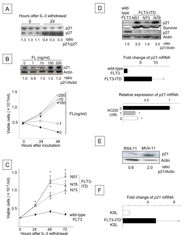
regulate FLT3-ITD signaling, similar to Survivin. We first investigated the association between p21 expression and cell proliferation in response to normal FLT3 and FLT3-ITD signaling. In IL-3-dependent Ba/F3 cells expressing wild-type human FLT3 (Ba/F3-FLT3), IL-3 withdrawal,
which generally induces G1arrest, resulted in down-regulation of the p21Cdkn1aprotein and
up-regulation of p27Cdkn1b, a protein that normally inhibits the G1/S transition [33,34] (Fig
1A). The stimulation of Ba/F3-FLT3 cells with FLT3 ligand (FL) concomitantly increased p21 protein levels and total cell number (Fig 1B). The ectopic expression of FLT3-ITD in Ba/F3
cells (FLT3-ITD+Ba/F3) significantly enhanced cell proliferation (Fig 1C). These changes were
coincident with the up-regulation of p21 and Survivin levels and a reduction in p27Cdkn1b
lev-els compared to cells expressing wild-type FLT3 (Fig 1D, upper panel). Consistent with the
up-regulation of the p21 protein,p21mRNA expression was significantly increased in FLT3-ITD+
Ba/F3 cells compared to cells expressing wild-type FLT3 (Fig 1D, middle panel). However, the
selective FLT3-ITD inhibitor AC220 [24] significantly decreasedp21mRNA expression in
FLT3-ITD+Ba/F3 cells (Fig 1D, lower panel). Similarly, treatment of FLT3-ITD+cells with
AG1296, which inhibits FLT3 kinase [35], decreased p21 expression (not shown). Consistent
Fig 1. FLT3 Signaling Up-regulates p21Cdkn1aExpression in Ba/F3 Cells.(A) Expression of p21 and p27 in Ba/ F3-FLT3 cells before and after IL-3 withdrawal. The blots are from triplicate cultures. The total number of viable cells was enumerated at the time of harvest using the trypan blue exclusion assay, and lysates from 5x10⁵viable cells were loaded into each lane. The p21/p27 ratio was determined by densitometry and is shown beneath the blot. (B) The number of viable Ba/F3-FLT3 cells stimulated with or without FL. Following IL-3 withdrawal for 16 h, the cells were resuspended in 10% FBS/RPMI-1640 at a concentration of 5x10⁵cells/ml with FL concentrations ranging from 0 to 200 ng/ml. The number
of viable cells was counted 24 and 48 h after incubation. P21 protein expression was analyzed by Western blotting at 48 h (top). The data are representative of two experiments with identical results. (C) Proliferation of Ba/F3 cells expressing
FLT3-ITD+MV4-11 human acute leukemia cells compared to RS4;11 cells expressing
wild-type FLT3 (Fig 1E). The transduction ofFLT3-ITD into mouse bone marrow cells and culture
in the absence of hematopoietic growth factors for 14 days resulted in an expansion of c-kit+,
Sca-1+, lineage-negative (KSL) cells as previously reported [30] (not shown), along with a
sig-nificant up-regulation of thep21mRNA in KSL cells (Fig 1F). These data indicate that p21
expression is up-regulated by FLT3 signaling, and similar to Survivin, the increase is associated with enhanced or accelerated cell proliferation.
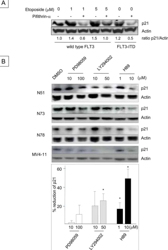
Because p21 is one of the major transcriptional targets of p53 [1,36], we next investigated whether p53 is involved in the up-regulation of p21 induced by FLT3-ITD (Fig 2A). Incuba-tion of BaF3 cells with etoposide, which activates p53 [37], increased p21 protein levels, indi-cating that p53 is functional in Ba/F3 cells. Etoposide-enhanced p21 expression was abrogated
by the p53 inhibitor pifithrin-α[1,38], and FLT3-ITD+Ba/F3 cells treated with pifithrin-α
also down-regulated p21 expression in the absence of etoposide. These results suggest that the
up-regulation of p21 in FLT3-ITD+Ba/F3 cells is mediated by p53. To identify additional
sig-naling pathways that regulate the p21 expression induced by FLT3-ITD, Ba/F3 cells express-ing several different human FLT3-ITD constructs were incubated with inhibitors of
mitogen-activated protein kinase (MAPK)p42/p44(PD98059), phosophoinositide-3 (PI3) kinase
(LY294002) and protein kinase A (PKA) (H89). Although the MAPK inhibitor had little effect on p21 protein expression, the PI3 kinase and PKA inhibitors reduced p21 levels. Consistent
with the results obtained using FLT3-ITD+Ba/F3 cells, the PI3 kinase and PKA inhibitors
reduced p21 expression in human FLT3-ITD+MV4-11 leukemia cells (Fig 2B). These data
indicate that the p53, PI3 kinase and PKA pathways mediate the FLT3-ITD-induced up-regu-lation of p21.
FLT3-ITD attenuates the growth factor-independent proliferation and cell
cycle progression of hematopoietic progenitor cells by up-regulating
p21
Cdkn1aexpression
The concurrent enhancement of cell proliferation and p21 up-regulation by FLT3-ITD sug-gested that p21 may positively regulate FLT3-ITD-potentiated cell proliferation, which was similar to our previous observations for the endogenous anti-apoptotic protein Survivin [30]. Therefore, we compared the functional roles of p21 and Survivin in the FLT3-ITD-induced growth factor-independent proliferation of HPCs. Although wild-type FLT3 failed to support the proliferation of colony-forming cells (CFCs), FLT3-ITD overexpression in control Survi-vinfx/fxor p21+/+bone marrow cells resulted in CFC proliferation in the absence of
type or FLT3-ITD. The cells were washed three times and cultured for 72 h in 1% FBS/RPMI-1640, and the numbers of viable Ba/F3 cells expressing wild-type or FLT3-ITD derived from 3 different patients (N51, N73 and N78) were counted every 24 h (*: P<0.05). The data shown are from one of three experiments that were performed in triplicate with identical results. (D) The upper panel shows the expression of the p21, Survivin and p27 proteins, as analyzed by Western blotting. The level of p21 relative to actin is shown beneath the blot. The middle panel shows the relativep21mRNA expression levels in Ba/F3 cells expressing wild-type or N51-FLT3-ITD, which were determined using quantitative RT-PCR. The presented data are the averages of two independent experiments (*: P<0.05). The lower panel shows thep21mRNA expression levels in FLT3-ITD (N51) Ba/F3 cells incubated with 1 or 2 nM AC220 for 48 h compared to the control cells, which were incubated with dimethyl sulfoxide (DMSO) alone (N = 3,*: P<0.05). (E) The blot shows p21 protein expression in RS4;11 cells and MV4-11 cells, as determined by Western blot analysis. (F) Thep21mRNA expression levels in FLT3-ITD+KSL cells compared to freshly isolated bone marrow KSL cells. C57BL/6 mouse bone marrow cells
were retrovirally transduced with N51-FLT3-ITD using the MSCV-IRES-EGFP vector and were cultured for 2 weeks without hematopoietic growth factors. The GFP+KSL cells were sorted by FACS, andp21mRNA expression was compared with freshly isolated KSL cells from the same donor. The presented data are the averages of three independent experiments (*: P<0.005).
Fig 2. Up-regulation of p21 Expression Induced by FLT3-ITD is Mediated through the p53, PI3 Kinase and PKA Pathways.(A) Ba/F3 cells expressing wild-type FLT3 were incubated with 20μM pifithrin-αfor 24
h, followed by exposure to 1 or 5μM etoposide for 2.5 h. Ba/F3 cells expressing N51-FLT3-ITD were also
incubated with pifithrin-αfor 24 h. The cell lysates were subjected to Western blot analysis to determine the
p21 protein expression levels. (B) Ba/F3 cells containing N51, N73 and N78 FLT3-ITD were incubated with the indicated concentrations of DMSO, PD98059, LY294002 or H89 (all of which were diluted in DMSO) in 10% FBS/RPMI-1640 supplemented with 0.1 ng/ml rmIL-3 for 24 h. Human MV4-11 leukemia cells harboring endogenousFLT3-ITD were treated in the same manner and were incubated in 10% FBS/RPMI-1640. The
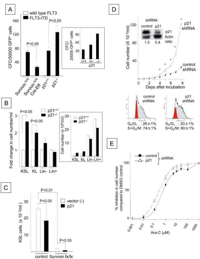
hematopoietic growth factors (Fig 3A). Conditional deletion of Survivin reduced the growth
factor-independent proliferation ofFLT3-ITD-transduced CFCs, as previously reported
(Cre-ER Survivinfx/fxinFig 3A) [30]. In contrast, deletion of thep21gene increased the
prolifera-tion of CFCs transduced withFLT3-ITD (Fig 3A). An intermediate number of CFCs was
gen-erated by p21+/-cells transduced withFLT3-ITD, indicating that the effect of p21 was gene
dosage-dependent (Fig 3A, inset). Similar to CFCs, the growth factor-independent
prolifera-tion of FLT3-ITD+KSL and c-kit+, lineage-negative (KL) cells was further enhanced by the
deletion of p21 (P<0.05,Fig 3B). In contrast, the loss of p21 did not significantly affect cell
number or the proportion of lineage-positive and lineage-negative cells. Although no
differ-ence was observed in the number of apoptotic cells between the FLT3-ITD+p21+/+KL and
FLT3-ITD+p21-/-KL cells, the enhancement of FLT3-ITD+cell proliferation by p21 deletion
was associated with a marginal but significant increase in the number of cells in S+G2/M
phase (18.2 ± 0.5%) compared to p21+/+cells (15.3 ± 0.8%, P<0.05, N = 3), concomitant
with increased levels of Ki-67, a marker for cycling cells (1.5 ± 0.1-fold increase, P<0.01,
N = 3). In contrast, ectopic p21 overexpression decreased the number of growth
factor-inde-pendent FLT3-ITD+KSL cells derived from the control cells compared to cells expressing
FLT3-ITD alone (P<0.05). The loss of Survivin (Cre-ER Survivinfx/fx) further reduced the
number of FLT3-ITD+KSL cells overexpressing p21 (Fig 3C). Similarly, treatment with ap21
-specific shRNA increased the growth factor-independent proliferation of FLT3-ITD+Ba/F3
cells compared to treatment with a control shRNA and was coincident with a marginal but
significant increase in the number of cells in S+G2/M phase (Fig 3D). Moreover, p21 silencing
increased the sensitivity of FLT3-ITD+Ba/F3 cells to the cell cycle-specific chemotherapeutic
agent cytarabine (Ara-C) (Fig 3E) [39]. The 50% inhibitory concentrations (IC50s) of Ara-C
were 0.6 and 0.27μM in the control FLT3-ITD+Ba/F3 cells and those transduced with p21
shRNA, respectively. These data suggest that the FLT3-ITD-mediated up-regulation of p21
inhibits FLT3-ITD+cell proliferation and confers resistance to chemotherapy by delaying cell
cycle progression.
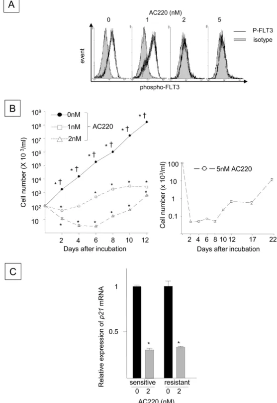
Blocking p21 by FLT3-ITD inhibition facilitates the emergence of
FLT3-ITD
+cells that are refractory to the FLT3-ITD inhibitor AC220
The poor efficacy of FLT3-ITD inhibitors for FLT3-ITD+AML [22,23] underscores the need
to investigate the mechanism responsible for the drug-resistant phenotype of FLT3-ITD+
AML. The capacity of the FLT3-ITD-mediated up-regulation of p21 expression to inhibit the
proliferation of FLT3-ITD+HPCs suggests that FLT3-ITD signaling attenuates the
prolifera-tion and cell cycle progression of FLT3-ITD+HPCs. However, the p21 down-regulation in
FLT3-ITD+cells triggered by inhibitors of FLT3-ITD, p53, PI3 kinase, and PKA suggests that
the sustained inhibition of FLT3-ITD or other molecular pathways downstream of FLT3-ITD may block endogenous anti-growth signaling, possibly contributing to the recurrence of
FLT3-ITD+AML. To determine whether inhibiting p21 expression in FLT3-ITD+cells by
antagonizing FLT3-ITD is associated with the proliferation and/or emergence of FLT3-ITD+
cells that are refractory to a FLT3-ITD inhibitor, the association between p21 expression and
cell proliferation was investigated in FLT3-ITD+cells treated with a FLT3-ITD inhibitor.
FLT3-ITD+Ba/F3 cells were cultured in the absence of cytokines with 2 or 5 nM AC220, a
p21 expression levels (relative to actin) were quantitated by Western blot analysis and densitometry. The histogram beneath the blot indicates the percentages by which p21 expression was reduced by the inhibitors, which were averaged in 4 wells and determined by a comparison with the DMSO control (*: P<0.05).
Fig 3. FLT3-ITD Attenuates the Growth Factor-independent Proliferation and Cell Cycle Progression of
Hematopoietic Progenitor Cells by Up-regulating p21Cdkn1aExpression.(A) Growth factor-independent proliferation of
CFCs in mouse bone marrow cells lacking p21 or Survivin and transduced withFLT3-ITD. Bone marrow cells from Survivinfx/ fx, Cre-ER Survivinfx/fx, p21+/+or p21-/-mice were retrovirally transduced with wild-typeFLT3or N51-FLT3-ITD. A total of
500,000 GFP+cells were plated on either 0.3% agar or methylcellulose medium containing 30% FBS in the absence of
hematopoietic growth factors. To delete theSurvivingene, cells from Cre-ER Survivinfx/fxor Survivinfx/fxmice were cultured in the presence of 1μM 4-hydroxy tamoxifen at the time of plating. The colonies were enumerated after 14 days. The presented
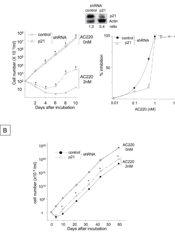
selective inhibitor of FLT3-ITD, for 24 hours, and FLT3 phosphorylation was inhibited (Fig 4A). Although the reduced FLT3 phosphorylation was not evident after 24 hours of incuba-tion with 1 nM AC220, prolonged incubaincuba-tion with 1, 2 or 5 nM AC220 significantly inhibited
the growth-factor independent proliferation of FLT3-ITD+Ba/F3 cells compared to the
con-trols, suggesting that AC220 antagonizes the growth-stimulatory function of FLT3-ITD (Fig 4B). In contrast, the same treatment significantly decreasedp21mRNA expression in the
FLT3-ITD+cells (Fig 4C), implying that FLT3-ITD inhibition disrupts growth-inhibitory
sig-naling pathways, and not just pathways that stimulate cell proliferation. When the FLT3-ITD+
Ba/F3 cells were incubated with 1 or 2 nM AC220 for more than 8 days, cell proliferation began to increase despite the presence of AC220 (Fig 4B, left panel). Similarly, the number of viable cells declined immediately after incubation with 5 nM AC220, but cells began to prolif-erate after 21 days in the presence of 5 nM AC220 (Fig 4B, right panel), indicating the
devel-opment of FLT3-ITD+cells that were refractory to AC220, as previously reported [30].
Incubating the AC220-refractory cells with AC220 down-regulated p21 expression (Fig 4C), suggesting that AC220 can also disrupt growth-inhibitory signaling in AC220-refractory
FLT3-ITD+cells. However, p21 overexpression in the FLT3-ITD+Ba/F3 cells significantly
delayed the emergence of AC220-resistant FLT3-ITD+cells (Fig 5A, left panel) and inhibited
the proliferation of AC220-resistant FLT3-ITD+cells cultured in the presence of 2 nM AC220
for up to 60 days (Fig 5B). The medium was replaced every 5 days, and fresh AC220 was added at a concentration of 2 nM; therefore, the emergence of resistant cells was not due to
the loss of AC220 activity. The IC50 for the control FLT3-ITD+cells was 0.77 nM, whereas
that in the cells expressing ectopic p21 was 0.65 nM on day 10 (Fig 5A, right panel). In
con-trast,p21silencing with an shRNA significantly delayed the reduction in the number of viable
FLT3-ITD+Ba/F3 cells induced by AC220 (Fig 6A, left panel) and accelerated the
develop-ment and proliferation of FLT3-ITD+cells refractory to 2 nM AC220 up to 60 days (Fig 6B).
The IC50 of AC220 for the FLT3-ITD+cells containing the control shRNA was 0.35 nM on
day 10, whereas p21 silencing increased the IC50 to 0.7 nM (Fig 6A, right panel). These find-ings suggest that the down-regulation of p21 resulting from FLT3-ITD inhibition facilitates
proliferation and the development of FLT3-ITD+cells that are refractory to AC220.
data are the averages of 9 experiments for p21 deletion and 2 experiments for Survivin deletion (P<0.05). The numbers of CFCs generated by p21+/-cells transduced withFLT3-ITD compared to those generated by p21+/+and p21-/-cells are shown
in the lower panel. The data shown were obtained from one of two experiments that yielded identical results. (B) The proliferation of bone marrow c-kit+, Sca-1+, lineage-(KSL), c-kit+, lineage-(KL), lineage-(Lin-) or lineage+(Lin+) cells
transduced withFLT3-ITD derived from p21+/+and p21-/-mice. Following transduction, the GFP+cells were sorted and cultured in 10% FBS/IMDM in the absence of hematopoietic growth factors. The cells were enumerated 7 days after culture. The fold change in the number of FLT3-ITD+p21-/-cells compared to the FLT3-ITD+p21+/+cells is shown. The presented
data are the averages of three experiments (P<0.05). The cell number from one representative experiment is shown in the lower panel. (C) The effect of ectopic p21 expression on FLT3-ITD+KSL cell proliferation. Bone marrow cells from control Survivinfx/fxmice or Cre-ER Survivinfx/fxmice were transduced with N51-FLT3-ITD andp21using a MIEG3 vector or a
negative MIEG3 vector control. Following transduction, the GFP+cells were cultured for 2 weeks in 10% FBS/IMDM
containing 1μM 4-hydroxy tamoxifen at a density of 5 x 10⁵cells/ml. The cells were stained for c-kit, Sca-1 and lineage
markers to quantify the number of KSL cells. The data shown were obtained from one of two experiments that were analyzed in triplicate. (D) Ba/F3 cells expressing FLT3-ITD (N51) were transduced with either ap21shRNA or a control shRNA and were cultured in 1% FBS/RPMI without any growth factors. The viable cells were enumerated using the trypan blue exclusion assay. The data shown were obtained from one of two experiments that were performed in quadruplicate with identical results (*: P<0.01). The expression levels of the p21 protein in both cell lines are shown in the inset. The lower panel shows a cell cycle histogram of FLT3-ITD+Ba/F3 cells transduced with a control shRNA (left plot) and the p21 shRNA (right plot). The
histogram represents one of three experiments with identical results. (E) Dose-dependent growth inhibition by Ara-C in N51-FLT3-ITD-Ba/F3 cells transduced with thep21shRNA and control cells. The cells were cultured in the presence of DMSO alone (control) or increasing doses of Ara-C in 1% FBS/RPMI-1640 without any growth factors for 24 hours. The y-axis represents the % inhibition of viable cells incubated with Ara-C compared to DMSO. The data shown represent one of three experiments with identical results.
Fig 4. Blocking FLT3-ITD Using AC220 Decreases p21 Expression Coincident with Emergence of FLT3-ITD+Cells Refractory to AC220.(A) The panel shows FLT3 phosphorylation in N51-FLT3-ITD-Ba/F3 cells incubated with 1, 2 or 5 nM AC220 for 24 hours. The cells were stained for phospho-FLT3 using a rabbit monoclonal antibody raised against Tyr591 of human FLT3, followed by staining with a PE-conjugated anti-rabbit IgG secondary antibody and flow cytometry analyses. (B) The left panel indicates the numbers of viable N51-FLT3-ITD-Ba/F3 cells incubated with 1 or 2 nM AC220 compared to the DMSO control (0 nM). Cells plated at a density of 1 x 10⁵cells/ml were incubated with 1 or 2 nM AC220 or control DMSO for 12
The enhanced proliferation of FLT3-ITD-transformed HPCs following
p21 deletion is associated with up-regulation of Pbx1 expression
In addition to regulating the cell cycle and apoptosis [1,2,6,7,13], p21 can modulate the
activ-ity of a number of transcription factors and co-activators [14–18], suggesting that it may
regu-late cell fate by influencing gene expression [19]. To identify the potential mechanisms by
which p21 attenuates the proliferation of FLT3-ITD+HPCs, we compared the gene expression
profiles of p21+/+and p21-/-FLT3-ITD+KSL cells. In three separate experiments, p21 deletion
in FLT3-ITD+KSL cells resulted in changes in the expression of 111 distinct mRNAs
com-pared to the p21+/+FLT3-ITD+KSL cells, which were either unaffected or differentially
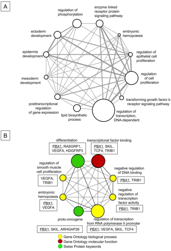
regu-lated in normal bone marrow KSL cells (Table 1andS1 Table). The 111 genes were
functionally classified based on the biological process subclasses of the Gene Ontology data-base (Fig 7A). The top 3 functional groups that were associated with p21 include the regula-tion of transcripregula-tion (DNA-dependent), cell proliferaregula-tion and phosphorylaregula-tion. Among the 111 genes, 12 were deregulated in human AML stem cells (AML-LSC) (Table 1) [40]. These 12 genes were further classified into 8 functional groups based on the Gene Ontology terms and the Swiss Protein database (Fig 7B). The transcription factor Pbx1, which is associated with HSC and HPC function [28,29] and is down-regulated in AML-LSC [40], was
up-regu-lated in FLT3-ITD+KSL cells upon p21 deletion. However, Pbx1 was not affected by p21
dele-tion in normal KSL cells (see below). Pbx1 itself was classified in 7 of the funcdele-tional groups (Fig 7B).
Although the loss of p21 did not significantly affectPbx1expression in normal KSL cells
(1.0 ± 0.3-fold change compared to p21+/+KSL cells, NS), the quantitative RT-PCR analysis
confirmed thatPbx1was up-regulated by 2.1 ± 0.5-fold in FLT3-ITD+p21-/-KSL cells
com-pared to FLT3-ITD+p21+/+KSL cells (P<0.001,Fig 8A, left panel). BecausePbx1expression is
transcriptionally regulated by the transcription factor Evi-1 [41] and FLT3-ITD and because
p21 altered Pbx1 expression, we hypothesized that the changes inPbx1expression induced by
FLT3-ITD and/or p21 are mediated by Evi-1. Therefore, we investigated theEvi-1levels in the
freshly isolated KSL cells and the FLT3-ITD-transformed KSL cells in the presence or absence
of p21. However, p21 deletion had no effect onEvi-1expression in normal or
FLT3-ITD-trans-formed KSL cells (Fig 8A, right panel). ThePbx1mRNA levels in the p21-/-KSL cells in G0and
G1phases were equivalent to those in the p21+/+cells (1.0 ± 0.3- and 1.4 ± 0.3-fold changes,
respectively), whereasPbx1expression in p21-/-KSL cells in S phase was elevated by
6.1 ± 1.1-fold (P<0.05, N = 3) compared to the p21+/+cells (Fig 8B). BecausePbx1expression
was not modulated by p21 in the absence of FLT3-ITD, we next determined the effect of
FLT3-ITD on Pbx1 expression. FLT3-ITD+Ba/F3 cells significantly down-regulatedPbx1
mRNA levels compared to FLT3-ITD-Ba/F3 cells (Fig 8C, left and middle panels). Conversely,
the FLT3-ITD inhibitor AC220 significantly up-regulatedPbx1mRNA expression in the
The medium was replaced every 5 days and contained 5 nM fresh AC220. The cells were enumerated using the trypan blue exclusion assay. The y-axis represents the cell number on the log scale. The data shown represent one of the three experiments that were analyzed in triplicate with identical results (*: P<0.05 compared to time 0,†: P<0.05 compared to 1 or 2 nM AC220). (C) The relative expression of p21 in N51-FLT3-ITD-Ba/F3 cells that were sensitive or refractory to AC220 and incubated with 2 nM AC220 compared to those incubated with the DMSO control. The refractory cells, which were established by incubation with 2 nM AC220, were washed and cultured for 48 hours in fresh medium without AC220. The cells were subsequently incubated with 2 nM AC220 for 48 hours. Similarly, parental cells that were sensitive to AC220 were incubated with 2 nM AC220 for 48 hours. Thep21mRNA levels were quantitated before and after incubation with AC220 (*: P<0.05, N = 3).
Fig 5. Overexpressing p21 Delays the Emergence and Proliferation of FLT3-ITD+Cells Refractory to AC220.
(A) The numbers of viable N51-FLT3-ITD-Ba/F3 cells transfected with the murinep21cDNA or control plasmid were quantitated in the presence or absence of 2 nM AC220. Cells were plated at a density of 1x10⁵cells/ml and incubated
with 2 nM AC220 or control DMSO for 10 days. The viable cells were enumerated using the trypan blue exclusion assay. The data shown represent one of three experiments that were analyzed in triplicate with identical results (*: P<0.05 compared to the control plasmid). The expression levels of the p21 protein in the control cells and those transfected with thep21cDNA are shown above the histogram. The right panel indicates the AC220 dose-dependent inhibition of the control FLT3-ITD+cells containing empty vector and those overexpressing p21. The cells were
incubated with different concentrations of AC220 for 10 days, and the percent inhibition of viable cells was calculated compared to the control cells incubated with DMSO. (B) The panel indicates the numbers of viable FLT3-ITD+cells
containing the empty vector or p21 cDNA that were cultured in the presence of 2 nM AC220 for 60 days. The cells were plated at a density of 1x10⁵cells/ml and were incubated with 2 nM AC220 or the DMSO control for 60 days. The
medium was replaced every 5 days and contained 2 nM fresh AC220. The y-axes of the histograms are shown in linear but different scales (*: P<0.05 compared to the control).
doi:10.1371/journal.pone.0158290.g005
FLT3-ITD+Ba/F3 cells (Fig 8C, right panel). Although the up-regulation of Pbx1 expression
induced by the loss of p21 in FLT3-ITD+KSL cells was independent of Evi-1 expression, the
transduction ofFLT3-ITD into primary mouse bone marrow resulted in significant decreases in
thePbx1andEvi-1mRNA levels compared to their levels in freshly isolated KSL cells (P<0.01,
N = 3,Fig 8D, upper panel). In addition, we found that thePbx1andEvi-1expression levels
were significantly reduced in the FLT3-ITD+AML cells (N = 78) compared to FLT3-ITD-AML
cells (N = 190, P<0.05,Fig 8D, middle and lower panels), which are listed in the GSE1159
dataset (www.ncbi.nlm.nih.gov/geo/) [42]. These results suggest that both p21 and FLT3-ITD down-regulate Pbx1 expression, although the effect of p21 on Pbx1 is dependent on FLT3-ITD. Consistent with the concomitant up-regulation of p21 expression and the down-regulation of
Pbx1 expression, we found thatp21expression (CDKN1A; 202284_s_at in GSE1159) was
inversely correlated withPbx1expression, but not withEvi-1expression, in FLT3-ITD+AML
subjects, particularly those with the FAB M1 subtype (r = -0.46, P<0.01, 212151_at in
GSE1159,Fig 8E, top panel), although this correlation was not evident in other AML subgroups.
In contrast, the association betweenp21andPBX1was not observed in either FLT3-ITD-AML
cells (r = -0.13, GSE1159,Fig 8E, bottom panel) or normal human CD34+cells (GSE2666,
HG-U133A and GSE30029,www.ncbi.nlm.nih.gov/geo/) [43].
quantitated in the presence or absence of 2 nM AC220. The cells were plated at a density of 1x10⁵cells/ml and incubated with 2 nM AC220 or control DMSO for 10 days. The viable cells were enumerated using the trypan blue exclusion assay. The data shown represent one of three experiments that were analyzed in triplicate with identical results (*: P<0.05 compared to the control shRNA). The expression levels of the p21 protein in both cell populations are shown in the inset (the same blot is shown inFig 3D). The right panel indicates the AC220 dose-dependent inhibition of the control FLT3-ITD+cells containing empty vector and those with shRNA for p21. The
cells were incubated with different concentrations of AC220 for 10 days, and the percent inhibition of viable cells was calculated compared to the control cells incubated with DMSO. (B) The panel indicates the numbers of viable FLT3-ITD+cells containing the empty vector or p21 shRNA that were cultured in the presence of 2 nM AC220 for 60 days. The cells were plated at a density of 1x10⁵cells/ml and were incubated with 2 nM AC220 or the DMSO
control for 60 days. The medium was replaced every 5 days and contained 2 nM fresh AC220.
doi:10.1371/journal.pone.0158290.g006
Table 1. Fold Changes in the Genes Regulated by p21 Deletion in the FLT3-ITD-transformed KSL Cells.
Gene symbol
Name Fold change after p21 deletion in FLT3-ITD+KSL cells
Number of functional classification hits (as shown inFig 7A)
Pbx1 Pre-B cell leukemia transcription factor 1
2.1 7
Vegfa Vascular endothelial growth factor A 1.8 4
Trib1 Tribbles homolog 1 (Drosophila) 2.3 4
Skil SKI-like 1.6 3
Tcf4 Transcription factor 4 2.1 2
Rasgrp1 RAS guanyl-releasing protein 1 -2.5 1
Hdgfrp3 Hepatoma-derived growth factor-related protein 3
1.7 1
P21 deletion in FLT3-ITD+KSL cells resulted in the modulation of the expression levels of 111 mRNAs that were either unaffected or differentially regulated
in normal bone marrow KSL cells. Of these 111 genes, 12 were deregulated in human AML-LSC [40]. These genes were classified based on Gene Ontology terms and the Swiss Protein database (as shown inFig 7B). Pbx1 was classified into 7 of the functional groups. The fold changes in the expression levels of the genes listed inFig 7Bare shown inTable 1.
doi:10.1371/journal.pone.0158290.t001
Fig 7. Enhanced Proliferation of FLT3-ITD+KSL Cells by p21 Deletion is Associated with Alteration of
Expression of Genes Linked to Various Biological Functions.(A) The functional classification of the 111 genes regulated by p21 in FLT3-ITD+KSL cells. These 111 genes were classified based on the Biological
Pbx1 knockdown abrogates the enhanced proliferation of FLT3-ITD
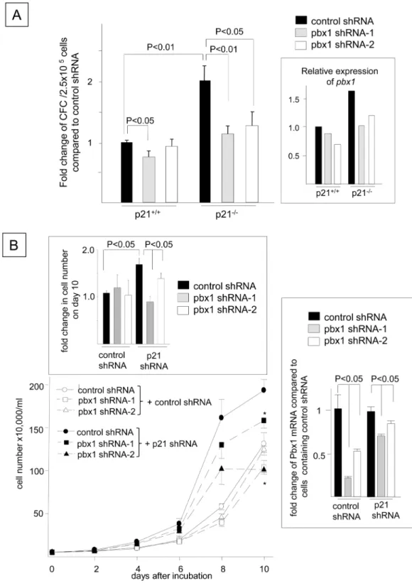
+HPCs induced by p21 deletion
Because the enhanced proliferation of FLT3-ITD+HPCs induced by the loss of p21 was
accom-panied by the up-regulation of Pbx1 expression, we next examined whether Pbx1 mediates the
increased proliferation induced by p21 deletion. FLT3-ITD+p21-/-bone marrow cells
trans-duced with a control shRNA showed a 1.9 ± 0.3-fold increase in the number of growth
factor-independent CFCs compared to p21+/+cells (P<0.01,Fig 9A), which coincided with the
up-regulation ofPbx1expression (Fig 9A, inset). The transduction of two different shRNAs
target-ingPbx1into FLT3-ITD+p21-/-bone marrow cells resulted in reducedPbx1mRNA levels (Fig
9A, inset) and CFC proliferation in the absence of hematopoietic growth factors compared to
the control shRNA-transduced FLT3-ITD+p21-/-cells (Fig 9A, shRNA-1: 43 ± 4% reduction,
P<0.01, N = 3; shRNA-2: 41 ± 7% reduction, P<0.05, N = 3). The number of CFCs resulting
from the transduction ofPbx1shRNA into FLT3-ITD+p21-/-bone marrow cells was
compara-ble to that generated by FLT3-ITD+p21+/+bone marrow cells that were transduced with the
control shRNA (NS, N = 3). Similarly, transduction of thePbx1shRNAs partially but
signifi-cantly decreased the Pbx1 mRNA levels in the FLT3-ITD+Ba/F3 cells containing the control
shRNA, along with those harboring the p21 shRNA (Fig 9B, inset, right panel). The
enhance-ment of the growth factor-independent proliferation of FLT3-ITD+Ba/F3 cells induced by the
p21shRNA was partially but significantly reduced by transducing these FLT3-ITD+Ba/F3 cells
with aPbx1shRNA (N = 3,Fig 9Band the inset on the top). These data indicate that Pbx1
mediates the enhanced proliferation of growth factor-independent FLT3-ITD+cells induced
by p21 deletion.
Discussion
The present study shows thatFLT3-ITD mutations produce proliferative and anti-growth
sig-nals (Fig 10A). FLT3-ITD concomitantly up-regulated p21 expression and down-regulated Pbx1 expression. The loss of p21 enhanced the growth factor-independent proliferation and
cell cycle progression of FLT3-ITD+cells and their sensitivity to Ara-C, suggesting that the
enhanced expression of p21 attenuates cell cycle progression and confers chemoresistance to
FLT3-ITD+cells. Furthermore, the p21 deficiency promoted the selective up-regulation of
Pbx1 expression in FLT3-ITD+KSL cells, whereas silencing Pbx1 expression partially
abro-gated the enhanced proliferation of FLT3-ITD+HPCs induced by p21 deletion, indicating that
the p21-mediated inhibition of the proliferation of FLT3-ITD+HPCs is mediated, at least in
part, by Pbx1. Importantly, the p21 down-regulation resulting from FLT3-ITD inhibition by
AC220 accelerated proliferation and development of FLT3-ITD+cells that were refractory to
AC220, suggesting that treatments targeting FLT3-ITD can eradicate growth-inhibitory signals
by inhibiting p21 expression, thereby potentially contributing to FLT3-ITD+AML progression
(Fig 10B & 10C).
lists and functional categories are shown inS1 Table. The array data have been deposited in the NCBI Gene Expression Omnibus (GSE75200). (B) The functional classifications of the 12 genes that were regulated by p21 in FLT3-ITD+KSL cells and deregulated in human AML-LSC [40]. The modulated genes were classified based
on their biological processes and molecular functions, as defined by Gene Ontology terms and Swiss Protein keywords using the DAVID program [44]. Five of the 12 molecules were not classified. The 8 significantly enriched functional categories (P<0.05) were connected and visualized using Cytoscape [45]. PBX1 was identified in 7 of 8 functional categories. The size of the circle indicates the number of genes in a given category, and the thickness of the bar represents the redundancy between the groups. The genes that were classified in each functional category are shown in the box. PBX1 is underlined. The fold changes in the expression levels of the listed genes are shown inTable 1.
doi:10.1371/journal.pone.0158290.g007
Fig 8. The Enhanced Proliferation of FLT3-ITD-transformed HPCs Following p21 Deletion is Associated with the Up-regulation of Pbx1 Expression.(A) The effects of p21 deletion on thePbx1andEvi-1mRNAs. The left panel shows the expression levels ofPbx1mRNA in freshly isolated KSL cells or FLT3-ITD+KSL cells, which were derived from p21
Our results indicate that growth factor withdrawal reduces p21Cdkn1aexpression in Ba/
F3-FLT3 cells while increasing p27Cdkn1bexpression, and FLT3 ligand and FLT3-ITD
up-regu-late p21 expression but down-reguup-regu-late p27Cdkn1bexpression. These data clearly support the
differential regulation of p21 and p27 expression in response to FLT3 signaling. The up-regula-tion of p21 expression by FLT3-ITD was mediated through the p53, PI3 kinase and PKA path-ways. Our data showed that FLT3-ITD up-regulated p21 expression in HPCs and Ba/F3 cells, concomitant with their enhanced proliferation. However, the loss of p21 enhanced the
prolifer-ation of FLT3-ITD+HPCs and Ba/F3 cells, whereas p21 overexpression resulted in the opposite
effect, indicating that although FLT3-ITD enhances HPC proliferation, the concomitant increase in p21 expression attenuates their proliferation. Therefore, FLT3-ITD harbors both growth-inhibitory and growth-stimulatory activities (Fig 10A). FLT3-ITD AML cells are
char-acterized by their high proliferation rate and leukocytosis in patients [20,21]. The presence of
growth-inhibitory signaling downstream of FLT3-ITD appears to be contradictory to the
increased proliferation potential of FLT3-ITD+AML cells. The majority of the signaling
com-ponents that exist downstream of FLT3-ITD are known to contribute to cell proliferation [20,21,30], suggesting that growth-stimulatory signaling dominates the p21-mediated inhibi-tory activity. The primitive KSL hematopoietic population exhibited the maximal increase in
FLT3-ITD+cell proliferation in response to the loss of p21. In contrast, the effect of the loss of
p21 was minimal in differentiated cells, suggesting that p21 attenuates the proliferation of early undifferentiated cells, which resemble AML stem cells. This result is in good agreement with the increased cell cycle progression in HSCs, without altering the marrow cellularity or periph-eral blood cell counts in p21-deficient mice [8]. A previous report showed that the induction of cell cycle entry eliminated AML stem cells in Ara-C-treated mice [39]. The enhanced cytotoxic
effect of Ara-C in FLT3-ITD+Ba/F3 cells induced by p21 silencing suggests that the
up-regula-tion of p21 expression by FLT3-ITD may confer resistance to Ara-C by inducing quiescence in
FLT3-ITD+AML cells in the patients.
The down-regulation of p21 expression by AC220-mediated FLT3-ITD inhibition
acceler-ated the development of FLT3-ITD+cells that were refractory to AC220. AC220
down-regu-lated p21 expression in FLT3-ITD+cells; however, p21 overexpression in FLT3-ITD+cells
significantly delayed the emergence of FLT3-ITD+cells that were refractory to AC220, whereas
p21 silencing resulted in the opposite effect. Our data suggest that the deregulation of p21
expression in FLT3-ITD+cells contributes to chemoresistance in two distinct ways. FLT3-ITD+
cells undergo quiescence by up-regulating p21 expression, which enhances their resistance to cells transduced with N51-FLT3-ITD were stained with antibodies for Sca-1, c-kit, and lineage markers, along with Hoechst 33342 and Pyronin Y. The relative expression levels of thePbx1mRNA in FACS-sorted KSL cells in G0, G1and
S phases compared to p21+/+cells in G
0phase are shown (N = 3). The sorting gate for cell cycle fractionation is shown in
the inset. (C) The relative expression levels ofPbx1mRNA in Ba/F3 cells transduced with N51-FLT3-ITD compared to those transduced with wild-typeFLT3. The left column showsPbx1expression in cells without IL-3. The middle histogram presents thePbx1mRNA levels in cells cultured with 0.1 ng/ml rmIL-3. The right histogram shows thePbx1mRNA expression levels in N51-FLT3-ITD+Ba/F3 cells incubated with 1 or 2 nM AC220 for 48 hours compared to the DMSO
controls (*: P<0.05, N = 3). (D) The upper panel shows thep21,Pbx1andEvi-1mRNA expression levels in FLT3-ITD+
p21+/+KSL cells compared to normal KSL cells, as determined by quantitative RT-PCR. The percent changes in
expression compared to the control KSL cells are shown (*: P<0.01, N = 3). The middle and lower panels show thePBX1
andEVI-1expression levels in human AML cells with or without FLT3-ITD. ThePBX1(middle) andEVI-1(lower) expression levels in human FLT3-ITD+AML cells (N = 78) were compared to those in the FLT3-ITD-AML cells (N = 190)
listed in GSE1159 (excluding patients withFLT3-TKD mutations,www.ncbi.nlm.nih.gov/geo/,*: P<0.05). (E) The relationship betweenp21andPBX1in the AML M1 cases with (upper panel) or without (lower panel) FLT3-ITD that are listed in GSE1159 (www.ncbi.nlm.nih.gov/geo/) was analyzed using Microsoft Excel. The x-axes are displayed in linear and log scales in the top and bottom panels, respectively. Correlation values of r>0.4 or<-0.4 were considered to indicate significant correlations betweenp21andPBX1(P<0.05).
doi:10.1371/journal.pone.0158290.g008
Fig 9. Pbx1 Knockdown Abrogates the Enhanced Proliferation of FLT3-ITD+HPCs Induced by p21
Deletion.(A) Bone marrow cells from p21+/+and p21-/-mice were transduced withFLT3-ITD. Following the sorting of FLT3-ITD+cells (using GFP as a marker) by FACS, the cells were transduced with a control shRNA or
Pbx1shRNA (shRNA-1 or shRNA-2) using a pSINsi-mU6 plasmid, and the number of growth factor-independent CFCs grown in methylcellulose was quantified 14 days later. The data are shown as the fold changes in the number of CFCs compared to those of FLT3-ITD+p21+/+cells transduced with the control shRNA (N = 3). Identical results were obtained after 1 week of culture. Quantitative RT-PCR was performed to comparePbx1
conventional chemotherapeutic drugs, such as Ara-C. In contrast, the disruption of the expres-sion and/or function of p21 resulting from FLT3-ITD inhibition contributes to the emergence
of FLT3-ITD+cells that are refractory to FLT3-ITD inhibitors. This result suggests that
treat-ments that antagonize p21 function can sensitize FLT3-ITD+cells to chemotherapy, whereas
treatments that activate p21 may aid in inhibiting the development of FLT3-ITD+cells that are
refractory to FLT3-ITD inhibitors. Similar to FLT3-ITD-transformed cells, cells containing Bcr-Abl [46], AML1-ETO [4] and N-RasE12 [47], all of which regulate the proliferation of myeloid leukemia cells, exhibit increased p21 expression. The loss of p21 facilitates AML1-ETO-induced leukemogenesis [4] and increases the proliferation of Bcr-Abl-trans-formed hematopoietic cells [46], suggesting that these oncogenes also contain signaling com-ponents that negatively modulate the proliferation of leukemia cells. Moreover, the FLT3 ligand and stem cell factor provided by the bone marrow microenvironment [48] up-regulate
p21 expression in Ba/F3-FLT3 cells and human AML cells [10,11], respectively. Taken
together, these data indicate that p21 expression that is up-regulated by the oncogenes and/or hematopoietic growth factors derived from the marrow microenvironment may contribute to the chemoresistance of AML cells.
Our data also indicate that the enhanced proliferation of FLT3-ITD+cells induced by the
loss of p21 was accompanied by the up-regulation of Pbx1 expression. Pbx1 is a product of a proto-oncogene that was originally discovered in acute leukemia and critically regulates organ-ogenesis and hematopoiesis [28]. Pbx1 regulates the self-renewal of hematopoietic stem cells by maintaining their quiescence, whereas it facilitates the expansion of HPCs by regulating the
function of common myeloid progenitor cells [28,29]. The hyperproliferation of FLT3-ITD+
HPCs and Ba/F3 cells induced by the concomitant loss of p21 and increased Pbx1 expression was partially abrogated by Pbx1 silencing, indicating that the enhanced proliferation of
FLT3-ITD+cells resulting from p21 deletion was due, at least in part, to the up-regulation of
Pbx1 expression induced by p21 deletion. The up-regulation of Pbx1 expression induced by
the p21 deletion implies that p21 down-regulates Pbx1 expression in FLT3-ITD+cells.
Interest-ingly,Pbx1silencing had little effect on the proliferation of FLT3-ITD+cells containing the
control shRNA. The data suggest that the involvement of Pbx1 in FLT3-ITD+cells is likely
more specific for the p21-mediated function, but not necessarily for the proliferation of
FLT3-ITD+cells in general. The finding that the p21 deletion had no effect on Pbx1 expression
in normal KSL cells suggests that the modulation of Pbx1 expression by p21 is dependent on FLT3-ITD. Consistent with the FLT3-ITD-dependent modulation of Pbx1 expression
follow-ing the loss of p21, FLT3-ITD decreasedPbx1mRNA expression in KSL and Ba/F3 cells,
whereas AC220 up-regulated thePbx1mRNA levels in FLT3-ITD+Ba/F3 cells. Moreover,
PBX1expression was significantly reduced in the FLT3-ITD+AML cells compared to cells
shRNA (inset). (B) FLT3-ITD+Ba/F3 cells (N51) harboring ap21shRNA or control shRNA were transduced with aPbx1-targeted shRNA or control shRNA. The cells were transfected with a p21 shRNA or a control shRNA and selected in the presence of 3μg/ml puromycin for 2 weeks. The reduction in p21 expression was validated as
shown inFig 3D. The cells were then transfected with the control shRNA or two different shRNAs for Pbx1 (Pbx1 shRNA-1 and Pbx1 shRNA-2) and selected with 1 mg/ml G418 for 2 weeks. The reduction inPbx1mRNA expression was validated by Q-RT-PCR and shown in the inset (right). The cells were cultured in 1% FBS/RPMI without any growth factors for 10 days and enumerated using the trypan blue exclusion assay. The data shown represent one of three experiments that were performed in quadruplicate with identical results (*: P<0.01 compared to the control shRNA paired with thePbx1shRNA). The top inset indicates the fold change in the cell number on day 10 compared to the FLT3-ITD+Ba/F3 cells containing two control shRNAs. The inset on the right
demonstrates the fold change in thePbx1mRNA expression in the FLT3-ITD+Ba/F3 cells transduced with the
Pbx1shRNA compared to those transduced with the control shRNA paired with thePbx1shRNA (N = 3, P<0.05).
doi:10.1371/journal.pone.0158290.g009
Fig 10. Suggested Roles of P21-mediated Inhibitory Signaling Pathways that Contribute to the Development of FLT3-ITD+Cells that are Refractory to AC220.(A) FLT3-ITD stimulates various signaling molecules that promote cell
proliferation or survival, such as MAP kinase, Akt, Stat5 and Survivin [20,21,30]. The present study shows that FLT3-ITD also generates a growth-inhibitory signal through p21, which is partially mediated by Pbx1. Because the signaling activity that stimulates cell proliferation or survival dominates the p21-mediated inhibitory signal, the FLT3-ITD+cells proliferate in
the absence of a FLT3-ITD antagonist. (B) Antagonizing FLT3-ITD with AC220 can not only eradicate the growth-stimulatory signals but also disrupt the growth-inhibitory signals by inhibiting p21 expression. FLT3-ITD+cells do not
withoutFLT3mutations. Although FLT3-ITD down-regulated Evi-1, a transcriptional regula-tor of Pbx1 [41], p21 deletion had no effect on Evi-1 expression, irrespective of the FLT3-ITD status, suggesting that the p21-dependent modulation of Pbx1 expression is independent of
Evi-1. Because p21 deletion in FLT3-ITD+HPCs marginally increased the percentage of cells
in S+G2/M phase and the expression of Pbx1 was higher in S phase than in G1 phase, Pbx1
up-regulation might be a consequence of cell cycle alterations induced by p21. A search of the
chromosome 1 genomic contig of theMus musculusstrain C57BL/6J (MGSCv37 C57BL/6J: gi|
149245775) uncovered a potential non-coding sequence in the 5’region of the mousePbx1
gene. A transcription factor-binding analysis using up to 2.0 kb of the identified 5’non-coding
sequence revealed candidate binding sequences for c-Myc, C/EBP-αand estrogen receptor-α,
which are known to be directly regulated by p21 [15,17,18]. This result implies that c-Myc, C/
EBP-αor estrogen receptor-αmay be involved in the p21-induced transcriptional regulation of
Pbx1.
Conclusions
Our data demonstrate that FLT3-ITD attenuates FLT3-ITD+cell proliferation by modulating
the p21/Pbx1 axis. Although the FLT3-ITD-mediated increase in p21 expression increases resistance to Ara-C by reducing cell cycle progression, disruption of p21 expression using an
FLT3-ITD inhibitor accelerates the development of refractory FLT3-ITD+cells. This result
suggests that the deregulated expression of p21 and/or Pbx1 by FLT3-ITD likely contributes to
the refractory phenotype of FLT3-ITD+AML cells. P21 and/or PBX1 may represent additional
therapeutic targets for patients with FLT3-ITD+AML, particularly those who are refractory to
FLT3-ITD inhibitors.
Supporting Information
S1 Table. List of differentially regulated genes following p21 deletion in FLT3-ITD+KSL cells compared to freshly isolated KSL cells.The genes that were exclusively up- and
down-regulated by p21 deletion in the FLT3-ITD+KSL cells but not the freshly isolated KSL cells are
listed. The functional classifications of these genes based on Gene Ontology biological pro-cesses and molecular functions and KEGG pathways are also listed.
(XLSX)
Acknowledgments
The authors thank Professor T. Urano of the Department of Biochemistry at Shimane Univer-sity School of Medicine for critically reading the manuscript and Mrs. Midori Furui for provid-ing technical assistance.
cells to re-proliferate, indicating that the cells acquire resistance to AC220, which is most likely mediated by the activation of stimulatory signals and/or disrupting the growth-inhibitory signals. Our data show that antagonizing FLT3-ITD with AC220 can decrease p21 expression and functions as a growth-inhibitory signal. Although p21 inactivation is not sufficient to initiate proliferation and other pro-survival pathways are required, the lack of p21 facilitates the development and
proliferation of AC220-refractory cells. The mechanism responsible for the activation of the pro-survival pathways includes additional mutations of the FLT3-ITD gene, microenvironment-mediated resistance, or autocrine or paracrine stimulation of FLT3-ITD by the FLT3-ligand [22,23]. These data strongly suggest that the disruption of p21 expression by FLT3-ITD inhibition facilitates the development and proliferation of AC220-resistant FLT3-ITD+cells. Treatments targeting FLT3-ITD
can potentially contribute to the refractory phenotype of FLT3-ITD+AML cells toward FLT3-ITD inhibitors by eradicating the growth-inhibitory signals.
doi:10.1371/journal.pone.0158290.g010
Author Contributions
Conceived and designed the experiments: MA SF. Performed the experiments: MA LP PS TH CO JP TT SY SF. Analyzed the data: MA LP PS SF. Contributed reagents/materials/analysis tools: MA SF. Wrote the paper: MA LP SF.
References
1. el-Deiry W, Tokino T, Velculescu VE, Levy DB, Parsons R, Trent JM, et al. WAF1, a potential mediator of p53 tumor suppression. Cell. 1993; 75: 817–825. PMID:8242752
2. Wade Harper J, Adami GR, Wei N, Keyomarsi K, Elledge SJ. The p21 Cdk-interacting protein Cip1 is a potent inhibitor of G1 cyclin-dependent kinases. Cell. 1993; 75: 805–816. PMID:8242751
3. Rego MA, Harney JA, Mauro M, Shen M, Howlett NG. Regulation of the activation of the Fanconi ane-mia pathway by the p21 cyclin-dependent kinase inhibitor. Oncogene. 2012; 31: 366–375. doi:10. 1038/onc.2011.237PMID:21685936
4. Peterson LF, Yan M, Zhang DE. The p21Waf1 pathway is involved in blocking leukemogenesis by the t (8;21) fusion protein AML1-ETO. Blood. 2007; 109: 4392–4398. PMID:17284535
5. Santos MA, Faryabi RB, Ergen AV, Day AM, Malhowski A, Canela A, et al. DNA-damage-induced dif-ferentiation of leukaemic cells as an anti-cancer barrier. Nature. 2014; 514: 107–111. doi:10.1038/ nature13483PMID:25079327
6. Overton KW, Spencer SL, Noderer WL, Meyer T, Wang CL. Basal p21 controls population heterogene-ity in cycling and quiescent cell cycle states. Proc Natl Acad Sci U S A. 2014; 111: E4386–E4393. doi: 10.1073/pnas.1409797111PMID:25267623
7. Suzuki D, Sahu R, Leu NA, Senoo M. The carboxy-terminus of p63 links cell cycle control and the prolif-erative potential of epidermal progenitor cells. Development. 2015; 142: 282–290. doi:10.1242/dev. 118307PMID:25503409
8. Cheng T, Rodrigues N, Shen H. Hematopoietic stem cell quiescence maintained by p21cip1/waf1. Sci-ence. 2000; 287: 1804–1808. PMID:10710306
9. Fukuda S, Mantel CR, Pelus LM. Survivin regulates hematopoietic progenitor cell proliferation through p21WAF1/Cip1-dependent and -independent pathways. Blood. 2004; 103: 120–127. PMID:12969960 10. Mantel C, Luo Z, Canfield J, Braun S, Deng C, Broxmeyer HE. Involvement of p21cip-1 and p27kip-1 in
the molecular mechanisms of steel factor-induced proliferative synergy in vitro and of p21cip-1 in the maintenance of stem/progenitor cells in vivo. Blood. 1996; 88: 3710–3719. PMID:8916935
11. Lee Y, Mantel C, Anzai N, Braun SE, Broxmeyer HE. Transcriptional and ERK1/2-dependent synergis-tic upregulation of p21(cip1/waf1) associated with steel factor synergy in MO7e. Biochem Biophys Res Commun. 2001; 280: 675–683. PMID:11162574
12. Braun SE, Mantel C, Rosenthal M, Cooper S, Liu L, Robertson KA, et al. A positive effect of p21cip1/ waf1 in the colony formation from murine myeloid progenitor cells as assessed by retroviral-mediated gene transfer. Blood Cells Mol Dis. 1998; 24: 138–148. PMID:9628851
13. Mishra A, Doyle NA, Martin WJ. Bleomycin-mediated pulmonary toxicity: evidence for a p53-mediated response. Am J Respir Mol Cell Biol. 2000; 22: 543–549.
14. Coqueret O, Gascan H. Functional interaction of STAT3 transcription factor with the cell cycle inhibitor p21WAF1/CIP1/SDI1. J Biol Chem. 2000; 275: 18794–18800. PMID:10764767
15. Fritah A, Saucier C, Mester J, Redeuilh G, Sabbah M. p21WAF1/CIP1 selectively controls the transcrip-tional activity of estrogen receptor alpha. Mol Cell Biol. 2005; 25: 2419–2430. PMID:15743834 16. Garcia-Wilson E, Perkins ND. p21WAF1/CIP1 regulates the p300 sumoylation motif CRD1 through a
C-terminal domain independently of cyclin/CDK binding. Cell Cycle. 2005; 4: 1113–1119. PMID: 16082198
17. Harris TE, Albrecht JH, Nakanishi M, Darlington GJ. CCAAT/enhancer-binding protein-alpha cooper-ates with p21 to inhibit cyclin-dependent kinase-2 activity and induces growth arrest independent of DNA binding. J Biol Chem. 2001; 276: 29200–29209. PMID:11369759
18. Kitaura H, Shinshi M, Uchikoshi Y. Reciprocal regulation via protein-protein interaction between c-Myc and p21(cip1/waf1/sdi1) in DNA replication and transcription. J Biol Chem. 2000; 275: 10477–10483. PMID:10744738
20. Grunwald MR, Levis MJ. FLT3 tyrosine kinase inhibition as a paradigm for targeted Drug development in acute myeloid leukemia. Semin Hematol. 2015; 52: 193–199. doi:10.1053/j.seminhematol.2015.03. 004PMID:26111466
21. Gilliland DG, Griffin JD. The roles of FLT3 in hematopoiesis and leukemia. Blood. 2002; 100: 1532– 1542. PMID:12176867
22. Grunwald MR, Levis MJ. FLT3 inhibitors for acute myeloid leukemia: a review of their efficacy and mechanisms of resistance. Int J Hematol. 2013; 97: 683–694. doi:10.1007/s12185-013-1334-8PMID: 23613268
23. Daver N, Cortes J, Ravandi F, Patel KP, Burger JA, Konopleva M, et al. Secondary mutations as media-tors of resistance to targeted therapy in leukemia. Blood. 2015; 125: 3236–3245. doi: 10.1182/blood-2014-10-605808PMID:25795921
24. Zarrinkar PP, Gunawardane RN, Cramer MD, Gardner MF, Brigham D, Belli B, et al. AC220 is a uniquely potent and selective inhibitor of FLT3 for the treatment of acute myeloid leukemia (AML). Blood. 2009; 114: 2984–2992. doi:10.1182/blood-2009-05-222034PMID:19654408
25. Smith CC, Wang Q, Chin C, Salerno S, Damon LE, Levis MJ, et al. Validation of ITD mutations in FLT3 as a therapeutic target in human acute myeloid leukaemia. Nature. 2012; 485: 260–263. doi:10.1038/ nature11016PMID:22504184
26. Takahashi S, Harigae H, Kaku M, Sasaki T, Licht JD. Flt3 mutation activates p21WAF1/CIP1 gene expression through the action of STAT5. Biochem Biophys Res Commun. 2004; 316: 85–92. PMID: 15003515
27. Spiekermann K, Dirschinger RJ, Schwab R. The protein tyrosine kinase inhibitor SU5614 inhibits FLT3 and induces growth arrest and apoptosis in AML-derived cell lines expressing a constitutively activated FLT3. Blood. 2003; 101: 1494–1504. PMID:12406902
28. DiMartino JF, Selleri L, Traver D. The Hox cofactor and proto-oncogene Pbx1 is required for mainte-nance of definitive hematopoiesis in the fetal liver. Blood. 2001; 98: 618–626. PMID:11468159 29. Ficara F, Murphy MJ, Lin M, Cleary ML. Pbx1 regulates self-renewal of long-term hematopoietic stem
cells by maintaining their quiescence. Cell Stem Cell. 2008; 2: 484–496. doi:10.1016/j.stem.2008.03. 004PMID:18462698
30. Fukuda S, Singh P, Moh A, Abe M, Conway EM, Boswell HS, et al. Survivin mediates aberrant hemato-poietic progenitor cell proliferation and acute leukemia in mice induced by internal tandem duplication of Flt3. Blood. 2009; 114: 394–403. doi:10.1182/blood-2008-11-188714PMID:19411632
31. Kelly LM, Liu Q, Kutok JL. FLT3 internal tandem duplication mutations associated with human acute myeloid leukemias induce myeloproliferative disease in a murine bone marrow transplant model. Blood. 2002; 99: 310–318. PMID:11756186
32. Fukuda S, Broxmeyer HE, Pelus LM. Flt3 ligand and the Flt3 receptor regulate hematopoietic cell migration by modulating the SDF-1alpha(CXCL12)/CXCR4 axis. Blood. 2005; 105: 3117–3126. PMID: 15618475
33. Cheng M, Olivier P, Diehl JA. The p21(Cip1) and p27(Kip1) CDK 'inhibitors' are essential activators of cyclin D-dependent kinases in murine fibroblasts. EMBO J. 1999; 18: 1571–1583. PMID:10075928 34. Cheng T, Rodrigues N, Dombkowski D, Stier S, Scadden DT. Stem cell repopulation efficiency but not
pool size is governed by p27(kip1). Nat Med. 2000; 6: 1235–1240. PMID:11062534
35. Ozeki K, Kiyoi H, Hirose Y. Biologic and clinical significance of the FLT3 transcript level in acute mye-loid leukemia. Blood. 2004; 103: 1901–1908. PMID:14604973
36. Waldman T, Kinzler KW, Vogelstein B. p21 is necessary for the p53-mediated G1 arrest in human can-cer cells. Cancan-cer Res. 1995; 55: 5187–5190. PMID:7585571
37. Fritsche M, Haessler C, Brandner G. Induction of nuclear accumulation of the tumor-suppressor protein p53 by DNA-damaging agents. Oncogene. 1993; 8: 307–318. PMID:8426740
38. Komarov PG, Komarova EA, Kondratov RV. A chemical inhibitor of p53 that protects mice from the side effects of cancer therapy. Science. 1999; 285: 1733–1737. PMID:10481009
39. Saito Y, Uchida N, Tanaka S, Suzuki N, Tomizawa-Murasawa M, Sone A, et al. Induction of cell cycle entry eliminates human leukemia stem cells in a mouse model of AML. Nat Biotechnol. 2010; 28: 275– 280. doi:10.1038/nbt.1607PMID:20160717
40. Majeti R, Becker MW, Tian Q, Lee T-M, Yan X, Liu R, et al. Dysregulated gene expression networks in human acute myelogenous leukemia stem cells. Proc Natl Acad Sci U S A. 2009; 106: 3396–3401. doi: 10.1073/pnas.0900089106PMID:19218430
41. Shimabe M, Goyama S, Watanabe-Okochi N, Yoshimi A, Ichikawa M, Imai Y, et al. Pbx1 is a down-stream target of Evi-1 in hematopoietic stem/progenitors and leukemic cells. Oncogene. 2009; 28: 4364–4374. doi:10.1038/onc.2009.288PMID:19767769
42. Valk PJM, Verhaak RGW, Beijen MA, Erpelinck CAJ, van Doorn-Khosrovani SBvW, Boer JM, et al. Prognostically useful gene-expression profiles in acute myeloid leukemia. N Engl J Med. 2004; 350: 1617–1628. PMID:15084694
43. de Jonge HJM, Woolthuis CM, Vos AZ, Mulder A, van den Berg E, Kluin PM, et al. Gene expression profiling in the leukemic stem cell-enriched CD34+ fraction identifies target genes that predict prognosis in normal karyotype AML. Leukemia. 2011; 25: 1825–1833. doi:10.1038/leu.2011.172PMID:
21760593
44. Huang DW, Sherman BT, Lempicki RA. Systematic and integrative analysis of large gene lists using David bioinformatics resources. Nat Protoc. 2008; 4: 44–57.
45. Cline MS, Smoot M, Cerami E, Kuchinsky A, Landys N, Workman C, et al. Integration of biological net-works and gene expression data using Cytoscape. Nat Protoc. 2007; 2: 2366–2382. PMID:17947979 46. Forster K, Obermeier A, Mitina O, Simon N, Warmuth M, Krause G, et al. Role of p21(WAF1/CIP1) as
an attenuator of both proliferative and drug-induced apoptotic signals in bcr-abl-transformed hemato-poietic cells. Ann Hematol. 2008; 87: 183–193. PMID:17960378
47. Tokunaga M, Ezoe S, Tanaka H, Satoh Y, Fukushima K, Matsui K, et al. Bcr-abl but not JAK2 V617F inhibits erythropoiesis through the Ras signal by inducing p21CIP1/WAF1. J Biol Chem. 2010; 285: 31774–31782. doi:10.1074/jbc.M110.118653PMID:20663870