http://dx.doi.org/10.1590/s2175-97902017000216105
A
r
*Correspondence: M.C. Santos-Silva. Departamento de Análises Clí-nicas. Centro de Ciências da Saúde. Universidade Federal de Santa Catarina. Campus Trindade - 88040-900 - Florianópolis - SC, Brasil. E-mail: [email protected]
Analysis of the presence of
FLT3
gene mutation and association
with prognostic factors in adult and pediatric acute leukemia
patients
Graciele Burnatt
1,
Marley Aparecida Licínio
1, Pâmela Cristina Gaspar
1, Arthur Schveitzer
Ferreira
2, Manoela Lira Reis
1, Ana Carolina Rabello de Moraes
1,2, Thaís Cristine Marques
Sincero
1,2, Maria Cláudia Santos-Silva
1,2*1Postgraduate Program in Pharmacy, Department of Clinical Analyses, Health Sciences Centre, Federal University of Santa
Catarina, Florianópolis, SC, Brazil, 2Department of Clinical Analyses, Health Sciences Centre, Federal University of Santa
Catarina, Florianópolis, SC, Brazil
When the FLT3 gene is mutated, it originates a modiied receptor with structural changes, which give
survival advantage and malignant hematopoietic cell proliferation. Thus, the presence of mutations in this gene is considered an unfavorable prognostic factor. A total of 85 consecutive samples of newly diagnosed untreated patients with AL were included in the study after they provided their informed consent. FLT3 gene mutations were detected by PCR. For the pediatric group, a positive correlation
was observed between WBC count and the presence of FLT3-ITD in patients with AML and ALL.
Furthermore, children with AML who had the FLT3-ITD mutation showed a tendency to express CD34
in blast cells. In the adult group, the AML patients with FLT3-ITD who expressed CD34 in blast cells
had a tendency to worse progression. The present data indicate no association between the prognostic factors evaluated and FLT3 gene mutations in adult with AL. Yet, the presence of FLT3-ITD mutation was signiicantly related with WBC count in the pediatric group. These indings demonstrate that FLT3
gene mutations can be considered as independent poor prognostic factors.
Uniterms: FLT3-ITD/mutation. Acute leucemia/prognostic factors. Acute leucemia/adults patients. Acute leucemia/pediatric patients. AML. ALL. FLT3-D835.
INTRODUCTION
Acute leukemia (AL) is a group of heterogeneous diseases characterized by fast and uncontrolled proliferation of hematopoietic progenitor cells in bone marrow, which eventually reach the peripheral circulation and can iniltrate other organic systems(McCulloch, 1982). In recent years, progress in the understanding of genetics and cell-signaling pathways has provided useful tools to stratify acute leukemia patients into diferent prognostic subgroups based on cytogenetic and molecular characteristics (Liersch
et al., 2014). Prognostic factors in malignant diseases
are positively or negatively associated with one or more
outcome parameters. Thus, they prove to be useful in deciding upon appropriate therapy (Liersch et al., 2014). According to current diagnostic criteria of the World
Health Organization (WHO) (Swerdlow et al., 2008),
immunophenotyping, cytogenetics, and molecular genetic
characteristics, among other prognostic correlations
such as white-blood-cell (WBC) count, patient age, and
percentage of blasts are related to prognosis and contribute
to the stratification of AL (Swerdlow et al., 2008). The
risk stratiication into favorable, intermediate, and adverse is based upon cytogenetic risks and molecular genetic
alterations such as FLT3, KIT, NPM1,and CEPBPA. Among these, mutations on the gene that transcribes the
The FLT3 gene is located in chromosome 13q12
and encodes a transmembrane protein that plays a crucial
role in normal hematopoiesis (Baldus et al., 2007). The
two main types of mutation in this gene are an internal
tandem duplication (FLT3-ITD) in exons 14 and 15,
and a missense point mutation in codon 835 in exon 20
(FLT3-D835). Both mutations produce a constitutively
active tyrosine-kinase receptor, despite the absence of its
ligant, leading to disturbance in the normal hematopoiesis (Baldus et al., 2007; Small, 2006; Gale, Green, Allen,
2008).
In this context, the objective of the present study is to evaluate the association between the presence of FLT3
gene mutations in adults and children with AL and other
prognostic factors or clinical progression.
PATIENTS AND METHODS
A total of 85 consecutive samples of bone marrow and/or peripheral blood were collected from patients
with AL. The subjects comprised adult patients from University Hospital of Federal University of Santa
Catarina, Florianopolis, Brazil, and pediatric patients from Children Hospital Joana de Gusmão of Santa Catarina,
Florianopolis, Brazil. The samples were collected and
evaluated for the presence of FLT3 mutations in the period
of January 2012 to September 2014. All the patients were included in the study after they provided their informed
consent.
Due to diferences in outcome between children and adults with AL, patients were divided into pediatric and adult groups according to age. Patients below 16 years old were considered as belonging to the pediatric group, while patients 16 years old or more were included in the adult group. A total of 45 patients (22 females and 23 males; median age of 41 years, ranging from 16 to 87 years old) were included in the adult group, while 40 children (17 female and 22 male, median age of 4.5 years, ranging from one month to 15 years old) were included in the pediatric group (Table I).
TABLE I - Clinical and laboratory characteristics of patients diagnosed with LA at the time of diagnosis
ADULT GROUP PEDIATRIC GROUP
Parameters N (45) % N (40) %
Gender
Female Male
22
23 35.764.3 17 23 42.5 57.5
Subtipo de leucemia AML
LLA 27 18
60.0
30.0 12 28
30.0 70.0
Age (years)
Mediana Interval
41.0 16 – 87
4.5 0.1 - 15
CD34
Positive Negative
28 17
62.2
37.8 3505
87.5 12.5
WBC (x103/mm3) Median Interval
6.7 0.4 – 288.7
24.5 2.0 - 450.0
LDH (U/L)
Median Interval
327.5 61 - 6.500
967.0 322 - 14.559 Blasts (%)
Median Interval
38,0 0,0 - 97,0
84,5 21.0 – 98.0
FLT3 Mutation
Positive Negative
07
38 08 32 20.0 80.0
Diagnosis and subclassiication of AL were based on morphological evaluation, immunophenotyping and molecular genetic studies [t(15;17), t(8;21), t(16;16) and t(9;22)] according to the criteria of the World Health Organization Classiication of Tumors of Hematopoietic and Lymphoid Tissue (Swerdlow et al., 2008). Because
many patients did not have karyotype request or because patient’s samples did not have cell growth, it was not possible to access all karyotypes results from all patients included in this study. Thus, only the results of genetic alterations investigated by molecular biology were considered for AL classification. A total of 39 patients were diagnosed with AML (27 adults and 12 children) and 46 patients with acute lymphoblastic leukemia (ALL - 18 adults and 28 children). The distribution was as follows: AML (5 cases; 5.9%), AML without maturation (5 cases; 5.9%), AML with maturation (6 cases; 7.0%), AML with t(8;21) (1 case; 1.2%), acute promyelocytic leukemia with t(15;17) (14 cases; 16.5%), acute myelomonocytic leukemia (1 case; 1.2%), acute monoblastic leukemia (5 cases; 5.9%), acute monocytic leukemia (1 case; 1.2%), acute megakaryoblastic leukemia (1 case; 1.2%), B-derived ALL (35 cases; 41.2%), B-derived ALL with t(9;22) (2 cases; 2.3%) and T-derived ALL (9 cases; 10.5%).
For the analysis of correlation between the presence
of FLT3 mutations (ITD or D835) and age or WBC count,
the adult and pediatric groups were divided according to prognosis. For the adult group, age above 50 years for AML and over 60 years for ALL were considered poor prognosis. Regarding WBC count, AML and ALL were considered of poor prognosis when counts were greater than 20.0x10³/mm3 and 10.0x10³/mm3, respectively. For
the pediatric group, age above nine years and WBC count greater than 50.0x10³/mm3 were included in the poor
prognosis group. Moreover, the association between the
presence of FLT3 gene mutations and the progression of
patients after six months of diagnosis was also evaluated; the patients were divided into four groups: remission, relapse, death in remission, and death without remission.
Genomic DNA extraction
The peripheral blood was use only in the cases that WBC was above 20.0x10³/mm3 and perceptual of
blast cells were about 20% at diagnosis. Due the fact that the method of detecting the FLT3 mutation with polymerase chain reaction (PCR) has a limit of detection of approximately 1 in 100 cells (Murphy et al., 2003), when the peripheral blood samples did not match this criterion,
the bone marrow sample was used. DNA from peripheral
blood or bone marrow was obtained using the commercial QIAmp DNA kit (QIAGEN, USA). Briely, 200 μL of the sample was mixed with 20 μL QIAGEN protease in a 1.5 mL microtube. Cells were lysed using 200 μL of AL bufer
in the sample, homogenized for 15 seconds and incubated
at 56 °C for 10 minutes. Absoluted ethanol (96-100%) was added and again the samples were homogenized for 15 seconds. The obtained solution was transferred to the QIAamp column and centrifuged at 6,000 x g for 1 minute.
The iltrate was discarded and 500 μL AW1 wash solution was added and the column was centrifuged at 6,000 x g for
1 minute. The column was washed by adding 500 μL of a second wash bufer (AW2) and centrifuged at 20-20,000 x g for 3 minutes. The purified DNA was eluted from
the spin columns with 100 µL of bufer AE or 100 μL of distilled water. Subsequently, the column was incubated
for 5 minutes at room temperature and centrifuged at 6,000
x g for 1 minute. The obtained DNA was stored at -20 °C
to be used in the next PCR reaction.
Detection of FLT3-ITD mutation
The Polymerase Chain Reaction (PCR) technique was employed to detect FLT3-ITD mutation. The
primers 5’-GCAATTTAGGTATGAAAGCCAGC-3’ and 5’-CTTTCAGCATTTTGACGGCAACC-3’ used to amplify exons 14 and 15 of the FLT3 gene were previously
described by Meshinchi et al. (2001) (Meshinchi et al., 2006). The PCR products were visualized in 12%
polyacrylamide denaturing gel with ethidium bromide staining under 320 nm UV light transilluminator. The
presence of ITD mutation in the FLT3 gene was determined
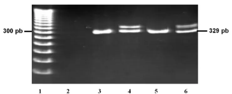
by visualization of a single band larger than 329 bp, wild-type gene fragment (homozygous mutation), or by the visualization of 329 bp wild-type gene fragment and one or more fragments larger than 329 bp (heterozygous mutation) as demonstrated in Figure 1. All PCR reagents were purchased from Invitrogen (Carlsbad, CA, USA).
Detection of FLT3-D835 mutation
The PCR technique followed by the restriction fragment length polymorphism (RFLP) technique described by Liang et al. (2003) were used to detect the FLT3-D835 mutation (Liang et al., 2003). First, the
region of exon 20 of the FLT3 gene was ampliied using specific primers (5’-CCGCCAGGAACGTGCTTG-3’
320 nm UV light transilluminator. The samples without mutation underwent complete enzymatic digestion, which resulted in the presence of two bands (68 bp/46 bp). Heterozygous samples for FLT3-D835 mutation showed
three bands (114 bp/68 bp/46 bp) after digestion, while homozygous samples showed a completely undigested band of 114 bp (Figure 2). All PCR reagents were purchased from Invitrogen (Carlsbad, CA, USA).
Statistical analysis
The association between the presence of FLT3
mutations and nominal variables such as gender, CD34 expression, WBC count at diagnosis, and patient outcome after six months of diagnosis were analyzed
using the Chi-square test. Numeric variables such as lactate dehydrogenase activity (LDH) and percentage of blasts were associated with the group of interest using Mann-Whitney U test. A p-value ≤0.05 was considered statistically signiicant for all analyses. Statistical analyses were done with the software SPSS (version 17.0).
RESULTS
During the period of this study, 85 samples of bone marrow and/or peripheral blood from patients with AL were evaluated for the presence of FLT3 mutations.
As displayed in Table II, the FLT3-ITD mutation
was observed in three patients in the adult group (6.7%)
and FLT3-D835 mutation in ive patients (11.1%). In the pediatric group, FLT3-IDT mutation was observed in
seven patients (17.5%) and FLT3-D835 mutation in just
one patient (2.5%).
According to the subtype in the adult group, it was observed that, of all cases of AML, 7.4% (two cases) and 14.8% (four cases) had FLT3-ITD and FLT3-D835,
respectively. In adult patients with ALL, 5.6% (one case) had either FLT3-ITD or FLT3-D835. These results show
that there was no correlation between AL subtype and presence of ITD or D835 mutations in adult patients.
The same was observed with the pediatric group, where 33.3% (four cases) of the patients with AML had
the FLT3-ITD mutation, while 10.7% (three cases) of the
patients with ALL showed the same mutation. Although
the FLT3-ITD mutation was not associated with the AL
subtypes (AML or ALL; P=0.168), it was observed that this mutation was more prevalent in children with AML.
Mutation FLT3-D835 was detected only in one child
(8.3%) with AML.
Regarding gender, FLT3 mutations were present in
20.8% (ive cases) of males and 13.6% (three cases) of females in the adult group. In the pediatric group, 17.4% (four cases) and 23.5% (four cases) of males and females, respectively, showed FLT3 mutations. No correlation was
found between gender and the presence of FLT3 mutations in either group.
As presented in the Table II, there was no correlation between the presence of FLT3-ITD or FLT3-D835 and prognostic factors such as age, WBC count, percentage
of blasts, CD34 expression, and LDH activity in the adult group. However, it is important to note that the patients who had FLT3-ITD mutation showed WBC count (median
29.4 x10³/mm3) and LDH activity (median 875.0 U/L)
above the upper reference values (11.0x10³/mm3 and 190.0
U/L, respectively).
As demonstrated in Table III, for the pediatric group, a
FIGURE 1 - PCR products of FLT3-ITD, analyzed by gel electrophoresis. Reverse view of ethidium-bromide-stained 12% polyacrylamide gel FLT3-ITD was detected as extra-bands in addition to normal band of 329 bp. Line 1- Molecular weight marker (50 bp); Line 2-Water control; Line 3-Patient sample wild type; Line 4- Patient sample with FLT3-ITD mutation; Line 5-wild type control; Line 6-) FLT3-ITD mutation control.
positive correlation was observed between WBC count and
the presence of FLT3-ITD in patients with AML (p=0.007)
and ALL (p=0.005). Furthermore, children with AML who
had the FLT3-ITD mutation showed a tendency to express
CD34 (p=0.078). Similar to the adult group, children who
had FLT3-ITD mutation showed LDH activity (median
1,292.0 U/L) above the maximum reference value. There was no correlation between FLT3-D835 mutation and the prognostic factors for the pediatric group. The association
between the presence of FLT3 gene mutations and the
patient progression after six months of diagnosis was also evaluated. In the adult group, the AML patients with FLT3-ITD who expressed CD34 tended to worse progression
(p=0.067). Regarding the pediatric group, there were no signiicant correlations between patient progression and
presence of FLT3 gene mutations.
DISCUSSION
Treatment of patients with FLT3 mutation represents a great clinical challenge. Patients with these mutations carry a particularly poor prognosis, with greater relapse rate, and inferior overall survival when compared to
patients with AL without this mutation (Taketani et al., 2004; Fedders et al., 2015; Wellmann et al., 2015). Given
the poor prognosis of patients with AL and FLT3 mutation, mainly FLT3-ITD, it has become a common practice to do allogeneic bone marrow transplantation during irst remission in patients with these mutations. However, there remains a signiicant relapse rate following transplant for FLT3-ITD, and it is possible that maintenance therapy with FLT-3 inhibitors will reduce the recurrence rates (Taketani
et al., 2004; Bornhauser et al., 2007; Schlenk et al., 2008; Dezern et al., 2011; Hassanein et al., 2016). Recently,
FLT3 inhibitors have emerged as a new option of treatment for these patients. Several FLT3 inhibitors have been tried as monotherapy or combined with other chemotherapeutic
agents (Hassanein et al., 2016).
Nevertheless, it is still unclear how and when these agents should be use during AML treatment
(Hassanein et al., 2016). Because of the impact of FLT3 mutation on prognosis and treatment protocols, the
guidelines from various major organizations, such as National Comprehensive Cancer Network and European LeukemiaNet, recommend FLT3 mutation analysis on
diagnosis.
TABLE II - Values of age, percentage of blasts, WBC count, LDH activity, CD34 expression, and progression of patients with AL
according to the absence or presence of FLT3 mutation and group
ADULT GROUP Presence of FLT3 mutation (n=7) Absence of FLT3 mutation (n=38)
Median Limits Median Limits
Age (years) 41.0 25.0 - 73.0 41.0 15.0 - 87.0
Blasts (%) 50.0 27.0 - 96.0 46.0 0.0 - 98.0
WBC (x103/mm3) 12.9 1.6 - 288.7 5.9 0.4 - 177.2
LDH (U/L) 875.0 259.0 - 5,189.0 300.0 61.0 - 6,500.0
N % N %
CD34 Expression 4 57.2 24 63.1
Death 3 42.8 18 47.4
Remission 4 57.2 20 52.6
PEDIATRIC GROUP Presence of
FLT3 mutation (n=8) Absence of FLT3 mutation (n=32)
Median Limits Median Limits
Age (years) 6.5 0.10 - 14.0 4.0 0.3 - 14.0
Blasts (%) 83.0 80.0 - 96.0 83.0 21.0 - 98.0
WBC (x103/mm3) 51.3 27.5 - 220.0 14.8 2.0 - 450.0
LDH (U/L) 1,227.0 786.0 - 13,087.0 955.0 322.0 - 14,559.0
N % N %
CD34 Expression 7 87.5 28 87.5
Death 5 62.5 8 25.0
Remission 3 37.5 24 75.0
Worldwide, the reported incidence of FLT3-ITD
in adults with AML is 15-35% (Nakao et al., 1996; Abu-Duhier et al., 2000; Kottaridis et al., 2001; Xu et al.,
2005; Amir, Yi-Bin, 2011). Additionally, the FLT3-D835 mutation incidence is 8-12% in AML cases (Small, 2006). Lucena-Araujo et al. demonstrated that the incidence of FLT3-ITD in Brazilian patients with AML is similar to the other countries, but the FLT3-TKD is lower. This
reinforces the idea that there are geographic diferences in FLT3 mutation frequency. Considering that Brazil
has continental proportions and multiethnic population,
it is expected that the incidence of these mutations are
different among Brazilian regions. The incidence and
clinical relevance of FLT3 mutations in AL patients from Santa Catarina are unknown, and it is important to
create regional data to establish public health policies that
improve AL with FLT3 mutation treatments in this region (Lucena-Araujo et al., 2010).
The results of this study showed a lower frequency
of FLT3-ITD mutation (7.4%). This discrepancy can be
explained by one of the two facts: i) the small number of patients included in the study, or ii) the intrinsic
characteristics of the population studied. Furthermore,
it should be noted that, as mentioned by Bao et al.
(2006), the incidences reported in other studies might be overestimated because the mutation analysis was usually performed on archived samples from patients who usually have leukocytosis, which is generally present in AML carrying FLT3 mutations (Bao et al., 2006). On the other hand, the incidence of the FLT3-ITD mutation in adults
with ALL described in other studies (5 - 10%)is similar
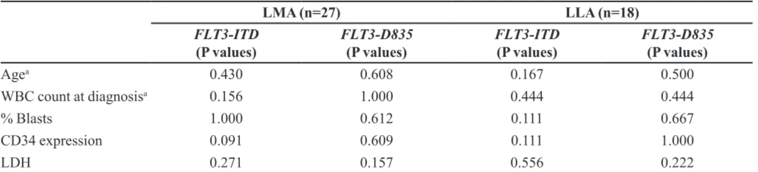
to that found in the present study (5.6%) (Kottaridis et TABLE III - Association between prognostic factors and presence of FLT3-IDT and FLT3-D835 mutations in adults with acute
leukemia
LMA (n=27) LLA (n=18)
FLT3-ITD
(P values)
FLT3-D835
(P values)
FLT3-ITD
(P values)
FLT3-D835
(P values)
Agea 0.430 0.608 0.167 0.500
WBC count at diagnosisa 0.156 1.000 0.444 0.444
% Blasts 1.000 0.612 0.111 0.667
CD34 expression 0.091 0.609 0.111 1.000
LDH 0.271 0.157 0.556 0.222
WBC - White-blood-cell; LDH – Lactate dehydrogenase; a For this analysis the variables age and WBC count at diagnosis were divided into two groups according to prognosis, as follows: Age - poor prognosis is ≥50 years old for AML and ≥60 years old for ALL, good prognosis is <50 years old for AML and <60 years old for ALL; WBC - poor prognosis is ≥20,000/mm3 for AML and ≥10,000/mm3 for ALL, good prognosis is <20,000/mm3 for AML and <10,000/mm3 for ALL. * P≤0.05 is considered to be statistically signiicant.
TABLE IV - Association between prognostic factors and presence of FLT3-ITD and FLT3-D835 mutations in children with acute
leukemia
LMA (n=12) LLA (n=28)
FLT3-ITD
(P values)
FLT3-D835
(P values)
FLT3-ITD
(P values)
FLT3-D835
(P values)
Agea 0.295 0.417 0.904
-WBC count at diagnosisa 0.007* 1.000 0.005*
-% Blasts 0.093 0.167 0.856
-CD34 expression 0.078 1.000 0.124
-LDH 0.111 0.667 0.514
al., 2001; Amir, Yi-Bin, 2011). However, because only
one case was identified in this study, it was difficult to assess properly the relation between this mutation and the
prognostic factors.
The incidence of FTL3-ITD in children with AML
is lower than in adults (5-16%) (Gale, Green, Allen,
2008). Regarding this aspect, the results obtained in this study showed higher percentages (33.3%). To the best of our knowledge, Al-Tonbary et al. (2009) conducted
the only available study on the presence of FLT3-ITD in children with ALL. Unlike the present study, which found an incidence of 10.7%, Al-Tonbary et al. reported that
children with ALL did not show the FLT3-ITD mutation.
As in the case of the adult group, this diference can be explained by the small number of patients included in this study and by the intrinsic characteristics of the population
studied.
In this study, FLT3-D835 incidence rate was 14.8% in AML cases, which is higher than the rates reported in other works (Small, 2006; Lucena-Araujo et al., 2010).
In ALL cases, our results showed FLT3-D835 frequency of 5.6%, with contrast with the literature results, that demonstrated a frequency of 1-3% (Abu-Duhier et al.,
2001; Yamamoto et al., 2001; Thiede et al., 2002). Concerning the FLT3-D835 mutations in the
pediatric group, there was only one case of AML (8.3%) and no ALL case positive for the mutation. The frequency of this mutation in AML children is in agreement with that mentioned in other pieces of research, which report it as rare (7 - 10%) (Meshinchi et al., 2006; Markova et al.,
2009). However, the reported incidence of FLT3-D835
in ALL children is 7.5% (Wang et al., 2010), which is
higher than that found in the present study. Based on
the incidences described for FLT3-ITD and FLT3-D835
mutations, a variation was observed among studies, which indicates that there are diferences in the frequency of these
mutations according to geographical areas.
In relation to the distribution of FLT3 mutations
according to gender, the present study is consistent with other indings where there is no signiicant correlation between these two variables (Al-Tonbary et al., 2009; Karabacak et al., 2010).
The prevalence of FLT3 mutations increases with age, but the presence of this mutation seems to have less
impact as a prognostic factor in patients above 60 years old than other adverse prognostic factors (Foran, 2010). In this study, no relation was observed between patient age
and the presence of FLT3 mutations in either group, which
is in agreement with other studies, which also found no association between these variables (Parcell et al., 2006; Wang et al., 2010).
WBC count, LDH activity, and cytogenetic
abnormalities at diagnosis are important prognostic
factors (Stone, 2009), as are the mutations in the FLT3
gene (Betz, Hess, 2010). The indings related to whether there is a correlation between the mutations in FLT3 gene and WBC count are controversial. While some groups
reported an association between these two variables in AML patients (Kottaridis et al., 2001; Thiede et al., 2002; Emerenciano et al., 2008; Peng et al., 2008; Wang et al., 2010), others found no such relationship (Schlenk et al., 2008; Karabacak et al., 2010). According to the
classiication of genetic abnormalities, mutations in the
FLT3 gene are considered class I mutations, which confer a survival advantage and increase cell proliferation (Betz,
Hess, 2010). This may explain why so many studies found an association between mutations in the FLT3 gene and
WBC count. In the present study, this relationship was not statistically signiicant in the adult group, but it was observed that the total WBC count was higher for patients who had the FLT3-ITD mutation. In the pediatric group, a
signiicant association was observed between high WBC
and presence of FLT3-ITD mutation.
In patients with AL, an increase in LDH activity is considered a poor prognostic factor (Varma, Varma, 2008). A study with adult AL patients showed an association between the presence of FLT3 gene mutations and increase
in LDH activity (Peng et al., 2008). However, this same
observation was not found in another study (Karabacak et al., 2010), as well as in the present one. Nevertheless, in
both the adult and the pediatric groups, patients with FLT3
gene mutations showed higher LDH activity (875.0 U/L and 1,227.0 U/L, respectively) than the highest reference limit (190.0 U/L).
Although the patients with FLT3 gene mutations
included in this study had high percentages of blast cells in both groups, as reported by Liu et al. (2007), the presence of FLT3 gene mutations and percentage of blasts were not
statistically related.
Another relevant prognostic factor is the CD34 expression in blast cells. A study by Zhu et al. (2013)
showed that CD34 expression is an FLT3-ITD-independent predictor of poor prognosis. In contrast, other studies
reported an association between FLT3-ITD mutation
and the presence of the CD34 marker in AML patients
(Barragán et al., 2011; Zhu et al., 2013). In the present
study, no correlation between FLT3 gene mutations and
CD34 expression were found. However, four of the ive
disease-free survival clearly associated with leukocytosis. These indings classify FLT3-ITD as a poor-prognostic
mutation (Zhu et al., 2013). Mathews et al. (1991)suggest
that there is an inverse relationship between the rate of molecular remission and patients with the presence of
FLT3-ITD mutation. In the present study, no association
was found between the presence of FLT3-ITD mutation
and a poor outcome (death or relapse) in adult patients. Nevertheless, the AML pediatric group showed a tendency of association between FLT3-ITD and death, which characterizes it as an indicator of poor prognosis. Studies
have not shown a signiicant relation between FLT3-D835 mutation and complete remission, disease-free survival, or
overall survival (Matthews et al., 1991; Wang et al., 2010).
These indings are consistent with the results presented in this study.
CONCLUSIONS
In conclusion, the present analysis showed that there is no association between the evaluated prognostic factors such as age, LDH activity, and CD34 expression and FLT3
gene mutations in adult and pediatric patients with AL.
Nevertheless, the presence of FLT3-ITD mutation was
signiicantly related to WBC count in the pediatric group.
These results suggest that the FLT3 gene mutations are independent poor prognostic factors.
ACKNOWLEDGEMENTS
This research was supported by grants and fellowships from the National Counsel of Technological and Scientific Development – CNPq (Brazil), the
Coordination for the Improvement of Higher Education
Personnel – CAPES (Brazil), and Fundação de Amparo à Pesquisa e Inovação do Estado de Santa Catarina –
FAPESC. We thank Professor Dr Maria Luiza Bazzo for allowing us the use of her molecularbiology laboratory
facilities.
REFERENCES
ABU-DUHIER, F.M.; GOODEVE, A.C.; WILSON, G.A.; GARI, M.A.; PEAKE, I.R.; REES, D.C.; VANDENBERGHE, E.A.; WINSHIP, P.R.; REILLY, J.T. FLT3 internal tandem duplication mutations in adult acute myeloid leukaemia deine a highrisk group. Br. J. Haematol., v.111, n.1, p.190-195, 2000.
ABU-DUHIER, F.M.; GOODEVE, A.C.; WILSON, G.A.; CARE, R.S.; PEAKE, I.R.; REILLY, J.T. Identification of novel FLT-3 Asp835 mutations in adult acute myeloid leukaemia. Br. J. Haematol., v.113, n.1, p.983-988, 2001.
AL-TONBARY, Y.; MANSOUR, A.K.; GHAZY, H.; ELGHANNAM, D.M.; ABD-ELGHAFFAR, H.A. Prognostic significance of foetal-like tyrosine kinase 3 mutation in Egyptian children with acute leukaemia. Int. J. Lab. Hematol., v.31, n.3, p.320-326, 2009.
AMIR, F.T; YI-BIN, C. Treatment of FLT3-ITD acute myeloid leukemia. Blood Res., v.1, n.2, p.175-189, 2011.
BALDUS, C.D.; MRÓZEK, K.; MARCUCCI, G.; BLOOMFIELD, C.D. Clinical outcome of de novo acute myeloid leukaemia patients with normal cytogenetics is afected by molecular genetic alterations: a concise review.
Br. J. Haematol., v.137, n.1, p.387-400, 2007.
BAO, L.; WANG, X.; RYDER, J.; JI, M.; CHEN, Y.; CHEN, H.; SUN, H.; YANG, Y.; DU, X.; KERZIC, P.; GROSS, S.A.; YAO, L.; LV, L.; FU, H.; LIN, G.; IRONS, R.D. Prospective study of 174 de novo acute myelogenousleukemias according to the WHO classiication: Subtypes, cytogenetic features and FLT3 mutations. Eur. J. Haematol., v.77, n.1, p.35-45, 2006.
BARRAGÁN, E.; MONTESINOS, P.; CAMOS, M.; GONZÁLEZ, M.; CALASANZ, M.J.; ROMÁN-GÓMEZ, J.; GÓMEZ-CASARES, M.T.; AYALA, R.; LÓPEZ, J.; FUSTER, Ó.; COLOMER, D.; CHILLÓN, C.; LARRAYOZ, M.J.; SÁNCHEZ-GODOY, P.; GONZÁLEZ-CAMPOS, J.; MANSO, F.; AMADOR, M.L.; VELLENGA, E.; LOWENBERG, B.; SANZ, M.A. Prognostic value of FLT3 mutations in patients with acute promyelocytic leukemia treated with all-trans retinoic acid and anthracycline monochemotherapy. Haematologica, v.96, n.10, p.1470-1477, 2011.
BETZ, B.L.; HESS, J.L. Acute Myeloid Leukemia Diagnosis in the 21st Century. Arch. Pathol. Lab. Med., v.134, n.1,
p.1427-33, 2010.
BORNHÄUSER, M.; ILLMER, T.; SCHAICH, M.; SOUCEK, S.; EHNINGER, G.; THIEDE, C. Improved outcome after stem-cell transplantation in FLT3/ITD-positive AML. Blood,
DEZERN, A.E.; SUNG, A.; KIM, S.; SMITH, B.D.; KARP, J.E.; GORE, S.D.; JONES, R.J.; FUCHS, E.; LUZNIK, L.; MCDEVITT, M.; LEVIS, M. Role of allogeneic transplantation for FLT3/ITD acute myeloid leukemia: outcomes from 133 consecutive newly-diagnosed patients from a single institution. Biol. Blood. Marrow Transplant, v.17, n.9, p.1404-1409, 2011.
EMERENCIANO, M.; MENEZES, J.; VASQUEZ, M.L.; ZALCBERG, I.; THULER, L.C.S.; POMBO-DE-OLIVEIRA, M.S.; THE BRAZILIAN COLLABORATIVE STUDY GROUP OF INFANT ACUTE LEUKEMIA. Clinical relevance of FLT3 gene abnormalities in Brazilian patients with infant leukemia. Leuk. Lymphoma, v.49, n.12, p.2291-2297, 2008.
FEDDERS, H.; SCHEWE, MARTIN, D.; ZIMMERMANN, M.; ALSADEQ, M.; MOERICKE, A.; STADT, U.Z.; HORSTMANN, A.; PIETERS, R.; SCHRAPPE, M.; CARIO, G.; STANULLA, M. Constitutive activation of FLT3 is a positive prognostic factor in infants with MLL-rearranged acute lymphoblastic leukemia. Blood, v.126, p.1417, 2015.
FORAN, J.M. New prognostic markers in acute myeloid leukemia: perspective from the clinic. Hematol. Am. Soc. Hematol. Educ. Program, v.2010, n.1, p.47-55, 2010.
GALE, R.E.; GREEN, C.; ALLEN, C. The impact of FLT3 internal tandem duplication mutant level, number, size, and interaction with NPM1 mutations in a large cohort of young adult patients with acute myeloid leukemia. Blood, v.111, n.5, p.2776-2784, 2008.
HASSANEIN, M.; ALMAHAYNI, M.H.; AHMED S.O.; GABALLA, S.; FAKIH, R. E. FLT3 inhibitors for treating acute myeloid leucemia. Clin. Lymphoma, Myeloma Leuk., v.16, n.10, p.543-549, 2016.
KARABACAK, B.H.; ERBEY, F.; BAYRAM, I.; YILMAZ, S.; ACIPAYAM, C.; KILINC, Y.; TANYEL, A. Fms-like tyrosine kinase 3 mutations in childhood acute leukemias and their association with prognosis. Asian. Pac. J. Cancer Prev. v.11, n.4, p.923-927, 2010.
KOH, Y. ; PARK, J.; AHN, K.S.; KIM, I.; BANG, S.M.; LEE, J.H.; YOON, S.S.; LEE, D.S.; LEE, Y.Y.; PARK, S.; KIM, B.K. Diferent clinical importance of FLT3 internal tandem duplications in aml according to FAB classiication: possible existence of distinct leukemogenesis involving monocyte diferentiation pathway. Ann. Hematol., v.88, n.11, p.1089-1097, 2009.
KOTTARIDIS, P.D.; GALE, R.E.; FREW, M.E.; HARRISON, G.; LANGABEER, S.E.; BELTON, A.A.; WALKER, H.; WHEATLEY, K.; BOWEN, D.T.; BURNETT, A.K.; GOLDSTONE, A.H.; LINCH, D.C. The presence of a FLT3 internal tandem duplication in patients with acute myeloid leukemia (AML) adds important prognostic information to cytogenetic risk group and response to the irst cycle of chemotherapy: analysis of 854 patients from the United Kingdom Medical Research Council AML 10 and 12 trials.
Blood, v.98, n.6, p.1752-1759, 2001.
LIANG, D.C.; SHIH, L.Y.; HUNG, I.J.; YANG, C.P.; CHEN, S.H.; JAING, T.H.; LIU, H.C.; WANG, L.Y.; CHANG, W.H. FLT3-TKD mutation in childhood acute myeloid leucemia. Leukemia, v.17, n.5, p.883-886, 2003.
LIERSCH, R.; MÜLLER-TIDOW, C.; BERDELW. E.; KRUG, U. Prognostic factors for acute myeloid leukaemia in adults– biological signiicance and clinical use. Brit. J. Haematol.,
v.165, n.1, p.17-38, 2014.
LIU, H.; YU, H.; JIA, H.Y.; ZHANG, W.; GUO, C.J. Detection of FLT3 gene mutation in hematologic malignancies and its clinical signiicance. Zhongguo Shi Yan Xue Ye Xue Za Zhi, v.15, n.4, p.709-713, 2007.
LUCENA-ARAUJO, A.R.; SOUZA, D.L.; MORATO DE OLIVEIRA, F.; BENICIO, M.T.; FIGUEIREDO-PONTES, L.L.; SANTANALEMOS, B.A.; DOS SANTOS, G.A.; JACOMO, R.H.; DINARTESANTOS, A.R.; YAMAMOTO, M.; SILVA-JR, W.A.; DE LOURDES CHAUFFAILLE, M.; REGO, E.M. Results of FLT3 mutation screening and correlations with immunophenotyping in 169 Brazilian patients with acute myeloid leukemia. Ann. Hematol., v.89, n.2, p.225-228, 2010.
MATTHEWS, W.; JORDAN, C.T.; WIEGAND, G.W.; PARDOLL, D.; LEMISCHKA, I.R. A receptor tyrosine kinase speciic to hematopoietic stem and progenitor cell-enriched populations. Cell, v.65, n.7, p.1143-52, 1991.
MCCULLOCH, E.A. Stem cells in normal and leukemic hemopoiesis (Henry Stratton Lecture). Blood, v.62, n.1, p.1-13, 1982.
MESHINCHI, S.; WOODS, W.G.; STIREWALT, D.L.; SWEETSER, D.A.; BUCKLEY, J.D.; TJOA, T.K.; BERNSTEIN, I.D.; RADICH, J.P. Prevalence and prognostic signiicance of FLT3 internal tandem duplication in pediatric acute myeloid leukemia. Blood, v.97, n.1, p.89-94, 2001.
MESHINCHI, S.; ALONZO, T.A.; STIREWALT, D.L.; ZWAAN, M.; ZIMMERMAN, M.; REINHARDT, D.; KASPERS, G.J.; HEEREMA, N.A.; GERBING, R.; LANGE, B.J.; RADICH, J.P. Clinical implications of FLT3 mutations in pediatric AML. Blood, v.108, n.12,
p.3654-3661, 2006.
MURPHY, K.M.; LEVIS, M.; HAFEZ, M.J.; GEIGER, T.; COOPER, L.C.; SMITH, B.D.; SMALL, D.; BERG, K.D. Detection of FLT3 internal tandem duplication and D835 mutations by a multiplex polymerase chain reaction and capillary electrophoresis assay. J. Molec. Diagn., v.5, n.2, p.96-108, 2003.
NAKAO, M.; YOKOTA, S.; IWAI, T.; KANEKO, H.; HORIIKE, S.; KASHIMA, K.; SONODA, Y.; FUJIMOTO, T.; MISAWA, S. Internal tandem duplication of the lt3 gene found in acute myeloid leukemia. Leukemia, v.10, n.12, p.1911-1918, 1996.
PARCELL, B.W.; IKEDA, A.K.; SIMMS-WALDRIP, T.; MOORE, T.B.; SAKAMOTO, K.M. FMS-like tyrosine kinase 3 in normal hematopoiesis and acute myeloid leukemia. Stem Cells, v.24, n.1, p.1174-1184, 2006.
PENG, H.L.; ZHANG, G.S.; GONG, F.J.; SHEN, J.K.; ZHANG, Y.; XU, Y.X.; ZHENG, W.L.; DAI, C.W.; PEI, M.F.; YANG, J.J. Fms-like tyrosine kinase (FLT)3 and FLT3 internal tandem duplication in diferent types of adult leukemia: analysis of 147 patients. Croat. Med. J., v.49, n.1, p.650-659, 2008.
SCHLENK, R.F.; DÖHNER, K.; KRAUTER, J.; FRÖHLING, S.; CORBACIOGLU, A.; BULLINGER, L.; HABDANK, M.; SPÄTH, D.; MORGAN, M.; BENNER, A.; SCHLEGELBERGER, B.; HEIL, G.; GANSER, A.; DÖHNER, H.; GERMAN-AUSTRIAN ACUTE MYELOID LEUKEMIA STUDY GROUP. Mutations and treatment outcome in cytogenetically normal acute myeloid leukemia. N. Engl. J. Med., v.358, p.1909-1918, 2008.
SMALL, D. FLT3 mutations: biology and treatment. ASH Educ. Program, v.2006, n.1, p.178-184, 2006.
STONE, R.M. Prognostic factors in AML in relation to (ab) normal karyotype. Best Pract. Res. Clin. Haematol., v.22, n.1, p.523-528, 2009.
SWERDLOW, S.H.; CAMPO, E.; HARRIS, N.L.; JAFFE, E.S.; PILERI, S.A.; STEIN, H.; THIELE, J.; VARDIMAN, J.W. WHO classiication of tumours of haematopoietic and
lymphoid tissues. 4.ed. Geneva: WHO Press, 2008. 439p.
TAKETANI, T.; TAKI, T.; SUGITA, K.; FURUICHI, Y.; ISHII, E.; HANADA, R.; TSUCHIDA, M.; SUGITA, K.; IDA, K.; HAYASHI, Y. FLT3 mutations in the activation loop of tyrosine kinase domain are frequently found in infant ALL with MLL rearrangements and pediatric ALL with hyperdiploidy. Blood, v.103, n.3, p.1085-8, 2004.
THIEDE, C.; STEUDEL, C.; MOHR, B.; SCHAICH, M.; SCHÄKEL, U.; PLATZBECKER, U.; WERMKE, M.; BORNHÄUSER, M.; RITTER, M.; NEUBAUER, A.; EHNINGER, G.; ILLMER, T. Analysis of FLT3-activating mutations in 979 patients with acute myelogenous leukemia: association with FAB subtypes and identification of subgroups with poor prognosis. Blood, v.99, n.12,
p.4326-35, 2002.
VARMA, N.; VARMA, S. Proliferative indices, cytogenetics, immunophenotye and other prognostic parameters in myelodysplastic syndromes. Indian J. Pathol. Microbiol., v.51, n.1, p.97-101, 2008.
WANG, W.; WANG, X.Q.; XU, X.P.; LIN, G.W. Prevalence and prognostic significance of FLT3 gene mutations in patients with acute leukaemia: analysis of patients from the Shanghai Leukaemia Cooperative Group. J. Int. Med. Res., v.38, n.2, p.432-442, 2010.
WELLMANN, S.; MODEREGGER, E.; ZELMER, A.; BETTKOBER, M.; STACKELBERG, A.V.; HENZE, G.; SEEGER, K. FLT3 mutations in childhood acute lymphoblastic leukemia at irst relapse. Leukemia, v.19, p.467-468, 2005.
XU, F.; TAKI, T.; YANG, H.W.; HANADA, R.; HONGO, T.; OHNISHI; KOBAYASHI M; BESSHO, F.; YANAGISAWA, M.; HAYASHI, Y. Tandem duplication of the FLT3 gene is found in acute lymphoblastic leukaemia as well as acute myeloid leukaemia but not in myelodysplastic syndrome or juvenile chronic myelogenousleukaemia in children. Br. J. Haematol., v.105, n.1, p.155-162, 2005.
YAMAMOTO, Y.; KIYOI, H.; NAKANO, Y.; SUZUKI, R.; KODERA, Y.; MIYAWAKI, S.; ASOU, N.; KURIYAMA, K.; YAGASAKI, F.; SHIMAZAKI, C.; AKIYAMA, H.; SAITO, K.; NISHIMURA, M.; MOTOJI, T.; SHINAGAWA, K.; TAKESHITA, A.; SAITO, H.; UEDA, R.; OHNO, R.; NAOE, T. Activating mutation of D835 within the activation loop of FLT3 in human hematologic malignancies. Blood, v.97, n.8, p.2434-39, 2001.
ZHU, H-H.; LIU, Y-R.; JIANG, H.; LU, J.; QIN, Y-Z.; JIANG, Q.; BAO, L.; RUAN, G-R.; JIANG, B.; HUANG, X. CD34 expression on bone marrow blasts is a novel predictor of poor prognosis independent of FlT3-ITD in acute myeloid leukemia with the NPM1-mutation. Leukemia Res., v.37,
n.1, p.624-630, 2013.