Antibodies to yeast Sm motif 1 cross-react with human Sm core
polypeptides
Diana Bahia1, Josep Font2, Amina Khaouja1, NarcõÂs Carreras1, Ruth Espuny1, Regina Maria Barretto Cicarelli3, Miguel Ingelmo2and Montse Bach-Elias1
1IIBB-CSIC, Barcelona, Spain;2Systemic Autoimmune Disease Unit, Hospital ClõÂnico y Provincial de Barcelona, Spain; 3Faculdade de CieÁncias FarmaceÃuticas, UNESP, Araraquara, SP, Brazil
Two regions common to all UsnRNP core polypeptides have been described: Sm motif 1 and Sm motif 2. Rabbits were immunized with a 22 amino-acid peptide containing one segment of Sm motif 1 (YRGTLVSTDNYFNLQL-NEAEEF, corresponding to residues 11±32) from yeast F protein. After immunization, the rabbit sera contained antibodies that not only reacted specifically with the peptide from yeast F protein but also cross-reacted with Sm polypeptides from mammals; that is, with purified human U1snRNPs. The results suggest that the peptide used and human Sm polypeptides contain a common feature recognized by the polyclonal antibodies. A large collection of human systemic lupus erythematosus sera was assayed using the yeast peptide as an antigen source. Seventy per cent of systemic lupus erythematosus sera contain an antibody specificity that cross-reacts with the yeast peptide.
Keywords: autoimmunity; epitope mapping; peptide-ELISA; Sm motif 1; UsnRNPs.
Core polypeptides of UsnRNPs are recognized by the sera of a subclass of rheumatic patients suffering from systemic lupus erythematosus (SLE). Autoantibodies against the UsnRNP-core polypeptides are detected in 10±30% of SLE patients, depend-ing on the detection technique and source of antigen used [1]. Core polypeptides are found in UsnRNPs [1] and are highly conserved through evolution being found in both humans and yeast [2±5].
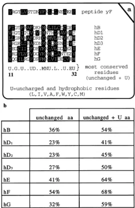
We focused on the Sm motifs of the UsnRNP-core polypeptides: Sm motif 1 (referred to as Motif-1) encompasses 32 amino acids and Sm motif 2 encompasses 14 amino acids [2± 5]. Both motifs are conserved in all human UsnRNP core polypeptides B0, B, D
1±3, E, F and G. Using Western blotting, it
has been demonstrated that B0, B and D
1±3 polypeptides are
recognized by sera from SLE patients (SLE sera) [6]. However, E, F and G autoantibodies are rarely detected in SLE sera [7]. This is particularly surprising because human D1 and D3
polypeptides present lower homologies to the consensus sequence than E, F and G polypeptides [2±5]. Neither epitope mapping studies nor peptide assays have given clear auto-immune data on the region that contains the UsnRNP Sm motif 1 and Sm motif 2 [8±17]. Only truncated versions of Sm motifs were used. In previous studies, Rokeach et al. [10] used two fusion proteins: one containing a complete Sm motif 1 and the other containing a truncated version of the Sm motif 1 plus a complete Sm motif 2. Both gave negative results with Western blotting against 19 anti-(Sm) sera. Therefore, the techniques used may explain why not all the UsnRNP-core polypeptides have been detected with anti-(Sm) sera. In this regard, Brahms
and colleagues [18] have recently shown that E, F and G proteins are recognized as a tri-polypeptide complex under native conditions, but are not recognized in their denatured states. Other authors have reported positive reactions for anti-(Sm) sera against truncated peptides of Sm motifs [8,11± 13,16,17], thus indicating that this region probably contains an epitope that reacts with anti-(Sm) sera. As the Sm motifs have been clearly described from sequence comparisons [2,4,5], it now is possible to study and analyze one complete Sm motif. Sm motifs 1 and 2 are interesting regions as they are found both in yeast and human proteins. There is some degree of conservation between the different Sm motifs, but it is not evident whether they show similar cross-reactivity to antibodies obtained against one particular Sm motif. We focused our interest on Sm motif 1 of the yeast F protein [2,4,5], and obtained polyclonal antibodies against the Sm motif 1. Immunoaffinity purified polyclonal antibodies from immunized animals cross-reacted extensively with human B0, B, D
1and D3
Sm proteins. This indicates that the Sm motifs from the yeast F protein and human B0, B, D
1and D3Sm polypeptides contain a
common feature which is recognized by the polyclonal sera.
M AT E R I A L S A N D M E T H O D S
Patients and sera from normal donors
Two-hundred and fourteen sera from human SLE patients were tested in this study. The SLE patients were selected according to American Rheumatism Association (ARA) classification criteria for SLE; 95 sera from healthy donors were obtained from the blood bank.
Peptide choice and synthesis
We selected a Sm motif 1 from the partially sequenced yeast F protein, taking into account the following: a yeast peptide should render a better antibody response in mammals; the Sm
Correspondence toM. Bach-Elias, IIBB-CSIC, c/Jorge Girona Salgado 18-26, 08034 Barcelona, Spain. Fax: + 34 3 204 5904;
Tel: + 34 3 400 6134; E-mail: mbebmc@cid.csic.es
motif 1 is larger than the Sm motif 2 and contains more unchanged amino acids in the conserved region (Figs 1 and 2a); as the Sm motif 1 is not a clear or strong epitope recognized by anti-(Sm) sera, a Sm motif 1 from a nontypical Sm antigen was chosen, i.e. a Sm motif 1 different to those found in B0, B and
D1±3Sm polypeptides, as these proteins are highly reactive with
anti-(Sm) sera. In conclusion, any one of the three E, F and G polypeptides would have been suitable for our purpose, and we finally chose the Sm motif 1 of the yeast F polypeptide because its chemical synthesis was easier than that of the other two.
The Sm motif 1 from the yeast F protein has a homology of 91% with the Sm motif 1 consensus sequence (U.G.U.UD.MNU.L.U.EU, U being uncharged and hydrophobic residues; Figs 1 and 2a), and a homology of 68% with the Sm motif 1 of human F protein (counting unchanged amino acids and hydrophobic amino acids at the same position; Fig. 2b). The percentage homology of the selected yeast F peptide with the same region of each core protein is shown in Fig. 2b.
A second important factor is the purity of the peptide. Special care was taken using HPLC to obtain a peptide with a purity of
<95%. The peptide was conjugated to keyhole limpet
hemocyanin (KLH), which is a nonrelated protein that normally gives a low background in ELISA against rabbit and SLE sera (our nonpublished results). One cysteine was added to the C-terminus of the peptide to allow coupling with KLH. A second conjugate containing Motif 1-BSA was also obtained to carry out ELISA assays with the immunized rabbit sera, thus monitoring the efficiency of immunization with Sm motif 1-KLH (named Motif 1-KLH).
Motif 1±peptide conjugate
Sm±peptide conjugate containing the Sm amino acid motif 1 of the putative yeast F protein (YRGTLVSTDNYFNLQL-NEAEEFC, 22 amino-acids from the Sm motif 1 of the yeast F protein plus a cysteine added to the C-terminus) was obtained using solid-phase methods, such as C-terminal carboxamides on
p-methylbenzhydrylamine resin using tert-butyloxycarbonyl (Boc) chemistry at the Department of Organic Chemistry of the University of Barcelona. An extra tyrosine residue (Y) was
added to the N-terminus in order to increase the homology of the Sm motif 1 of the yeast F peptide with the Sm motif 1 of the human F peptide. After hydrofluoric acid (HF) cleavage and HPLC purification, the products were characterized by amino acid analysis and electrospray mass spectrometry. Coupling to KLH was carried out using the 3-maleimidobenzoyl-N -hydroxysuccinimide ester following Kitagawa and Aikawa [19]. Peptide-to-carrier molar ratios of 3517 to 1, were determined by amino acid analysis of the conjugates and the conjugate thus obtained was named Motif 1-KLH. A similar conjugate using BSA rather than KLH was also obtained using the same procedure.
Animal immunizations
Two New Zealand White rabbits, R328 and R329, were immunized with the Motif 1-KLH conjugate as follows: day 1, 150mg in NaCl/Pi/complete Freund adjuvant (NaCl/Pi: NaCl,
140 mm; K2H2PO4, 20 mm) (1 : 1, v/v); subsequent
immuniz-ations were carried out with 100mg in NaCl/Pi/incomplete
Freund adjuvant (1 : 1, v/v) on days 15, 30 and 60. On day 67, blood was taken from the animals' ears. Sera were obtained by
Fig. 1. Comparison of yeast and human Sm motifs.The amino acids in the black boxes represent fully conserved positions, whereas those in white boxes indicate sequences with 70±85% conservation. Sm motif 1 and Sm motif 2 are 32 and 14 amino acids long, respectively, according to the previously described nomenclature [2,4]. Gene accession numbers and initial references are as follows: (`h' indicates Homo sapiensand `y' represents
Saccharomyces cerevisiae) hB, X17567 [26]; hD2, U15008 [27]; hD1,
P13641 [28]; hD3, U15009 [27]; yD1L04669 [29]; yD3, M65144 [30]; hE,
X12466 [31]; hF, X85372 [2]; yF X82778 [4,32]; hG, X85373 [2]; and yG, L31794. The peptide chosen for this study corresponds to amino acids 11±32 of Sm motif 1 and is shown in the figure.
centrifugation of the blood and their titers were assayed using ELISA.
Purification of human UsnRNPs and Western blotting
Purification of human UsnRNPs from human extracts (HeLa cells) was carried out with anti-(trimethylguanosine) columns [20]. Further purification of the UsnRNPs using Mono Q FPLC columns gave purified U1snRNP [20]. Western blotting with purified human U1snRNPs was performed as described previously [21]. Two gel systems were used to resolve the U1snRNP proteins: 10% polyacrylamide/SDS/tricine gels [22] or 12.5% polyacrylamide/SDS/high-TEMED gels [23], depending on the purpose of the experiment (see Results). Western blotting with the peptide only was similarly performed by running 2mg
peptide´lane21in 10% polyacrylamide/SDS/tricine gels [22] and transferring the peptide to 0.2mm nitrocellulose membranes at
200 mA for 1 h in transfer buffer (Tris/HCl 25 mm pH 8.3,
glycine 192 mm, 10% methanol). Blot immunostaining was
performed with protein A±alkaline phosphatase and ECL (Amersham Life Science) Western detection reagents (chemo-luminescence), following the manufacturer's instructions.
Immunoprecipitation
Immunoprecipitation of the UsnRNPs from HeLa nuclear extracts was essentially performed as described previously [24] with some modifications: all incubation steps including binding of the polyclonal antibody to protein A±Sepharose and the UsnRNPs to the protein A±antibody matrix were performed overnight at 48C. HeLa nuclear extract samples were first incubated at 378C for 30 min, to improve the exposure of the epitopes to the antibodies, and then 50mL of this extract was
added to the protein A±antibody matrix to continue the immunoprecipitation.
Purification of the antibodies using immunoaffinity columns
The peptide containing the yeast Sm motif 1 was covalently bound to Sepharose using CNBr-activated beads, following the manufacturer's instructions. Sera from both immunized rabbits were saturated to 50% with ammonium sulfate and centrifuged. The pellets, which contained the antibodies, were dissolved and dialyzed against NaCl/Pi300 (same as NaCl/Pibut containing
300 mm NaCl) plus 0.5 mm PhCH2SO2F. After dialysis, the
antibody solution was applied slowly to the peptide column (previously equilibrated with NaCl/Pi300/PhCH2SO2F). After
washing with NaCl/Pi300/PhCH2SO2F (6 column vol.), the
bound antibodies were eluted with 100 mmglycine/HCl pH 2.5,
and the eluted fractions were immediately collected in one fraction-volume of 1mTris pH 8, and mixed to change the pH.
Protein fractions were dialyzed against NaCl/Pi/PhCH2SO2F and
then assayed using ELISA plates with wells coated with Motif 1-BSA. Using this column we have been able to purify 1±2% of the IgG fraction.
Anti-(Motif 1-KLH) ELISA
ELISA wells (Maxisorp, NUNC) were coated with 50mL of a
solution of Motif 1-KLH conjugate in NaCl/Pi(20 mmKH2PO4
pH 7.4, 140 mm NaCl, 0.02% NaN3) at a concentration of
10mg´mL21. The control wells were coated with the same
amount of KLH alone. The plates were covered with a wipe and incubated overnight at room temperature in order to dry the wells. Next day the plates were dried for 1 h at 378C and then
washed six times with NaCl/Pi and saturated with 100mL of
NaCl/Pi/2% teleostean gelatin for 1 h at 378C. Then the plates
were washed six times with NaCl/Pi, and 50mL of the human
sera diluted 1/400 in 2% teleostean gelatin/NaCl/Pi/T (NaCl/Pi
with 0.1% Tween 20) was added to each well (both the peptide± KLH and KLH alone wells) and then incubated for 90 min at 378C. The plates were washed six times with NaCl/Pi and
50mL of anti-(human specific IgG), (with minimal
cross-reactivity to either IgM or IgA, as these antibodies were purified by loading to human IgM and IgA columns), conjugated to alkaline phosphatase and diluted 1/2000 in 2% teleostean gelatin/NaCl/Pi/T, was added to all the wells, which were then
incubated for 1 h at room temperature. The plates were washed six times with NaCl/Pi, 80mL of substrate solution (50 mm
Na2CO3/NaHCO3 pH 9.5, 2 mm MgCl2 and 1 mg´mL21 p
-nitro-phenyl-phosphate) was added to each well and the plates were read at 405/450 nm 30 min later. The value obtained against KLH only is the background of the assay and was subtracted from the values obtained against Motif 1-KLH conjugate (the present work contains already subtracted data; i.e. absorbance value of the serum vs. Motif 1-KLH conju-gate ± absorbance value of the serum vs. KLH only). Owing to the high ratio of conjugation (3500 : 1) 30% of the sera was also assayed in parallel with 10mg KLH only, and similar
absorbance values were obtained with 5mg or 10mg of KLH
only. Absorbance values against KLH were low (the statistical evaluation of the data is shown in the legend to Fig. 1).
ELISA using rabbit sera was performed in a similar way, but using Motif 1-BSA to coat the wells (thus avoiding cross-reactivity with the carrier KLH) and with a specific anti-(rabbit) IgG.
Competitive inhibition ELISA
The ELISA wells (Maxisorp, NUNC) were prepared as described above but with 50mL of the solution of Motif
1-KLH conjugate and NaCl/Pi(20 mm KH2PO4 pH 7.4, 140 mm
NaCl, 0.02% NaN3), at a concentration of 5mg´mL21, and
incubated at room temperature. Sm motif 1 peptide was prepared in NaCl/Pi at 300 ng´mL21. The sera were diluted
1/400 in 2% teleostean gelatin/NaCl/Pi. A 50-mL aliquot of
the diluted sera was allowed to compete with 50mL of the
Motif 1-KLH conjugate solution at room temperature for 1 h in an assay tube. The control, which consisted of diluted serum without inhibitor, was prepared by adding 50mL of the diluted
incubated sera to 50mL of NaCl/Piunder the same conditions as
those used for the competition samples. The plates were then washed three times with NaCl/Pi/T and saturated with 100mL of
NaCl/Pi/2% teleostean gelatin for 30 min at room temperature.
The plates were then washed three times with NaCl/Pi, the
competition mixture (50mL of diluted serum plus 50mL of
peptide solution) and control incubation (50mL of diluted serum
plus 50mL of NaCl/Pi) were added to the corresponding wells
and the plates were incubated for 1 h at 378C. The plates were washed three times in NaCl/Pi/T and 50mL of specific
anti-(human) IgG (with minimal cross-reactivity to IgM, IgA conjugated to alkaline phosphatase, diluted 1/4000 in 2% teleostean gelatin/NaCl/Pi/T) was added to the wells, which
were then incubated for 90 min at room temperature. The plates were washed three times with NaCl/Pi/T and 80mL of substrate
solution (50 mm Na2CO3/NaHCO3 pH 9.5, 2 mm MgCl2 and
to the noncompeted wells. Competition ELISAs with rabbit sera were performed in a similar way, but using Motif 1-BSA conjugate to coat the wells (thus avoiding cross-reactivity with the carrier KLH) and with a specific anti-(rabbit) IgG. As a second set of control experiments, sera were also competed with 300 ng of BSA or KLH and no inhibition was seen.
R E S U LT S A N D D I S C U S S I O N
Antibodies obtained in rabbits against the yeast Sm motif 1 immunoprecipitated UsnRNPs from HeLa nuclear extracts
Two rabbits were immunized with the Motif 1-KLH conjugate. Sera from both animals showed titers higher than 1/5000 in ELISAs coated with the Motif 1-BSA conjugate, whereas the corresponding nonimmune sera did not react with the Motif 1-BSA conjugate. The immunoreaction of the sera from immunized animals with the Motif 1-BSA conjugate was inhibited in competition ELISAs with increasing concentrations of free peptide (results not shown). To show that the animals were not rendering an immune response to the adjuvants, two other rabbits were immunized with KLH only (same protein quantity and same immunization protocol). Any sera from the two latter rabbits showed reactivity with the Sm motif 1 peptide (not shown).
Both sera were studied in immunoprecipitation assays in order to see whether they were able to bind to native UsnRNPs from human extracts. Figure 3 shows that serum R328 immunoprecipitated native UsnRNPs (Fig. 3, lane 1), whereas the nonimmune serum from the same animal did not (Fig. 3, lane 2). Although serum from rabbit R329 has the same titer as serum R328, R329 failed to immunoprecipitate native UsnRNPs (Fig. 3, lane 3). Lane 4 in Fig. 3 shows a positive control of the immunoprecipitation with anti-(trimethylguanosine). Other bands also detected in the immunoprecipitation assay with serum R328 may correspond to small ribosomal RNAs (Fig. 3, lane 1). These additional bands were not studied further, as they were very faint, whereas UsnRNAs were clearly still present in immunoprecipitations performed using a smaller amount of nuclear extract (10mL instead of the customary 50mL;
data not shown). Immunoprecipitation with 50mL was
selected as it allowed better visualization using silver
staining. This experiment gave information about which UsnRNP was immunoprecipitated by the sera but did not indicate which UsnRNP polypeptide is recognized during the immunoprecipitation assay. To answer this question we performed assays with denatured polypeptides.
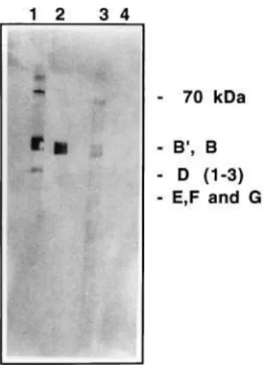
Antibodies obtained in rabbits against the yeast Sm motif 1 recognized Sm polypeptides from human UsnRNPs on Western blotting
The reactivity of both R328 and R329 sera against denatured human U1snRNP polypeptides was assayed using Western blotting. As the polyacrilamide gel system used (tricine-based gel, 12.5%) did not separate the three bands D1, D2and D3(see
below), we refer to the section of the gel containing D1±3as the
D1±3 region. Figure 4 shows that both sera recognized human
B0, B polypeptides and D
1±3 region (Fig. 4, lanes 1 and 2),
whereas the corresponding nonimmune serum of each animal did not (Fig. 4, lane 3 for R329 nonimmune serum and lane 4 for R328 nonimmune serum). In this assay, serum R329 showed less immunoreactivity to the D1±3 region than serum R328.
Detection of the 70-kDa band in lane 1 with serum R328 is discussed below. As B0, B and D
1±3polypeptides contain the Sm
motif 1, it may be concluded that the antibodies against the yeast Sm motif 1 cross-react with the human Sm B0, B polypeptides
and the D1±3region, presumably by recognizing the Sm motif 1
of all these polypeptides. Immunorecognition of E, F and G polypeptides was not obtained (Fig. 4 lanes 1 and 2); this is discussed further below. Interesting, R329 serum did not recognize native UsnRNPs (Fig. 3 lane 3) but did recognize denatured Sm polypeptides using Western blotting (Fig. 4 lane 2). This observation allows us to suggest that the denatured epitopes detected by R329 serum on Western blotting may be not accessible or hidden to the R329 polyclonal antibodies when native UsnRNPs are used as an antigen source, i.e. in immunoprecipitation assays. As R329 serum
Fig. 3. Immunoprecipitation of human UsnRNPs with antisera to the yeast peptide containing the Sm motif 1.RNAs extracted from the pellets after immunoprecipitation with protein A±Sepharose. The RNAs were electrophoresed in a 10% polyacrilamide/urea gel and stained with silver as described previously [20]. Lane 1, immunoprecipitated with serum R328; lane 2, immunoprecipitated with the same amount of nonimmune serum R328; lane 3, immunoprecipitated with serum R329; lane 4, immunopre-cipitated with anti-(trimethylguanosine) serum (R331) obtained in rabbit [21a]; and lane 5, immunoprecipitated with the same amount of non-immune serum R329 as in lane 3.
Fig. 4. Western blot of Sm polypeptides from human U1snRNP stained with crude sera from rabbits immunized with the yeast Sm motif 1 peptide.Lane 1, immunodetection with serum R328 (diluted 1/100); lane 2, immunodetection with serum R329 (diluted 1/100); lane 3, immunodetection with the nonimmune serum R329 (diluted 1/100); and lane 4, immunodetec-tion with the nonimmune serum R328 (diluted 1/100). Molecular weight of the polypeptides: 70 kDa; B0, 29 kDa; B, 28 kDa; D
1, 16 kDa; D2,
16.5 kDa; D3, 18 kDa; E, 12 kDa; F, 11 kDa; G, 9 kDa. The gel system
failed in the immunoprecipitation assays we further purified this sera by affinity columns in order to confirm the results seen in Western blotting.
Immunoaffinity-purified antibodies from serum R329 recognized both the yeast Sm motif 1 peptide and the Sm polypeptides from human UsnRNPs on Western blotting
In order to better discern whether the immunorecognition of the B0, B polypeptides and the D
1±3region by the rabbit sera (shown
in Fig. 4) is due to the binding of antibodies to the Sm motif 1 of each polypeptide, both sera were purified through an immuno-affinity column with the convalently bound peptide. Both sera bound to the peptide column, indicating that the polyclonal antibodies specifically recognized the peptide (data not shown). In this paper, we show the results obtained with the R329 serum as introduced before, but similar results were obtained with the purification of the R328 serum.
Immunoaffinity-purified antibodies recognized the yeast Sm motif 1 peptide (transferred alone to a nitrocellulose sheet) by Western blotting (Fig. 5, lane 2), whereas a nonrelated and immunoaffinity-purified antibody did not (Fig. 5 lane 3). Thus, the antibodies from serum R329 bound specifically to the peptide immunoaffinity column, and were also retained after elution of the immunoreactivity with the free Sm peptide.
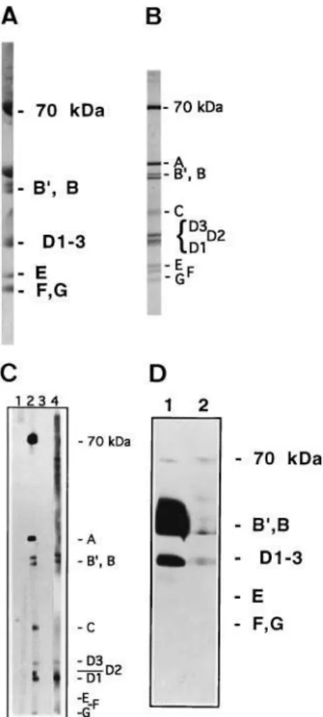
In order to better determine which polypeptides from the D1±3
region are recognized by the sera, we used Western blotting after running U1snRNP proteins in high-TEMED/SDS poly-acrilamide gels, which separate the D1±3 region into three
polypeptides: D1, D2and D3.As this is the first report to also use
tricine-Gly-based gels to separate UsnRNPs proteins, Fig. 6A (tricine-based gel) and Fig. 6B (high-TEMED-based gel) show a comparison of both systems. These figures show that tricine-based gels do not resolve the D1±3region as well as the
high-TEMED-based gels. Nevertheless, the tricine-based gels gave good resolution of the B0, B, E and F/G polypeptides. Using these Fig. 5. Western blot of the yeast Sm motif 1 peptide alone with the immunoaffinity-purified antibodies from rabbit R329.The peptide alone was blotted onto nitrocellulose sheets as described in the Materials and methods. Lane 1, blotted peptide stained with Amido Black; lane 2, blotted peptide immunostained with the immunoaffinity-purified R329 antibody; lane 3, peptide immunostained with a nonrelated immunoaffinity-purified antibody. This nonrelated immunoaffinity-purified antibody was a rabbit polyclonal anti-(m3G) (antinucleoside) purified through a m3G-affinity
column [19]. Lanes 2 and 3 were incubated with the same amount of immunoaffinity-purified antibody. The gel system used was tricine-Gly/SDS polyacrilamide (10%) mini-gel (5 cm long). The front of the gel was run for less time in order to have the peptide in the middle of the gel.
Fig. 6. Western blot of proteins extracted from human U1snRNP immunostained with immunoaffinity-purified R329 antibody. (A) U1snRNP proteins resolved in a long (15 cm) tricine-Gly/SDS polyacrila-mide gel (10%) and stained with Coomassie blue. (B) U1snRNP proteins resolved on a long (15 cm) high-TEMED/SDS polyacrilamide gel (12.5%) and stained with Coomassie blue. (C) U1snRNP proteins (8mg´lane21)
resolved in a long (15 cm) high-TEMED/SDS polyacrilamide gel (12.5%), blotted onto a nitrocellulose membrane and immunostained with: lane 1, human serum from a healthy donor (diluted 1/500); lane 2, anti-RNP/Sm serum from a MCTD (mixed connective tissue disease) patient (diluted 1/500); lane 3, U1snRNP proteins immunostained with a nonrelated immunoaffinity-purified antibody; lane 4, U1snRNP proteins immuno-stained with the immunoaffinity-purified R329 antibodies. This nonrelated immunoaffinity-purified antibody (used in lane 3) was a rabbit polyclonal anti-(m3G) (antinucleoside) purified through a m3G-affinity column [20].
Lanes 3 and 4 were incubated with the same amount of antibody. (D) U1snRNP proteins were overloaded (24mg´lane21) on a tricine-Gly/SDS
polyacrilamide gel (10%) mini-gel (5 cm long), blotted onto a nitrocellu-lose membrane and immunostained: lane 1, U1snRNP proteins immuno-stained with immunoaffinity-purified R329 antibody; lane 2, U1snRNP proteins immunostained with a nonrelated immunoaffinity-purified anti-body. This nonrelated immunoaffinity-purified antibody was a rabbit poly-clonal anti-(m3G) (antinucleoside) purified through a m3G-affinity column
results we selected the gel system depending on the purpose of the experiment: when the D1±3region was to be discriminated,
high-TEMED-based gels were used; and when low molecular weight polypeptides were to be assayed (e.g. the E, F and G polypeptides or the free yeast peptide) tricine-based gels were used.
Purified R329 antibodies were again used with denatured human U1snRNP analyzed by Western blotting with a high-TEMED gel. Figure 6C shows that the purified antibodies recognized the Sm polypeptides B0, B, D
1 and D3 (Fig. 6C,
lane 4), whereas a nonrelated immunoaffinity-purified antibody did not (Fig. 6C lane 3). The D2 polypeptide was not detected
using the immunoaffinity-purified antibodies. Similar results were obtained with purified R328 antibodies (not shown). Therefore, we conclude that the purified antibody cross-reacts with the Sm motif 1 of the human Sm polypeptides B0, B, D
1and
D3. Similar results were obtained previously with several sera
from SLE patients, which contained antibodies that cross-reacted with B0, B, D
1and D3polypeptides but not with D2polypeptide
[23]; this was further confirmed using immunoaffinity-purified antibodies [23].
The immunoaffinity-purified antibodies did not stain E, F and G polypeptides even after increasing the amount of protein loaded onto the gel and using tricine-based gels (Fig. 6D, lane 1). The crude sera were also unable to stain E, F and G polypeptides (Fig. 4 lanes 1 and 2). This was not expected, as the peptide used in the immunizations has a higher homology to the Sm motif 1 of human F protein than to human B0, B and D
1±3
polypeptides (Fig. 2b). Special care was taken to ensure that E, F and G polypeptides were present in our blots. After transfer, nitrocellulose sheets were stained with Ponceau S, Amido Black and Coomassie blue; in all the experiments, the staining of the blots indicated that both E, F and G were perfectly blotted and that the amount of polypeptides blotted was large enough to be immunodetected. Other membranes were also used to perform the transfer, such as poly(vinylidene difluoride) (PVDF) membranes (e.g. Immobilon-P and Immobilon-PSQ), which are particularly good at retaining low molecular weight polypeptides or small peptides. Using PVDF membranes the results obtained
were similar to those shown in Fig. 4 (data not shown). The immunostaining method was also reduced as much as possible (1 h) but no staining of E, F or G was obtained. All these control experiments indicated that there is no adverse event during transfer and immunostaining that could explain the failure to detect E, F and G polypeptides. Two other explanations are suggested to explain why E, F and G are not immunodetected. Post-translational modifications may play an important role in the immunorecognition ofbona fideB0, B and D
1polypeptides
by anti-(Sm) sera [16,25]. The Sm motif 1 in human E, F and G polypeptides could be post-translationally modified, and these modifications may disturb immunorecognition by our purified antibodies. Alternatively, protein folding may play an important role in immunorecognition of bona fide B0, B and D
1
polypeptides by anti-(Sm) sera [16,25]. Brahms et al. [18] described a major SLE autoantibody class that recognizes native E±F±G complexes, but not E, F and G denatured polypeptides (e.g. Western blotting). Brahmset al.suggested the existence of an important conformational epitope in E±F±G complexes, which is only detected in the complex when in the native form. The antibodies studied here may require an epitope with some type of protein folding not present in E, F and G polypeptides. With this reasoning we suggest that the conformation of the Sm motif 1 on the yeast peptide is more similar to that of the homologous region on human B0, B, D
1and D3proteins, but is
apparently different from the corresponding regions of D2, E, F
and G proteins.
Because of the high amount of protein loaded onto the gel (24mg´lane21), the immunoaffinity-purified antibodies begin to
stain the 70 kDa polypeptide nonspecifically (compare lanes 1 and 2 in Fig. 6D), this polypeptide is also recognized by the nonspecific serum; this nonrelated immunoaffinity-purified antibody was a rabbit polyclonal anti-(m3G), then an
antinucleo-side antibody, purified through a m3G-affinity column [20].
Further 70 kDa staining was not seen when the amount of
Fig. 7. Cross-reactivity of human SLE sera with the yeast Sm motif 1 peptide.ELISAs were performed as described in the Materials and methods. Statistical treatment of the data was carried out with Statview II program (anova). Number of sera: healthy (n= 95); SLE (n= 214). *A significant
reaction of the SLE sera with the peptide (P,0.05), as compared with data from healthy sera. Column A shows the results with all the SLE sera: 0.3^0.15 (mean ^SD). Column B shows the data with healthy sera: 0.06^0.06 (mean^SD). The definition of positive sera by ELISA was as follows: detection threshold = 2£mean^2£SD of the values obtained with the healthy donor sera. This definition indicated that 70% of the sera were positive, and only these are shown in column C: 0.37^0.12 (mean^SD).
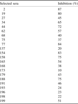
Table 1. Competition of the reaction between the antibodies and the Motif 1-KLH conjugate with 300 ng of free peptide.The assay was performed as described in Materials and Methods with 22 sera selected from column C in Fig. 7. As a negative control experiment sera were competed with 300 ng of BSA and 300 ng KLH and no inhibition was observed.
Selected sera Inhibition (%)
2 87
19 80
27 45
34 65
44 72
62 57
68 40
75 37
77 84
137 20
154 83
158 75
165 54
168 38
173 10
179 43
188 75
191 46
193 24
196 23
198 22
protein loaded onto the gel was reduced to 8mg´lane21
(Fig. 6C, lane 4). Nevertheless, it is not clear whether the 70 kDa staining in Fig. 4 lane 1 by R328 crude sera is a consequence of a secondary immunoreaction of the animal, e.g. epitope spreading, and this is currently being studied.
Interestingly, in human Sm polypeptides Sm motif 1 does not seem to be equally accessible to all antibodies. Comparing lane 1 with lane 4 in Fig. 3 shows that the amount of immuno-precipitated U1, U2, U4 and U5 snRNAs is very similar (lane 1), whereas the total amount of U1snRNA in the sample is higher [note the amount of U1snRNA in lane 4, when anti-(trimethylguanosine) were used]. This may indicate that in native U1snRNP the Sm motif 1 is more hidden or less accessible to the anti-(Sm motif 1) than in the other UsnRNPs. Finally we conclude that the Sm motifs 1 of the UsnRNPs contain a common feature that allows cross-reaction of antibodies between yeast and human Sm motifs 1.
Yeast Sm motif 1 peptide is specifically recognized by a SLE antibody
Our data indicate that antibodies raised against the yeast Sm motif 1 peptide cross-react with human Sm polypeptides. We were also interested in knowing whether a reversed experiment would be also successful, i.e. whether antibodies raised to human Sm motifs might cross-react with yeast Sm polypeptides. As a first approach, we tested a collection of SLE sera. The presence of anti-(Sm motif 1) that cross-reacted with yeast peptide containing the Sm motif 1 was assayed using a 214 SLE sera collection classified according to ARA criteria, and with 95 healthy donor sera. Figure 7 shows that 70% of the sera (150/ 214) presented statistically significant cross-reaction to the yeast Sm motif 1 peptide (P,0.05) when compared with data obtained using healthy sera (asterisk in Fig. 7 and its legend). The specificity of the reaction of the positive sera antibodies with the peptide was established by competitive inhibition: 22 sera from Fig. 7 were selected with absorbance units ranging between 0.25 and 0.5. Table 1 shows that the reaction of the sera to the Motif 1-KLH conjugate was inhibited when the free peptide was added to the assay, thus indicating that the antibody reaction is specific. Many sera from Table 1 were tested with different concentrations of the free competitor, and in all cases a higher level of inhibition was obtained when the concentration increased. Table 1 shows the levels of inhibition obtained at a free peptide concentration of 300 ng. The sera from Table 1 with an inhibition of ,50% with 300 ng of free peptide gave higher inhibition values when the concentration of the free peptide increased. This may represent affinity or quantitative differences in the antibodies. The assay did not determine whether this antibody specificity is an autoantibody (we intend to study this subject further using human peptides), but the results shown in Figs 3±6 indicated that antibodies to the yeast Sm motif 1 cross-reacted with human Sm polypeptides. Therefore, the antibodies found in SLE might recognize human Sm motifs. We randomly selected 30 sera from the positive population, i.e. sera that reacted with the yeast Sm motif 1 peptide, and in all of them we detected anti-(Sm) autoantibodies, i.e. antibodies that reacted with blotted human B0, B and D
1±3 polypeptides (not shown). We may therefore
presume that the sera which cross-reacted with the yeast Sm motif 1 peptide probably contain anti-(Sm) autoantibodies.
Taken together, these results allow us to suggest a hypothesis in which a Sm Motif 1-like protein, e.g. from an external agent, could initiate an immune reaction with the subsequent synthesis of antibodies that would cross-react with the host Sm
polypeptides by a process of mimicry, and thus generate an autoimmune disorder. We are currently performing longer immunizations using more rabbits in order to answer this question.
A C K N O W L E D G E M E N T S
We thank X. Jimenez for technical assistance, and all members of the M. Bach-Elias' group for their comments about the manuscript. This work was supported by PGC grant no. PB92-0004 and PETRI no. 94-0186. R.B. Cicarelli was the recipient of a fellowship from CAPES and Spanish MEC; D. Bahia was the recipient first of a MUTIS fellowship and later of a CNPq fellowship. A. Khaouja was recipient of a fellowship from the Moroccan government. We also thank MartõÂ Cullell for revising this manuscript.
R E F E R E N C E S
1. Tan, E.M. (1989) Antinuclear antibodies: diagnostic markers for autoimmune diseases and probes for cell biology.Adv. Immunol.44, 93±151.
2. Hermann, H., Fabrizio, P., Raker, V.A., Foulaki, K., Hornig, H., Brahms, H. & LuÈhrmann, R. (1995) snRNP Sm proteins share two evolutionarily conserved sequence motifs which are involved in Sm protein±protein interactions.EMBO J.14, 2076±2088.
3. Fabrizio, P., Esser, S., Kastner, B. & Luhrmann, R. (1994) Isolation of
S. cerevisiaeSnRNPs ± comparison of U1 and U4/U6.U5 to their human counterparts.Science264, 261±265.
4. Seraphin, B. (1995) Sm and Sm-like proteins belong to a large family: identification of proteins of the U6 as well as the U1, U2, U4 and U5 snRNPs.EMBO J.14, 2089±2098.
5. Cooper, M., Johnston, L.H. & Beggs, J.D. (1995) Identification and characterization of Uss1p (Sdb23p): a novel U6 snRNA-associated protein with significant similarity to core proteins of small nuclear ribonucleoproteins.EMBO J.14, 2066±2075.
6. Pettersson, I., Hinterberger, M., Mimori, T., Gottlieb, E. & Steitz, J.A. (1984) The structure of mammalian small nuclear ribonucleopro-teins. Identification of multiple protein components reactive with anti-(U1) ribonucleoprotein and anti-Sm autoantibodies. J. Biol. Chem.259, 5907±5914.
7. Reuter, R., Rothe, S., Habets, W., Van Venrooij, W.J. & LuÈhrmann, R. (1990) Autoantibody production against the U small nuclear ribonucleoprotein particle proteins E, F and G in patients with connective tissue diseases.Eur. J. Immunol.20, 437±440. 8. Barakat, S., Briand, J.P., Weber, J.C., van Regenmortel, M.H. &
Muller, S. (1990) Recognition of synthetic peptides of Sm-D autoantigen by lupus sera.Clin. Exp. Immunol.81, 256±262. 9. Sabbatini, A., Dolcher, M.P., Marchini, B., Bombardieri, S. &
Migliorini, P. (1993) Mapping of epitopes on the SmD molecule: the use of multiple antigen peptides to measure autoantibodies in systemic lupus erythematosus.J. Rheumatol.20, 1679±1683. 10. Rokeach, L.A., Jannatipour, M., Haselby, J.A. & Hoch, S.O. (1992)
Mapping of the immunoreactive domains of a small nuclear ribonucleoprotein-associated Sm-D autoantigen. Clin. Immunol. Immunopathol.65, 315±324.
11. James, J.A., Mamula, M.J. & Harley, J.B. (1994) Sequential autoantigenic determinants of the small nuclear ribonucleoprotein Sm D shared by human lupus autoantibodies and MRL lpr/lpr antibodies.Clin. Exp. Immunol.98, 419±426.
12. Ohosone, Y., Mimori, T., Fujii, T., Akizuki, M., Matsuoka, Y., Irimajiri, S., Hardin, J.A., Craft, J. & Homma, M. (1992) Autoantigenic epitopes of the B polypeptide of Sm small nuclear RNP particles ± identification of regions accessible only within the U1 small nuclear RNP.Arthritis Rheum.35, 960±966.
13. James, J.A. & Harley, J.B. (1992) Linear epitope mapping of an Sm B/B0polypeptide.J. Immunol.148, 2074±2079.
15. Elkon, K.B., Hines, J.J., Chu, J.L. & Parnassa, A. (1990) Epitope mapping of recombinant HeLa SmB and B0peptides obtained by the polymerase chain reaction.J. Immunol.145, 636±643.
16. Hirakata, M., Craft, J. & Hardin, J.A. (1993) Autoantigenic epitopes of the B and D polypeptides of the U1 snRNP ± analysis of domains recognized by the Y12-monoclonal anti-Sm antibody and by patient sera.J. Immunol.150, 3592±3601.
17. James, J.A., Gross, T., Scofield, R.H. & Harley, J.B. (1995) Immunoglobulin epitope spreading and autoimmune disease after peptide immunization: Sm B/B0-derived PPPGMRPP and PPPGIRGP induce spliceosome autoimmunity.J. Exp. Med.181, 453±461. 18. Brahms, H., Raker, V.A., van Venrooij, W.J. & LuÈhrmann, R. (1997) A
major, novel systemic lupus erythematosus autoantibody class recog-nizes the E, F, and G Sm snRNP proteins as an E±F±G complex but not in their denatured states.Arthritis Rheum.40, 672±682. 19. Kitagawa, T. & Aikawa, T. (1976) Enzyme-coupled immunoassay of
insulin using a novel coupling reagent.J. Biochem.79, 233±236. 20. Bach, M., Bringmann, P. & LuÈhrmann, R. (1990) Purification of small
nuclear ribonucleoproteins particles with antibodies against modified nucleosides of small nuclear RNAs.Methods Enzymol.181, 232±257. 21. Anderson, G.J., Bach, M., LuÈhrmann, R. & Beggs, J.D. (1989) Conservation between yeast and man of a protein associated with U5 small nuclear ribonucleoprotein.Nature342, 819±821.
21a. Espuny, R., Bahia, D., Cicarelli, R.M.B., Codony, C., Khaovja, A., AvinÄoÂ, A., Eritja, R. & Bach-Elias, M. (1999) Preparation of N2,N2,7-trimethylguanosine affinity columns.Nucleosides & Nucleo-tides18, 125±136.
22. SchaÈgger, H. & von Jagow, G. (1987) Tricine-sodium dodecyl sulphate-polyacrilamide gel electrophoresis for the separation of proteins in the range from 1 to 100 kDa.Analyt. Biochem.166, 368±379. 23. Lehmeier, T., Foulaki, K. & LuÈhrmann, R. (1990) Evidence for
three distinct D proteins, which react differentially with anti-Sm
autoantibodies, in the cores of the major snRNPs U1, U2, U4/U6 and U5.Nucleic Acids Res.18, 6475±6484.
24. Steitz, J. (1989) Immunoprecipitation of ribonucleoproteins using autoantibodies.Methods Enzymol.180, 468±481.
25. Ou, Y., Sun, D., Sharp, G.C. & Hoch, S.O. (1997) Screening of SLE sera using purified recombinant Sm-D1 protein from a baculovirus expression system.J. Immunol.83, 310±317.
26. van Dam, A., Winkel, I., Zijlstra-Baalbergen, J., Smeenk, R. & Cuypers, H.T. (1989) Cloned human snRNP proteins B and B0differ only in their carboxy-terminal part.EMBO J.8, 3853±3860. 27. Lehmeier, T., Raker, V., Hermann, H. & Luhrmann, R. (1994) cDNA
cloning of the Sm proteins D2 and D3 from human small nuclear ribonucleoproteins: Evidence for a direct D1±D2 interaction.Proc. Natl Acad. Sci. USA91, 12317±12321.
28. Rokeach, L.A., Haselby, J.A. & Hoch, S.O. (1988) Molecular cloning of a cDNA encoding the human Sm-D autoantigen.Proc. Natl Acad. Sci. USA85, 4832±4836.
29. Blanton, S., Srinivasan, A. & Rymond, B.C. (1992) PRP38 Encodes a yeast protein required for pre-messenger-RNA splicing and main-tenance of stable U6 small nuclear RNA levels.Mol. Cell. Biol.12, 3939±3947.
30. Preston, R., Manolson, M.F., Becherer, K.A., Weindnhammer, E., Kirkpatrick, D., Wright, R. & Jones, E.W. (1991) Isolation and characterization ofPEP3, a gene required for vacuolar biogenesis in
Saccharomyces cerevisae.J. Mol. Cell. Biol.11, 5801±5812. 31. Stanford, D.R., Kehl, M., Perry, C.A., Holicky, E.L., Harvey, S.E.,
Rohleder, A.M., Rehder, K.J., Luhrmann, R. & Wieben, E.D. (1988) The complete primary structure of the human snRNP E protein.
Nucleic Acids Res.16, 10593±10605.