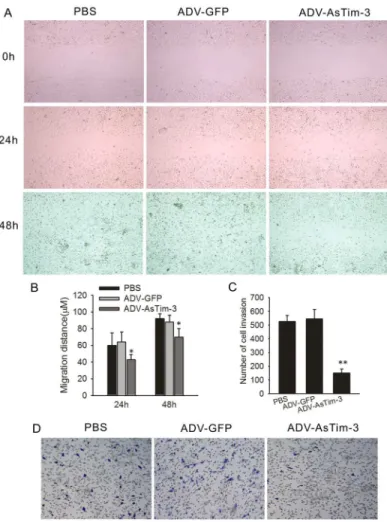
Correction: Tim-3 Expression in Cervical Cancer Promotes Tumor Metastasis.
Texto
Imagem
Documentos relacionados
(B) Graph represents percentage of viable B-1 cells present in culture after 24 and 72 hours when B-1 cells were cultivated alone, on BALB/c or BALB/ Xid peritoneal macrophages or
HeLa cells were left untreated (Medium, A) or transiently transfected with control RNAi (B) or CK18 RNAi (C).. After 24 h, cells were infected with tis- sue culture trypomastigotes
cells enriched from pancreatic cancer patient blood as in cancer cells and tumor tissues; (3) BC-15 + cells in cancer patients blood were CK + [20]; (4) BC-15 + /CD45 - cells in
Human HeLa and HOS cells, together with AGM Vero cells, were transfected with HIV-1 NL4.3 and NL4.3delVpu proviral plasmids, or an MLV Gag-Pol– expression vector, as indicated, in
In conclusion, our results demonstrated that both high CCR7 expression in tumor cells and increased intratumoral FOXP3 + Tregs were associated with worse OS in gastric
(C) HeLa cells transfected with either non-targeting siRNA or siRNA targeting CPSF6 were infected with VSV-G-pseudotyped GFP carrying CA mutants that lost the cell cycle independence
In this study, we found that both higher Gal-9 expression and negative Tim-3 expression were significantly associated with better overall survival ( P = 0.002, P = 0.010,
(C) HeLa cells stably expressing a centrin-GFP fusion (green) were either fixed and stained for endogenous b arr2 using either rARR or gBARR2 polyclonal antibodies (red) or
