Apoptosis by Up-Regulating DR4 and DR5 via a
MEK1-Dependent Pathway
Zhongfan Deng, Huijuan Yan, Jiajie Hu, Shengwei Zhang, Peng Peng, Qingzhen Liu*, Deyin Guo*
State Key Laboratory of Virology and The Modern Virology Research Centre, College of Life Sciences, Wuhan University, Wuhan, People’s Republic of China
Abstract
Background: Hepatitis C virus (HCV) is the leading cause of liver fibrosis, cirrhosis and hepatocellular carcinoma. It is believed that continuous liver cell apoptosis contributes to HCV pathogenesis. Recent studies have shown that HCV infection can sensitize host cells to TNF-related apoptosis-inducing ligand (TRAIL) induced apoptosis, but the mechanism by which HCV regulates the TRAIL pathway remains unclear.
Methods and Results:Using a sub-genomic replicon and full length virus, JFH-1, we demonstrate that HCV can sensitize host cells to TRAIL-induced apoptosis by up-regulating two TRAIL receptors, death receptor 4 (DR4) and death receptor 5 (DR5). Furthermore, the HCV replicon enhanced transcription of DR5 via Sp1, and the HCV-mediated up-regulation of DR4 and DR5 required MEK1 activity. HCV infection also stimulated the activity of MEK1, and the inhibition of MEK1 activity or the knockdown of MEK1 increased the replication of HCV.
Conclusions: Our studies demonstrate that HCV replication sensitizes host cells to TRAIL-induced apoptosis by up-regulating DR4 and DR5 via a MEK1 dependent pathway. These findings may help to further understand the pathogenesis of HCV infection and provide a therapeutic target.
Citation:Deng Z, Yan H, Hu J, Zhang S, Peng P, et al. (2012) Hepatitis C Virus Sensitizes Host Cells to TRAIL-Induced Apoptosis by Up-Regulating DR4 and DR5 via a MEK1-Dependent Pathway. PLoS ONE 7(5): e37700. doi:10.1371/journal.pone.0037700
Editor:Young-Min Lee, Utah State University, United States of America
ReceivedDecember 2, 2011;AcceptedApril 22, 2012;PublishedMay 25, 2012
Copyright:ß2012 Deng et al. This is an open-access article distributed under the terms of the Creative Commons Attribution License, which permits unrestricted use, distribution, and reproduction in any medium, provided the original author and source are credited.
Funding:This study was supported by China ‘‘973’’ program (grant no. 2009CB522506 and 2010CB911800) and the China Natural Science Foundation (grant #30921001). The funders had no role in study design, data collection and analysis, decision to publish, or preparation of the manuscript.
Competing Interests:The authors have declared that no competing interests exist.
* E-mail: [email protected] (QL); [email protected] (DG)
Introduction
TNF-related apoptosis-inducing ligand (TRAIL), also known as Apo2L, is a member of the tumor necrosis factor (TNF) super family [1]. However, in contrast to TNF-a and FasL, TRAIL typically induces apoptosis in transformed cells but not in healthy cells [2]. Furthermore, recent studies have shown that TRAIL also induces apoptosis in virus-infected cells, including cells infected with hepatitis B virus, hepatitis C virus (HCV), human immuno-deficiency virus, and respiratory syncytial virus [3,4,5,6,7]. Thus, TRAIL may function as an immune surveillance factor by selectively killing transformed and virus-infected cells. There are four specific TRAIL receptors on the cell membrane, death receptor 4 (DR4; also known as TRAIL-R1 or TNFRSF10A), death receptor 5 (DR5; also known as TRAIL-R2, KILLER, or TNFRSF10B), decoy receptor 1 (DcR1; also known as TRAIL-R3, TRID, or TNFRSF10C) and decoy receptor 2 (DcR2; also known as TRAIL-R4 or TNFRSF10D). DcR1 and DcR2 are two decoy receptors that contain a TRAIL-binding domain but lack a functional death domain. TRAIL can also bind to osteoprote-gerin, which is a soluble TNF receptor family member; however, it has low binding affinity at physiological conditions [8]. The binding of TRAIL to DR4 or DR5 results in receptor trimerization and cell apoptosis via the recruitment of
Fas-associated death domain (FADD) to the C terminus of the receptors. FADD then recruits an apoptosis-initiator caspase (caspase 8 or caspase 10) via its death effecter domain to form the death-inducing signaling complex, which allows for auto activation of caspases [9]. The downstream signaling of activated caspase 8 or 10 is dependent on the cell type. In type 1 cells, caspase 3 is activated and cleaves many cellular proteins to induce apoptosis. In type 2 cells, the apoptosis signal is augmented by the mitochondrial pathway, which involves the activation of caspase 9 following the loss of the mitochondrial membrane potential and Apaf-1 activation [10].
apoptosis is involved in the pathogenesis of HCV infection [14]. It is believed that liver damage, at least in part, causes fibrosis of the liver [15]. Although a direct cytopathic effect of a high HCV viral load has also been reported [16], the current prevailing view is that the apoptosis of liver cells in chronically infected HCV patients is initiated by the host innate and adaptive immune response. Many studies examining HCV and apoptosis have been reported. Several of these studies focused on a single HCV protein, and other studies used a cell culture replicon. However, the results from these studies have been conflicting, and there is currently no consensus regarding the role of HCV in liver cell apoptosis [17].
Previous studies have reported that TRAIL and its functional receptors: DR4 and DR5 are up-regulated in the liver of HCV-infected patients [18,19] and that the expression levels of DR4 and DR5 are elevated in some cases of HCV-related hepatocellular carcinoma (HCC) [20]. However, it has also been reported that the expression of DR4 or DR5 is not altered in the liver during HCV-mediated cirrhosis [21]. In cell culture system, it has been shown that the HCV core protein can enhance TRAIL-induced apoptosis in Huh7 cells, a hepatoma cell line normally insensitive to TRAIL [22]. Furthermore, it has also been recently shown that infection with the full-length virus JFH-1 can sensitize Huh7.5 cells to TRAIL-induced apoptosis without changing the expression level of the TRAIL receptors [4]. However, another study demonstrates that JFH-1 infection can trigger the expression of TRAIL and its functional receptors in a cell culture system [23]. Taken together, it still remains controversial if TRAIL receptors are elevated in HCV-infected cells, and the molecular mechanism that underlies the up-regulation of TRAIL receptors has not been examined to date.
A stable cell line, 9–13, that contains an HCV 1b sub-genomic replicon that lacks the genes for all structural proteins and the NS2 protein has been derived from Huh7 cells [24]. The first infectious HCV cell culture model, JFH-1, was developed by Dr. Wakita in 2005 [25]. These two HCV cell culture models have greatly aided HCV research. In the current study, we found that 9–13 cells were more sensitive to TRAIL-induced apoptosis than Huh7 cells and that the expression of DR4 and DR5 was significantly higher in 9– 13 cells at both the mRNA and protein levels when compared with Huh7 cells. Elimination of the HCV replicon in the 9–13 cells decreased the expression of DR4 and DR5. Similarly, the expression level of DR4 and DR5 was elevated in Huh 7.5.1 cells when infected with JFH-1. Furthermore, HCV increased the transcription of DR5 via the transcription factor Sp1. HCV replication also stimulated the activity of MEK1 and MEK1 activity was required for DR4 and DR5 up-regulation in both 9– 13 cells and JFH-1 infected Huh7.5.1 cells. Importantly, inhibition the activity or expression of MEK1 increased the replication of HCV. Together, these results demonstrate that HCV replication up-regulates DR4 and DR5 via a MEK1-dependent pathway, which results in the sensitization of host cells to TRAIL-induced apoptosis. These results suggest that MEK1 may be a negative regulator of HCV replication.
Results
HCV replicon-containing cells are more sensitive to TRAIL-induced apoptosis than naive cells
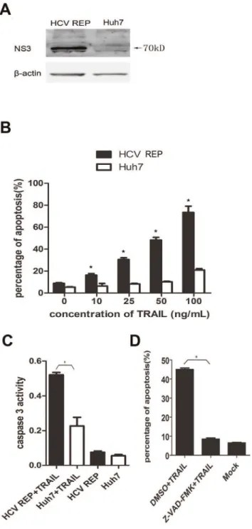
TRAIL has the ability to induce apoptosis in some transformed cells [2]. However, Huh7 cells, a HCC cell line, are resistant to TRAIL-induced apoptosis [22]. Therefore, we tested if 9–13 cells (designated ‘‘HCV REP’’ in all figures), which were derived from the Huh7 cell line and contain a HCV 1b sub genomic replicon (Fig. 1A), were sensitive to TRAIL. When exposed to 25–100 ng/
mL TRAIL for 2 hr, a large proportion of 9–13 cells underwent apoptosis, whereas only a small fraction of the TRAIL-treated Huh7 cells underwent apoptosis (Fig. 1B). Furthermore, the activity of caspase 3 was much greater in 9–13 cells than in Huh7 cells (Fig. 1C), indicating that the HCV replicon sensitized the host cells to TRAIL-induced apoptosis. The cell death induced by TRAIL was dependent on caspase activity because pre-treating 9– 13 cells with the pan-caspase inhibitor, Z-VAD-FMK, completely abolished TRAIL-induced apoptosis (Fig. 1D).
DR4 and DR5 are both up-regulated by HCV replication
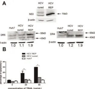
We evaluated the mRNA levels of the TRAIL receptors using real-time PCR and found that the mRNA levels of DR4 and DR5 were higher in 9–13 cells when compared with Huh7 cells. Moreover, there was no significant change in the mRNA levels of DcR1 and DcR2 (Fig. 2A). Western blot analyses confirmed that DR4 and DR5 were up-regulated in 9–13 cells (Fig. 2B). The activity of the DR4 and DR5 promoters was then tested in both cell lines. The DR4 and DR5 promoter activities were significantly higher in 9–13 cells than in Huh7 cells (Fig. 2C). To verify that the up-regulation of DR4 and DR5 was due to HCV replicon replication, we eliminated the HCV replicon from 9–13 cells and analyzed the expression of DR4 and DR5. We treated 9–13 cells with 100 IU/mL IFN-a and cultured the cells for four passages. As shown in Fig. 3A, the replication of HCV was completely inhibited after the IFN-a treatment, and HCV RNA was barely detected by using real-time PCR (data not shown). These
IFN-atreated cells were designated HCV-cured cells. The expression of DR4 and DR5 was significantly decreased in HCV-cured cells when compared with 9–13 cells (Fig. 3A). We analyzed the sensitivity of the HCV-cured cells to TRAIL-induced apoptosis and found that the HCV-cured cells were less sensitive to TRAIL when compared with 9–13 cells (Fig. 3B). These results demonstrate that HCV replication up-regulates DR4 and DR5 and sensitizes host cells to TRAIL-induced apoptosis. To further confirm the elevation of DR4 and DR5 levels is resulted from HCV infection, we infected Huh7.5.1 cells with JFH-1 and measured the expression of DR4 and DR5 1, 2, 3 days post-infection by western blotting. As shown in Fig. 4A, the expression of DR4 and DR5 was significantly up-regulated at the 3rd day post-infection. Similar up-regulation was observed at mRNA level at the 3rdday post-infection (Fig. 4B). The expression of DR4 and DR5 had a modest decrease at the 1stday post-infection (Fig. 4A), this result suggests that HCV may have some method to inhibit the expression of DR4 and DR5, in order to prevent host cell apoptosis at the early period of infection. However, more experiments are needed to confirm this observation in future study. In addition, the promoter activity of DR4 and DR5 were significantly higher in the JFH-1 infected cells (Fig. 4C). And correlated with the up-regulation of TRAIL receptors, the infected cells were more sensitive to TRAIL-induced apoptosis (Fig. 4D).
HCV-mediated up-regulation of DR4 transcription depends on the2464 to2384 bp region of the DR4 promoter
study has shown that2410/2404 is an AP-1 binding site that regulates the transcription of DR4 [26]. However, a point mutation in this AP-1 binding site (Fig. 5E) failed to influence the promoter activity of DR4 in 9–13 cells (Fig. 5B).
HCV replicon enhances transcription of DR5 via Sp1
To elucidate the mechanism underlying HCV-mediated regu-lation of DR5, we constructed several reporter plasmids containing different regions of the DR5 promoter (pDR5/2560, pDR5/ 2220, and pDR5/2115). The promoter activity of these plasmids was compared in 9–13 and Huh7 cells. As shown in Fig. 5C, the 2220 bp fragment of the DR5 promoter was sufficient for promoter activity. These results suggest that the increased DR5 expression caused by the HCV sub-genomic replicon is dependent on the DR5 promoter and that the 2220 bp fragment of the promoter is the minimal promoter. The2195/2189 and2149/ 2143 sites of the promoter have been shown to be two Sp1 binding sites, which are important for the transcription of DR5 [27]. A luciferase assay was then performed with the following mutant constructs: pDR5/2560/2195m, pDR5/2560/2149m, and pDR5/2560/dm (pDR5/2560/2195m,2149m) (Fig. 5E). Mutation of the each of the Sp1 binding sites decreased the activity of the promoter, and the pDR5/2560/dm had no promoter activity (Fig. 5C). To further confirm that Sp1 is required for transcription of DR5 in 9–13 cells, we transfected 9–13 cells with a siRNA targeting Sp1. The expression of DR5 was detected using real-time PCR and western blot analyses 2 days post-transfection. The RNA and protein levels of DR5 were decreased when compared with cells transfected with control siRNA (Fig. 5F). These results suggest that the transcription of DR5 is controlled by Sp1 in 9–13 cells and that the HCV sub-genomic replicon enhances the transcription of DR5 via Sp1. We then co-transfected a plasmid expressing individual HCV nonstructural protein and a plasmid containing either DR4/2632 or DR5/ 2560 into Huh7 cells. Expression of the NS3/4, NS4B or NS5A protein increased DR5 promoter activity, and expression of the NS3/4A or NS4B protein increased DR4 promoter activity. However, the luciferase activity was too low, suggesting that the expression of these individual nonstructural proteins barely activates the DR4 and DR5 promoters (Fig. 5G).
HCV-mediated up-regulation of DR4 and DR5 requires MEK1 activity
Sp1 is a member of a large family of transcription factors characterized by their affinity for GC-rich motifs. These transcription factors control the basal expression of housekeeping genes and genes lacking or containing a TATA box [28]. Sp1 activity is mainly regulated by post-translational modifications, with phosphorylation the most studied post-translational modifi-cation. Recent studies have shown that MAPKs, such as ERK and JNK, can phosphorylate Sp1 [29,30,31].
Macdonald and colleagues (2003, 2005) have reported that the HCV NS5A protein can inhibit AP-1 activity by perturbing Ras-ERK signaling [32,33]. Because the DR5 promoter contains two Sp1 binding sites, we studied the role of MAPKs in the HCV replicon-induced up-regulation of DR5. When compared with Huh7 cells, the AP-1 and Sp1 activity was increased in 9–13 cells, while the NF-kB activity was not changed (Fig. 6A). Furthermore, the over expression of MEK1 in Huh7 cells enhanced the transcription activity of AP-1 and Sp1, and the over expression of MEKK1 in Huh7 cells increased the transcription activity of AP-1 (Fig. 6B). These results suggest that HCV replication may activate MAPKs.
Figure 1. HCV replicon sensitizes host cells to TRAIL-induced and caspase-dependent apoptosis. (A) Cell lysates from HCV replicon-containing cells (9–13) and Huh7 cells were subjected to western blot analyses using a rabbit polyclonal antibody against HCV NS3/4A. (B) Huh7 and 9–13 cells were treated with TRAIL at different concentrations (10 ng/mL, 20 ng/mL, 50 ng/mL or 100 ng/mL) for 2 hr and stained with annexin V and PI. The proportion of apoptotic cells was measured using flow cytometry. (C) Huh7 and 9–13 cells were treated with 50 ng/mL TRAIL for 2 hr, and caspase 3 activity was measured using a Caspase 3 Activity Assay Kit. (D) 9–13 cells were treated with 20mM Z-VAD-FMK (a pan-caspase inhibitor) or DMSO for
MEK1 is a dual specificity kinase that phosphorylates and activates ERK-1 and ERK-2 [34], which can activate AP-1 [29]. Additionally, MEKK1 can activate JNK [35], which also activates AP-1. Therefore, we investigated the relationship between the MAPKs and the expression of the TRAIL receptors. We co-transfected pDR4/2464 or pDR4/2384 with plasmids expres-sing MEK1 or MEKK1 into Huh7 cells and found that over expression of MEK1 increased the activity of pDR4/2464, but did not affect the activity of pDR4/2384. Similarly, over expression of MEK1 increased the activity of the DR5 promoter pDR5/2560 but not the pDR5/2560dm. In contrast, MEKK1 only slightly increased the DR4 promoter activity and did not affect the DR5 promoter activity (Fig. 6C). These results suggest that MEK1/ERK, but not MEKK1/JNK, is involved in the regulation of DR4 and DR5 expression. Furthermore, the region between 2464 and 2384 in the DR4 promoter and the Sp1
binding sites in the DR5 promoter were required for MEK1-mediated regulation of DR4 and DR5 activity.
cells (Fig. 7B), and knockdown of MEK1 using siRNA prior to infection reversed the HCV-mediated up-regulation of DR4 and DR5 (Fig. 7E). Taken together, these data indicate that MEK1 has a critical role in the HCV-mediated up-regulation of DR4 and
DR5. To confirm that knockdown of MEK1 could decrease the sensitivity to TRAIL-induced apoptosis, we tested other four pairs of siRNA of MEK1, and choose two of them to perform the next experiment (Fig. 7F). As shown in Fig. 7G and 7H, knockdown of MEK1 weakened the apoptosis induced by TRAIL both in 9–13 cells and JFH-1 infected Huh7.5.1 cells.
Inhibition of MEK1 activity by its specific inhibitor, PD98059, can enhance the replication of HCV [37,38]. Therefore, we tested if inhibition of MEK1 enhanced HCV replication. As shown in Fig. 8, both PD98059 and a MEK1-specific siRNA increased JFH-1 replication in Huh7.5.JFH-1 cells. Together, these results suggest that MEK1 may be stimulated to inhibit HCV replication when cells are infected by HCV.
Discussion
The relationship between HCV infection and TRAIL and its receptors has been previously studied. Clinical evidence has shown that HCV infection is related to the up-regulation of DR4 and DR5. Saitou et al. (2005) have reported that TRAIL, DR4 and DR5 are over expressed in the cytoplasm and on the surface of hepatocytes from patients chronically infected with HCV [18]. Volkmann and colleagues (2007) reported that TRAIL alone triggers massive apoptosis and caspase activation in tissue explants from patients with liver steatosis or HCV infection and that the enhanced sensitivity of diseased liver is associated with the increased expression of DR4 and DR5 [19]. Yano et al. (2003) reported that some HCV-related HCC cases have increased caspase 3 activity and expression of DR4 and DR5 in the tumor tissue [20]. However, Mundt et al. (2003) reported that DR4 and DR5 are not up-regulated in chronic hepatitis-associated cirrhosis [21]. It also remains controversial if TRAIL receptors are up-regulated by HCV infection in a cell culture system. Lan et al. (2008) reported that DR4 and DR5 are not up-regulated in JFH-1 Figure 3. Up-regulation of DR4 and DR5 is HCV
replication-dependent.(A) Lysates from Huh7, 9–13 and HCV-cured cells were subjected to western blot analysis using rabbit polyclonal antibodies against HCV NS3/4A, DR4 or DR5. (B) Huh7, 9–13 and HCV-cured cells were treated with 25 and 50 ng/mL TRAIL for 2 hr, and the proportion of apoptotic cells was measured using flow cytometry after the cells were stained with annexin V and PI. The data are presented with the SD from three independent experiments, and statistical significance was calculated by two-way ANOVA, * indicates apvalue less than 0.05. doi:10.1371/journal.pone.0037700.g003
Figure 4. JFH-1 infection up-regulates the expression of DR4 and DR5.(A) Western blot analysis was performed to measure the expression of DR4 and DR5 in Huh7.5.1 cells infected with JFH-1 1, 2, 3 days post-infection (MOI 0.02). (B) Real-time PCR was performed to measure the mRNA levels of DR4 and DR5 in Huh7.5.1 cells infected with JFH-1 (MOI 0.02) 3 days post-infection. (C) The DR4 reporter plasmid (DR4/21156; 100 ng) or DR5 reporter plasmid (DR5/21192; 100 ng) was co-transfected with theRenillaluciferase reporter plasmid (50 ng) into Huh7.5.1 cells, 6 hr later, cells were infected with JFH-1 (MOI 0.5). After 3 days, the cells were harvested, and the luciferase activity was measured. The data from the infected cells were normalized to Huh7.5.1 cells to directly show the fold induction caused by HCV. (D) Huh7.5.1 cells were infected with JFH-1 (MOI 0.5), 3 days later, cells were treated with indicated concentration of TRAIL for 2 hr, and stained with annexin V and PI. The proportion of apoptotic cells was measured using flow cytometry. The data are presented with the SD from three independent experiments, and statistical significance was calculated byttest or two-way ANOVA, * indicates apvalue less than 0.05.
Figure 5. Transcriptional analysis of DR4 and DR5 in 9–13 cells.(A and C) Luciferase reporter plasmids (100 ng) containing different regions of the (A) DR4 or (C) DR5 promoter and theRenillaluciferase reporter plasmid (100 ng) were co-transfected into 9–13 or Huh7 cells. 2 days post-transfection, the cells were harvested, and luciferase activity was measured. (B and D) The indicated reporter plasmids (100 ng) illustrated in (E) were co-transfected with theRenillaluciferase reporter plasmid (100 ng) into 9–13 cells. 2 days post-transfection, the cells were harvested, and the luciferase activity was measured. (E) The mutations introduced into the AP-1 binding sites in pDR4/2632 and Sp1-binding sites in pDR5/2560 are illustrated. (F) The Sp1-specific siRNA or the control siRNA (100 pmol) was transfected into 9–13 cells cultured in 6-well plates. 2 days post-transfection, the Sp1 and DR5 RNA and protein levels were measured using real-time PCR and western blot analyses, respectively. (G) A plasmid expressing HCV NS3/4A, NS4B, NS5A or NS5B (600 ng) was individually co-transfected with either DR4/2632 or DR5/2560 (100 ng) and theRenilla luciferase reporter plasmid (100 ng) into Huh7 cells. 2 days post-transfection, the cells were harvested, and luciferase activity was measured. The data are presented with the SD from three independent experiments, and statistical significance was calculated byttest or two-way ANOVA, * indicates ap value less than 0.05.
infected Huh7.5 cells, whereas Zhu et al. (2007) reported that the expression of DR4 and DR5 is elevated in a JFH-1 infected hepatoma cell line, named by LH86 [4,23]. Therefore, the mechanism underlying the HCV-mediated regulation of the TRAIL receptors remains unclear.
MEK1 is a dual specificity kinase that phosphorylates and activates ERK1/2 by phosphorylating threonine and tyrosine residues [34], and ERK1/2 can phosphorylate Sp1 [29]. Schmitz and colleagues (2008) reported that the increased phosphorylation of ERK1/2 (pERK) is correlated with HCV-induced cirrhosis and
that approximately half of HCV-infected HCCs are pERK1/2 positive [39]. These findings are similar to the results in this study where phosphorylation of MEK1 was found to be increased in 9– 13 cells and JFH-1 infected Huh7.5.1 cells (Figs. 7A and 7B), suggesting that MEK1/ERK are activated by HCV. Moreover, the current study demonstrates that the inhibition of MEK1 activity or expression reverses the HCV-mediated up-regulation of DR4 and DR5 (Figs. 7C, 7D and 7E). JNK can also phosphorylate Sp1 to increase the stability of Sp1 [40]. The present study showed that the over expression of MEKK1, which can strongly activate JNK, did not increase DR5 transcription but slightly increased DR4 transcription (Fig. 6C). Bild et al. (2002) reported that the over expression of MEKK1 can induce expression of DR4 and that MEKK1 can increase DR4 expression at the RNA level approximately 1.5 fold [41]. In the present study, MEK1 increased the DR4 promoter activity to a greater extent. Treating 9–13 cells with the JNK inhibitor, SP600125, did not abolish the expression of DR4 or DR5. These results indicate that the HCV-induced up-regulation of DR4 was mediated by MEK1 but not MEKK1.
The results of the present study showed that Sp1 activity was critical for HCV-induced up-regulation of DR5, but we were unable to identify the transcription factor that controls DR4 transcription in 9–13 cells. Moreover, point mutations at the two putative AP-1 binding sites in the DR4 promoter (2410/2404 bp or 2552/2545 bp) did not influence the activity of the DR4 promoter (Fig. 5B). Previous studies have demonstrate that DR4 is regulated by both NF-kB and p53 [42,43]. However, NF-kB activity was not significantly different in 9–13 and Huh7 cells (Fig. 6A). Importantly, the Huh7 cell line carries a single p53 mutation (A:TRG:C at codon 220) and is p53 defective [44,45]. Thus, it is possible that other transcription factors are involved in the HCV-mediated up-regulation of DR4, and further research is needed to identify the transcription factors involved in HCV-regulated DR4 transcription.
Because both the sub genomic replicon and full-length virus increased the expression of DR4 and DR5, the structural proteins may not be involved in the regulation of the TRAIL pathway. We examined which HCV nonstructural proteins contributed to the up-regulation of DR4 and DR5. Although we found that the expression of NS3/4A or NS4B had a modest effect on the DR4 promoter activity and that the expression of NS3/4A, NS4B or NS5A had a similar modest up-regulation of the DR5 promoter activity, these effects were much weaker than those observed when the replicon was present (Fig. 5G). These results suggest that either the replication of the HCV genome or multiple HCV proteins may be needed to fully induce DR4 and DR5 expression.
Treating cells with the MEK1 inhibitor, PD98059, enhances HCV replication [37,38]. Moreover, the Ras-ERK pathway has an important role in the IFN-cand oxidative stress-induced anti-HCV effect, and activation of the Ras-ERK pathway either by EGF stimulation or over expression of Ras can suppress HCV replication [46,47]. Therefore, the Ras-ERK pathway is a negative regulator of HCV replication. However, Gretton et al. (2009) reported that different MEK1/2 inhibitors have different effects on HCV replication: PD98059 causes a modest increase in HCV replication, but U0126 and PD184352 inhibit HCV replication. Moreover, they showed that neither EGF nor a high concentration of PD98059 affects HCV NS5A protein expression in cells transfected with the HCV replicon and that the transfection of a dominant negative MEK1 mutant inhibits HCV replication [48]. These results demonstrate that low MEK/ERK activity is required for HCV replication because PD98059 is less effective than U0126 in inhibiting MEK/ERK activity. In this study, we found elevated MEK1 activity in HCV-infected cells (Fig. 7B). Figure 6. Over expression of MEK1 activates the DR4 and DR5
promoters. (A) The AP-1, Sp1 or NF-kB luciferase reporter plasmid (100 ng) was co-transfected with theRenillaluciferase reporter plasmid (100 ng) into 9–13 or Huh7 cells cultured in 24-well plates. 2 days post-transfection, luciferase activity was measured. (B and C) pMEK1-pRK or pMEKK1-pRK (600 ng) and the Renilla luciferase reporter plasmid (100 ng) were co-transfected with the (B) AP-1 or Sp1 reporter plasmid (100 ng) or (C) the indicated DR4 or DR5 reporter plasmid (100 ng) into Huh7 cells cultured in 24-well plates. 2 days post-transfection, luciferase activity was measured. The data are presented with the SD from three independent experiments, and statistical significance was calculated by ttest, * indicates apvalue less than 0.05.
Partial inhibition of MEK1 by PD98059 or a siRNA significantly increased the replication of JFH-1 (Fig. 8). Together, these results suggest that MEK1 with high activity is a negative regulator of HCV and is stimulated by HCV infection. This study demon-strates that HCV replication sensitizes host cells to TRAIL-induced apoptosis by up-regulating the DR4 and DR5 via a MEK1-dependent pathway. These findings may help to elucidate the mechanism underlying the HCV-mediated sensiti-zation of cells to TRAIL and, thus, may help to further unravel the pathogenesis of HCV infection and provide new therapeutic targets for the treatment of HCV infection.
Materials and Methods
Plasmids and reagents
Reporter plasmids were constructed using the pGL3 vector containing a firefly luciferase open reading frame (Promega). The human DR4 and DR5 promoters were cloned from DNA extracted from Huh7 cells using the PUREGENE DNA Purifi-cation System, as recommend by the manufacturer (Gentra). PCR for the DR4 promoter was performed with following sense primers: DR4/21156 (sense, 59 -GCAGATCTGCCCGGTC-GAAAAGAGTCTTTTCAA-39), DR4/2632 (sense, 59-GCCTCGAGCCAAAACAGTGAAACCCCCGTCTC-39), DR4/2541 (sense, 59-GCCTCGAGGCTGAGGCAG-GAAAATCGCTTGAAC-39), DR4/464 (sense, 59 -GCCTCGAGGGGCGACAGAGCTTGACTCCATCTC-39), DR4/2384 (sense, GCCTCGAGGGAGGCCG-TAAAAGCCTCTTAGAGG-39), and DR4/2349 (sense, 59-GCCTCGAGCAGTGGCCTCTGTGTCCTTCATTCC-39). The following antisense primer was used for all of the above
reactions for DR4: 59-GCAAGCTTCATCCTGCCAGGT-CAATCCAAGAAG-39. PCR for the DR5 promoter was performed with following sense primers: DR5/21192 (sense, 59 -GCCTCGAGCACCAGAAGGAAGAAACTCCGAACA-39), DR5/2560 (sense, 59-GCCTCGAGAGAGAAGGAGAGAACA-GAAGGGGCA-39), DR5/2220 (sense, 59 -GCCTCGA-GAGTTGCACATTGGATCTGATTCGCC), and DR5/2115 (sense, 59-ACCTCGAGGGCCGGAGAACCCCG-CAATCTCTGC). The following antisense primer was used for all of the above reactions for DR5: 59 -GCAAGCTTGGCGG-TAGGGAACGCTCTTATAGTC-39. Point mutations were in-troduced into pDR4/2632 and pDR5/2560 using PCR-based site-directed mutagenesis and the following primers: 59-ATCC-CAGTTACTTGGGAGGC-39 and 59 -GCCTCCCAAG-CAGCTGGGAT-39 for the 2552/2545 AP-1 binding site in DR4 promoter to generate pDR4/2632/2547m, 59-GGCAGGCTGAATTGCTCGCC-39 and 59-GGCGAG-CAATTCAGCCTGCC-39 for the 2410/2404 AP-1 binding site in DR4 promoter to generate pDR4/2632/2410m, 59 -ATTCGCCATGTACCGAATGA-39 and 59-TCATTCGGTA-CATGGCGAAT-39for the2195/2189 Sp1-binding site in the DR5 promoter to generate pDR5/2560/2195m and 59 -AGCCGCGATGATCCAAGTCA-39 and 59 -TGACTTGGTA-CATCGCGGCT-39for the2149/2143 Sp1-binding site in the DR5 promoter to generate pDR5/2560/2149m. The pDR4/ 2632/dm plasmid was mutated both at the 2552/2547 and 2410/2404 sites in the DR4 promoter and the pDR5/2560/dm plasmid was mutated both at the2195/2189 and2149/2143 sites in the DR5 promoter (the ATG translation initiation site of DR4 and DR5 was designed as+1). The Sp1 reporter plasmid was constructed by introducing five Sp1-binding sites (59 -Figure 7. HCV-mediated increased DR4 and DR5 expression is MEK1 dependent.Huh7 and 9–13 cell lysates (A) or Huh7.5.1 and JFH-1 infected Huh7.5.1 cell (MOI 0.02) lysates (B) were subjected to western blot analyses using antibodies against phospho-MEK1 or MEK1. (C) Untreated 9–13 cells or 9–13 cells treated with 100mM PD98059, 100mM SP600125 or DMSO for 2 days were harvested and subjected to western blot analyses using antibodies against DR4 or DR5. (D) The MEK1-specific siRNA or control siRNA (100 pmol) was transfected into 9–13 cells cultured in 6-well plates. 2 days post-transfection, the expression of MEK1, DR4 and DR5 was determined using western blot analyses. (E) Huh7.5.1 cells were transfected with the MEK1-specific siRNA or control siRNA and infected with JFH-1 (MOI 0.02) 6 hr post transfection. 3 days post infection, the expression of MEK1, DR4 and DR5 was determined using western blot analyses. (F) Huh7.5.1 cells were transfected with indicated siRNA, and the expression of MEK1 was measured by using western blot 2 days later. (G) 9–13 cells were transfected with MEK1 siRNA1, MEK1 siRNA2 or scramble RNA, 3 days post transfection, cells were treated indicated concentration of TRAIL for 2 hr, and stained with annexin V and PI. The proportion of apoptotic cells was analyzed by using flow cytometry. (H) Huh7.5.1 cells were transfected with MEK1 siRNA1, MEK1 siRNA2 or scramble RNA, 6 hr later, cells were infected with JFH-1 (MOI 0.5), 3 days post infection, cells were treated indicated concentration of TRAIL for 2 hr, and stained with annexin V and PI. The proportion of apoptotic cells was analyzed by using flow cytometry. The data are presented with the SD from three independent experiments, and statistical significance was calculated by two-way ANOVA, * indicates apvalue less than 0.05.
doi:10.1371/journal.pone.0037700.g007
Figure 8. Inhibition of MEK1 increases HCV replication.Huh7.5.1 cells were transfected with a MEK1-specific siRNA (A) or treated with 10mM PD98059 (B) and 6 hr post-transfection or treatment, infected with JFH-1 (MOI 0.02). 3 days post-infection, the expression of the HCV core protein was detected using western blot.
TGGGCGGGGC-39) into the pGL3 vector. The AP-1 reporter plasmid (pAP-1-pGL3), NF-kB reporter plasmid (pNF-kB-pGL3), MEK1 expression plasmid (pMEK1-pRK) and MEKK1 expres-sion plasmid (pMEKK1-pRK) were kindly provided by Dr. Hongbin Shu from Wuhan University. The MEK1 inhibitor, PD98059, was purchased from Cell Signaling Technology, and the JNK inhibitor, Sp600125, was purchased from Sigma.
Cell lines, virus and DNA transfection
The human hepatoma cell lines, Huh7 and 9–13, were kindly provided by Dr. Ralf Bartenschlager from Universita¨tsklinikum Heidelberg. Both cell lines were cultured in DMEM supplemented with 10% fetal bovine serum (GIBCO) at 37uC, and the 9–13 cells were maintained in medium supplemented with 250mg/mL G418 (Sigma). All transfection was performed using Lipofectamine 2000 (9–13 and Huh7 cells) (Invitrogen) or FuGENE HD (Huh7.5.1 cells) (Roche), as recommended by the manufacturer. JFH-1 and Huh7.5.1 cells were kindly provided by Prof. Hongbin Shu (Wuhan University, Wuhan, China). The virus titer was 104and 106ffu/mL, and Huh7.5.1 cells were infected at an MOI of 0.02 or 0.5.
Establishment of HCV-cured cells
HCV-cured cells were established according to the method described by Blight et al. [49]. Briefly, 9–13 cells were treated with 100 IU/mL IFN-a for four passages. Clearance of the HCV replicon was confirmed by the loss of G418 resistance and NS3/ 4A expression.
Caspase 3 activity assay
A Caspase 3 Activity Assay Kit (Beyotime) was used to measure caspase 3 activity. Huh7 and 9–13 cells were treated with 50 ng/ mL TRAIL for 2 hr, and the cells were then lysed. The caspase 3 substrate (Ac-DEVD-pNA) was then added to the samples, after incubation at 37uC for 2 hrcaspase 3 activity was measured by detecting the absorbance at 405 nm.
Luciferase activity assay
Cells cultured in 24-well plates were co-transfected with 50 or 100 ng of theRenillaluciferase reporter plasmid and 100 ng of the indicated Firefly luciferase reporter plasmids. 2 days post-trans-fection, cells were harvested and lysed in the a buffer containing 20 mM Tris-HCl (pH 7.4), 150 mM NaCl, 1 mM EDTA, 1% Triton X-100 and a protease inhibitor cocktail (Calbiochem).
Firefly and Renilla luciferase activity was detected using a Dual Luciferase Reporter Assay System (Promega), as recommended by the manufacturer.Fireflyluciferase activity was normalized using
Renillaluciferase activity.
Real-time PCR
Total cellular RNA was purified from cultured cells using TRIZOL (Invitrogen), as recommended by the manufacturer. Reverse transcription was performed using random primer and Moloney murine leukemia virus reverse transcriptase (Promega), and the cDNA was used as a template for real-time PCR. Real-time PCR was performed using a SYBR master mix (TOYOBO) and an ABI7300. The PCR conditions were as follows: 95uC for 60 s followed by 40 cycles of 95uC for 15 s, 60uC for 15 s, and 72uC for 45 s. The expression of DR4, DR5, Sp1 and GAPDH (as an internal control) was measured in 9–13 and Huh7 cells using the following primers: DR4 (sense, 59 -CTCGCAGTCCGCTTTCGTGT-39, and antisense, 59 -CAG-CATCAGAGTCGCAGTGG-39), DR5 (sense,
59-AA-GACCCTTGTGCTCGTTGT-39, and antisense, 59 -AGGTG-GACACAATCCCTCTG-39), DcR1 (sense, 59 -GGTGTGGATTACACCAACGCTTC-39, and antisense, 59 -CTGACACACTGTGTCTCTGGTC-39), DcR2 (sense, 59 -CTGCTGGTTCCAGTGAATGACG-39, and antisense, 59 -TTTTCGGAGCCCACCAGTTGGT), Sp1 (sense, 59 -ATT-GAGTCACCCAATGAGAACAG-39, and antisense, 59 -CAGC-CACAACATACTGCCC-39), and GAPDH (sense, 59 -CACT-CAGCCGCATCTTCTTT-39, and antisense, 59 -ACGACCTAATCCGTTCACTC-39).
The DR4, DR5, DcR1, DcR2 and Sp1 levels in each sample were normalized based on the GAPDH level in each sample.
RNA interference
The Sp1-specific siRNA was purchased from Santa Cruz (sc-29487). The MEK1-specific siRNA was purchased from Gene-pharma (sense, 59-GAGGUUCUCUGGAUCAAGUTT-39, and antisense, 59-ACUUGAUCCAGAGAACCUCTT-39) and QIA-GEN MEK1 siRNA1 (TTGTGAATAAATGCTTAATAA), siRNA2 (CTGGAAGAATTCCTGAACAAA). The siRNA (100 pmol/well) was transfected into cells cultured in 6-well plates using Lipofectamine 2000 (Invitrogen) or 37.5 ng/well into cells cultured in 24-well plates using Hiperfect transfection reagent (QIAGEN). 2 days post-transfection, the RNA and protein levels were measured using real-time PCR and western blot analyses, respectively.
Induction of apoptosis by TRAIL
Cells were seeded (60–70% confluent) in 12-well plates. Twenty-four hr after plating or 3 days post transfection and infection the cells were treated with recombinant human TRAIL (R&D Systems) for 2 hr. Alternatively, 9–13 cells were incubated in the presence or absence of 20mM Z-VAD-FMK (a pan-caspase inhibitor; R&D Systems) for 1 hr prior to treatment with TRAIL. Subsequently, the cells were harvested, washed with PBS and stained with annexin V (Bender MedSystems), as recommended by the manufacturer. The proportion of apoptotic cells was determined using flow cytometry.
Western blot analysis
Cells were lysed in a buffer described above in ‘‘Luciferase activity assay’’ section. After two freeze/thaw cycles, cell debris was removed by centrifugation. Equal amounts of the protein samples were resolved by 12% SDS-PAGE, and the proteins were transferred onto a nitrocellulose membrane (Amersham Bios-ciences). The membrane was probed using a polyclonal antibody against HCV NS3/4A protein (produced by our laboratory), core protein (Santa Cruz sc-57800), human DR4 (Abcam ab8414), DR5 (Sigma D3938), Sp1 (Santa Cruz sc-59), MEK1 (Cell Signaling Technology#2352), phospho-MEK1/2 (Cell Signaling Technology#9121) or a mouse monoclonal antibody againstb -actin (Santa Cruz). Densitometric analyses were performed in some results by showing the ratio tob-actin.
Acknowledgments
Author Contributions
Conceived and designed the experiments: DG QL ZD. Performed the experiments: ZD HY SZ PP. Analyzed the data: ZD QL. Contributed reagents/materials/analysis tools: JH. Wrote the paper: ZD QL.
References
1. Wiley SR, Schooley K, Smolak PJ, Din WS, Huang CP, et al. (1995) Identification and characterization of a new member of the TNF family that induces apoptosis. Immunity 3: 673–682.
2. Walczak H, Miller RE, Ariail K, Gliniak B, Griffith TS, et al. (1999) Tumoricidal activity of tumor necrosis factor-related apoptosis-inducing ligand in vivo. Nat Med 5: 157–163.
3. Liang X, Liu Y, Zhang Q, Gao L, Han L, et al. (2007) Hepatitis B virus sensitizes hepatocytes to TRAIL-induced apoptosis through Bax. J Immunol 178: 503–510.
4. Lan L, Gorke S, Rau SJ, Zeisel MB, Hildt E, et al. (2008) Hepatitis C virus infection sensitizes human hepatocytes to TRAIL-induced apoptosis in a caspase 9-dependent manner. J Immunol 181: 4926–4935.
5. Kotelkin A, Prikhod’ko EA, Cohen JI, Collins PL, Bukreyev A (2003) Respiratory syncytial virus infection sensitizes cells to apoptosis mediated by tumor necrosis factor-related apoptosis-inducing ligand. J Virol 77: 9156–9172. 6. Janssen HL, Higuchi H, Abdulkarim A, Gores GJ (2003) Hepatitis B virus enhances tumor necrosis factor-related apoptosis-inducing ligand (TRAIL) cytotoxicity by increasing TRAIL-R1/death receptor 4 expression. J Hepatol 39: 414–420.
7. Babu CK, Suwansrinon K, Bren GD, Badley AD, Rizza SA (2009) HIV induces TRAIL sensitivity in hepatocytes. PLoS One 4: e4623.
8. LeBlanc HN, Ashkenazi A (2003) Apo2L/TRAIL and its death and decoy receptors. Cell Death Differ 10: 66–75.
9. Johnstone RW, Frew AJ, Smyth MJ (2008) The TRAIL apoptotic pathway in cancer onset, progression and therapy. Nat Rev Cancer 8: 782–798. 10. Scaffidi C, Fulda S, Srinivasan A, Friesen C, Li F, et al. (1998) Two CD95
(APO-1/Fas) signaling pathways. Embo J 17: 1675–1687.
11. Lindenbach BD, Rice CM (2005) Unravelling hepatitis C virus replication from genome to function. Nature 436: 933–938.
12. Binder M, Kochs G, Bartenschlager R, Lohmann V (2007) Hepatitis C virus escape from the interferon regulatory factor 3 pathway by a passive and active evasion strategy. Hepatology 46: 1365–1374.
13. Feld JJ, Hoofnagle JH (2005) Mechanism of action of interferon and ribavirin in treatment of hepatitis C. Nature 436: 967–972.
14. Bantel H, Schulze-Osthoff K (2003) Apoptosis in hepatitis C virus infection. Cell Death Differ 10 Suppl 1: S48–58.
15. Canbay A, Friedman S, Gores GJ (2004) Apoptosis: the nexus of liver injury and fibrosis. Hepatology 39: 273–278.
16. Deng L, Adachi T, Kitayama K, Bungyoku Y, Kitazawa S, et al. (2008) Hepatitis C virus infection induces apoptosis through a Bax-triggered, mitochondrion-mediated, caspase 3-dependent pathway. J Virol 82: 10375–10385.
17. Fischer R, Baumert T, Blum HE (2007) Hepatitis C virus infection and apoptosis. World J Gastroenterol 13: 4865–4872.
18. Saitou Y, Shiraki K, Fuke H, Inoue T, Miyashita K, et al. (2005) Involvement of tumor necrosis factor-related apoptosis-inducing ligand and tumor necrosis factor-related apoptosis-inducing ligand receptors in viral hepatic diseases. Hum Pathol 36: 1066–1073.
19. Volkmann X, Fischer U, Bahr MJ, Ott M, Lehner F, et al. (2007) Increased hepatotoxicity of tumor necrosis factor-related apoptosis-inducing ligand in diseased human liver. Hepatology 46: 1498–1508.
20. Yano Y, Hayashi Y, Nakaji M, Nagano H, Seo Y, et al. (2003) Different apoptotic regulation of TRAIL-caspase pathway in HBV- and HCV-related hepatocellular carcinoma. Int J Mol Med 11: 499–504.
21. Mundt B, Kuhnel F, Zender L, Paul Y, Tillmann H, et al. (2003) Involvement of TRAIL and its receptors in viral hepatitis. Faseb J 17: 94–96.
22. Chou AH, Tsai HF, Wu YY, Hu CY, Hwang LH, et al. (2005) Hepatitis C virus core protein modulates TRAIL-mediated apoptosis by enhancing Bid cleavage and activation of mitochondria apoptosis signaling pathway. J Immunol 174: 2160–2166.
23. Zhu H, Dong H, Eksioglu E, Hemming A, Cao M, et al. (2007) Hepatitis C virus triggers apoptosis of a newly developed hepatoma cell line through antiviral defense system. Gastroenterology 133: 1649–1659.
24. Lohmann V, Korner F, Koch J, Herian U, Theilmann L, et al. (1999) Replication of subgenomic hepatitis C virus RNAs in a hepatoma cell line. Science 285: 110–113.
25. Wakita T, Pietschmann T, Kato T, Date T, Miyamoto M, et al. (2005) Production of infectious hepatitis C virus in tissue culture from a cloned viral genome. Nat Med 11: 791–796.
26. Guan B, Yue P, Lotan R, Sun SY (2002) Evidence that the human death receptor 4 is regulated by activator protein 1. Oncogene 21: 3121–3129.
27. Yoshida T, Maeda A, Tani N, Sakai T (2001) Promoter structure and transcription initiation sites of the human death receptor 5/TRAIL-R2 gene. FEBS Lett 507: 381–385.
28. Briggs MR, Kadonaga JT, Bell SP, Tjian R (1986) Purification and biochemical characterization of the promoter-specific transcription factor, Sp1. Science 234: 47–52.
29. Milanini-Mongiat J, Pouyssegur J, Pages G (2002) Identification of two Sp1 phosphorylation sites for p42/p44 mitogen-activated protein kinases: their implication in vascular endothelial growth factor gene transcription. J Biol Chem 277: 20631–20639.
30. Chuang JY, Wang YT, Yeh SH, Liu YW, Chang WC, et al. (2008) Phosphorylation by c-Jun NH2-terminal kinase 1 regulates the stability of transcription factor Sp1 during mitosis. Mol Biol Cell 19: 1139–1151. 31. Tan NY, Khachigian LM (2009) Sp1 phosphorylation and its regulation of gene
transcription. Mol Cell Biol 29: 2483–2488.
32. Macdonald A, Crowder K, Street A, McCormick C, Saksela K, et al. (2003) The hepatitis C virus non-structural NS5A protein inhibits activating protein-1 function by perturbing ras-ERK pathway signaling. J Biol Chem 278: 17775–17784.
33. Macdonald A, Chan JK, Harris M (2005) Perturbation of epidermal growth factor receptor complex formation and Ras signalling in cells harbouring the hepatitis C virus subgenomic replicon. J Gen Virol 86: 1027–1033. 34. Brott BK, Alessandrini A, Largaespada DA, Copeland NG, Jenkins NA, et al.
(1993) MEK2 is a kinase related to MEK1 and is differentially expressed in murine tissues. Cell Growth Differ 4: 921–929.
35. Yan M, Dai T, Deak JC, Kyriakis JM, Zon LI, et al. (1994) Activation of stress-activated protein kinase by MEKK1 phosphorylation of its activator SEK1. Nature 372: 798–800.
36. Alessi DR, Saito Y, Campbell DG, Cohen P, Sithanandam G, et al. (1994) Identification of the sites in MAP kinase kinase-1 phosphorylated by p74raf-1. Embo J 13: 1610–1619.
37. Murata T, Hijikata M, Shimotohno K (2005) Enhancement of internal ribosome entry site-mediated translation and replication of hepatitis C virus by PD98059. Virology 340: 105–115.
38. Ndjomou J, Park IW, Liu Y, Mayo LD, He JJ (2009) Up-regulation of hepatitis C virus replication and production by inhibition of MEK/ERK signaling. PLoS One 4: e7498.
39. Schmitz KJ, Wohlschlaeger J, Lang H, Sotiropoulos GC, Malago M, et al. (2008) Activation of the ERK and AKT signalling pathway predicts poor prognosis in hepatocellular carcinoma and ERK activation in cancer tissue is associated with hepatitis C virus infection. J Hepatol 48: 83–90.
40. Wang SA, Chuang JY, Yeh SH, Wang YT, Liu YW, et al. (2009) Heat shock protein 90 is important for Sp1 stability during mitosis. J Mol Biol 387: 1106–1119.
41. Bild AH, Mendoza FJ, Gibson EM, Huang M, Villanueva J, et al. (2002) MEKK1-induced apoptosis requires TRAIL death receptor activation and is inhibited by AKT/PKB through inhibition of MEKK1 cleavage. Oncogene 21: 6649–6656.
42. Mendoza FJ, Ishdorj G, Hu X, Gibson SB (2008) Death receptor-4 (DR4) expression is regulated by transcription factor NF-kappaB in response to etoposide treatment. Apoptosis 13: 756–770.
43. Zhou J, Lu GD, Ong CS, Ong CN, Shen HM (2008) Andrographolide sensitizes cancer cells to TRAIL-induced apoptosis via p53-mediated death receptor 4 up-regulation. Mol Cancer Ther 7: 2170–2180.
44. Hailfinger S, Jaworski M, Marx-Stoelting P, Wanke I, Schwarz M (2007) Regulation of P53 stability in p53 mutated human and mouse hepatoma cells. Int J Cancer 120: 1459–1464.
45. Liu X, Zhang S, Lin J, Zhang S, Feitelson MA, et al. (2008) Hepatitis B virus X protein mutants exhibit distinct biological activities in hepatoma Huh7 cells. Biochem Biophys Res Commun 373: 643–647.
46. Huang Y, Chen XC, Konduri M, Fomina N, Lu J, et al. (2006) Mechanistic link between the anti-HCV effect of interferon gamma and control of viral replication by a Ras-MAPK signaling cascade. Hepatology 43: 81–90. 47. Yano M, Ikeda M, Abe K, Kawai Y, Kuroki M, et al. (2009) Oxidative stress
induces anti-hepatitis C virus status via the activation of extracellular signal-regulated kinase. Hepatology 50: 678–688.
48. Gretton S, Hughes M, Harris M (2009) Hepatitis C virus RNA replication is regulated by Ras-Erk signalling. J Gen Virol 91: 671–680.