CORRECTION
Correction: Tim-3 Expression in Cervical
Cancer Promotes Tumor Metastasis
Yang Cao, Xiaoxi Zhou, Xiaoyuan Huang, Qinlu Li, Lili Gao, Lijun Jiang, Mei Huang,
Jianfeng Zhou
The authors would like to correct
Fig 5
, as errors were introduced in the preparation of this
fig-ure for publication. In
Fig 5D
, the assay image for ADV-GFP is derived from the same
micro-scopic image as the assay of PBS. The authors have provided a corrected version of
Fig 5
here.
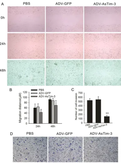
Fig 5. Effect of Tim-3 inhibition on Hela cell migration and invasion in vitro.(A) Cell migration capability was determined with a wound healing assay. Photographs were taken immediately (0 h), at 24 h and 48 h after wounding. (B) Quantification of wound closure. The data present the mean distance of cell migration to the wound area at 24 h and 48 h after wounding in three independent wound sites per group. (C) The ability of the cells to invade Matrigel was analyzed by the transwell invasion assay through a gel matrix. Hela cells were either infected with ADV-GFP or with ADV-antisense Tim-3, After 10 h viable invasive cells were fixed and counted. Values and error bars shown in this graph represent the averages and standard deviations respectively, of three independent experiments. (D) Representative images of the transwell invasion assay.
doi:10.1371/journal.pone.0152830.g001
PLOS ONE | DOI:10.1371/journal.pone.0152830 March 29, 2016 1 / 2
OPEN ACCESS
Citation:Cao Y, Zhou X, Huang X, Li Q, Gao L, Jiang L, et al. (2016) Correction: Tim-3 Expression in Cervical Cancer Promotes Tumor Metastasis. PLoS ONE 11(3): e0152830. doi:10.1371/journal. pone.0152830
Published:March 29, 2016
The authors confirm that these changes do not alter their findings. The authors have
pro-vided the underlying images for all figures in the original article as Supporting Information.
Supporting Information
S1 File. Underlying images for all figures.
(ZIP)
Reference
1. Cao Y, Zhou X, Huang X, Li Q, Gao L, Jiang L, et al. (2013) Tim-3 Expression in Cervical Cancer Pro-motes Tumor Metastasis. PLoS ONE 8(1): e53834. doi:10.1371/journal.pone.0053834PMID: 23335978