Abstract published online: July 7, 2009
Full paper published online: August 31, 2009 ISSN 1678-9199.
BRAIN AND LUNG CRYPTOCOCCOMA AND CONCURRENT Corynebacterium pseudotuberculosis INFECTION IN A GOAT: A CASE REPORT
Luvizotto MCR (1), Carreira VS (1), Ferrari HF (1),RibeiroD (2), Vallim MA (3), Azevedo V (2), Cardoso TC (1)
(1) Laboratory of Animal Pathology and Microbiology, School of Veterinary Medicine, São Paulo State University, UNESP, Araçatuba, São Paulo State, Brazil; (2) Laboratory of Cellular and Molecular Genetics, Federal University of Minas Gerais, UFMG, Belo Horizonte, Minas Gerais State, Brazil; (3) Department of Microbiology, Federal University of São Paulo, UNIFESP, São Paulo, São Paulo State, Brazil.
ABSTRACT: A four-year-old male goat with a history of neurological disorder was euthanized. It presented uncommon nodules in the brain and lungs associated with multiple abscesses, predominantly in the spleen and liver. Histological examination of brain and lung sections revealed yeast forms confirmed to be Cryptococcus gattii after a combination of isolation and polymerase chain reaction (PCR) procedures. Moreover,
Corynebacterium pseudotuberculosis infection was diagnosed by PCR of samples from the lung, spleen and liver. The present report highlights the rare concurrent infection of C. gatti and C. pseudotuberculosis in an adult goat from São Paulo state, Brazil, and indicates the necessity of surveillance in the treatment of goats with atypical pulmonary infections associated with neurological disorders.
KEY WORDS: Cryptococcus gattii, Corynebacterium pseudotuberculosis, meningoencephalitis, goat.
CONFLICTS OF INTEREST: There is no conflict.
CORRESPONDENCE TO:
TEREZA CRISTINA CARDOSO, Laboratório de Microbiologia, Departamento de Apoio, Produção e Saúde Animal, UNESP, Rua Clóvis Pestana, 793, Araçatuba, SP, 16.050-680, Brasil. Phone: +55 18 3636 1421. Fax: +55 18 36361403. Email:
INTRODUCTION
Cryptococcosis is an important cause of life-threatening meningoencephalitis both in human and other animals. Cryptococcus gattii is a basidiomycetes, a encapsulated yeast that is typically found in tropical and subtropical climate zones and predominantly infects immunocompetent hosts (1, 2). Traditionally, the natural habitat of C. gattii is mainly associated with several tree species (3). Moreover, cryptococcosis is characterized as an invasive fungal infection most commonly caused by one of two species normally defined as subacute or chronic, and often confused with viral or bacterial meningoencephalitis or other infections, including tuberculosis (3-5). Besides, there are substantial differences in the ecology that can influence directly on cryptococcosis epidemiology (3). Recently, C. gattii has been described to be associated with non-immunocompromised individual infection, resulting in a severe meningoencephalitis disorder in a domestic cat (5).
Corynebacterium pseudotuberculosis is the etiological agent of caseous lymphadenitis (CLA), a common disease in small ruminants throughout the world. This infection has been an important disease in the majority of sheep-rearing regions for over a century. Because of the chronic and often subclinical nature of CLA, it has proved to be difficult to control and its prevalence is high in many parts of the world, which, in turn, leads to significant economic losses for farmers (6, 7). Furthermore, to the best of our knowledge, the direct association between C. gatti and C. pseudotuberculosis infecting goats has never been described before.
CASE REPORT
A 4-year-old goat male, Boer breed, presenting a two-week history of gait and right head-tilt unresponsive to anti-inflammatory and antibiotic conventional treatment was sent to the Teaching Hospital of São Paulo State University, UNESP, in Araçatuba, SP, Brazil, and was killed for pathological examination.
samples using the sets of primers, as previously described (8). An m-PCR was also carried out to identify C. pseudotuberculosis through sets of primers targeting on 16S rRNA, rpoB and pld genes, as previously described (9). Total DNA was extracted from all clinical samples; the primer sequences employed are shown on Table 1 (8, 9).
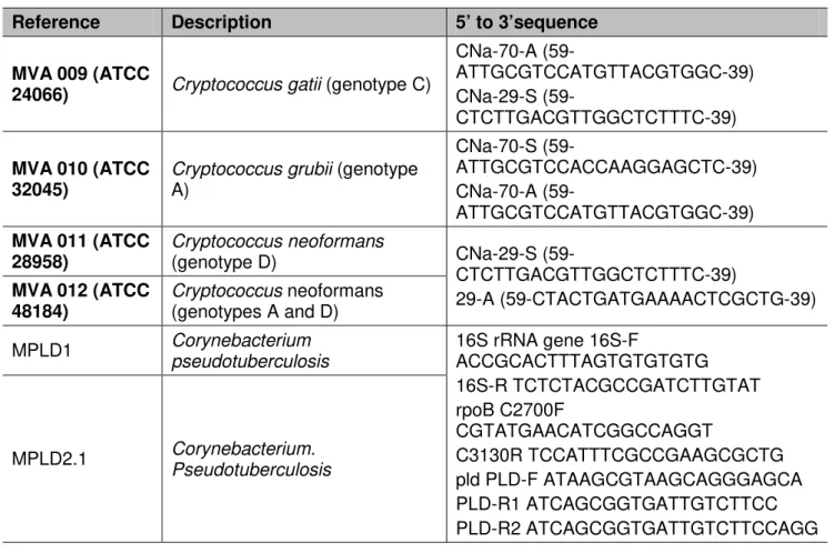
Table 1. Sequences of oligonucleotides employed to characterized the serotype of
Cryptococcus neoformans and C. gatti and molecular identification of Corynebacterium pseudotuberculosis
Reference Description 5’ to 3’sequence
MVA 009 (ATCC
24066) Cryptococcus gatii (genotype C)
CNa-70-A
(59-ATTGCGTCCATGTTACGTGGC-39) CNa-29-S
(59-CTCTTGACGTTGGCTCTTTC-39)
MVA 010 (ATCC 32045)
Cryptococcus grubii (genotype A)
CNa-70-S
(59-ATTGCGTCCACCAAGGAGCTC-39) CNa-70-A
(59-ATTGCGTCCATGTTACGTGGC-39)
MVA 011 (ATCC 28958)
Cryptococcus neoformans
(genotype D) CNa-29-S
(59-CTCTTGACGTTGGCTCTTTC-39)
29-A (59-CTACTGATGAAAACTCGCTG-39)
MVA 012 (ATCC 48184)
Cryptococcus neoformans (genotypes A and D)
MPLD1 Corynebacterium
pseudotuberculosis
16S rRNA gene 16S-F
ACCGCACTTTAGTGTGTGTG 16S-R TCTCTACGCCGATCTTGTAT rpoB C2700F
CGTATGAACATCGGCCAGGT
C3130R TCCATTTCGCCGAAGCGCTG pld PLD-F ATAAGCGTAAGCAGGGAGCA PLD-R1 ATCAGCGGTGATTGTCTTCC PLD-R2 ATCAGCGGTGATTGTCTTCCAGG
MPLD2.1 Corynebacterium.
Pseudotuberculosis
At necropsy, a 4.5 x 4.0-cm firm yellowish nodule filled with gelatinous substance associated with multiples 1 to 2-cm abscesses were found in the left lung lobe (Figure 1A). Ovoid abscesses and multiple coalescing translucent cysts in the spleen and liver were also observed. In addition, 2 to 3-mm translucent cysts were discovered dispersed in cerebral cortex, surrounded by necrosis (Figure 1B).
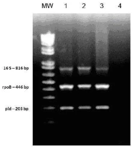
Figure 2. Multiplex polymerase-chain reaction (m-PCR). MW: molecular weight marker (1 kb plus ladder). Lines 1 and 2: samples from the lung and spleen positive for C. pseudotuberculosis; line 3: positive control (pure culture of C. pseudotuberculosis); line 4: negative control.
DISCUSSION
Natural CLA infections in goats demonstrate several similarities with the disease in sheep, especially when the same biotype of C. pseudotuberculosis is responsible. However, consideration of the differences in clinical presentation between sheep and goats may be helpful in respect of pathogenesis and specific risk factors (6, 9). In both sheep and goats, the major sites of infection are the superficial lymph nodes. However, in the present study no superficial lymph node was clinically observed. Furthermore, visceral lesions have been described as rare and found only in a minority of animals (7). Additionally, in sheep, visceral lesions, and particularly lung lesions, occur more frequently and in greater numbers than in goats (6, 7, 10, 11). Herein, it seems that primary infection with C.
pseudotuberculosis, in a different pathway, predisposed the animal to C. gattii infection. This is an important epidemiological aspect since C. gattii has been identified as a primary pathogen and led to other infections in both humans and other animals (2, 4, 5, 12-15). In addition, an association between C. neoformans and Mycobacterium bovis infection has already been described in goat, generating a granulomatous pneumonia (4).
There are few reports that describe C. gattii causing diseases in goats and it appears that chronic and often subclinical infections can predispose to C. gatti concurrent infections, even in non-immunocompromised individuals (16, 17). Different from CLA in goats, there are a small number of studies that report C. gatti infecting in this species. In Spain, the disease was described in outbreaks and caused severe pneumonia associated with cachexia in most affected animals (2). In Brazil, a survey on regional patterns of C. neoformans and C. gatti was carried out and found predominance of C. gatti in the northeast region (18). The present case seems to be the first one to report the occurrence of the disease in a Brazilian goat, caused by C. gatti serotype B. Since cryptococcosis is not well known in goats in the country, this infection may have predisposed to the concurrent infection by C. pseudotuberculosis, as it was herein presented. Both pathogens can be transmitted to non-immunocompromised hosts, both humans and other animals, and the surveillance of their natural occurrence should be stimulated aiming at preventing new infections.
ACKNOWLEDGEMENTS
REFERENCES
1. Cutsem JV, Rochette F. Mycoses in domestic animals. Belgium: Janssen Research Foundation; 1991. 226 p.
2. Baró T, Torrez-Rodriguez JM, De Mendoza MH, Morera Y, Alía C. First identification of autochthonous Cryptococcus neoformans var. gattii isolated from goats with predominantly severe pulmonary disease in Spain. J Clin Microbiol. 1998;36(2):458-61.
3. Nishikawa MM, Lazera MS, Barbosa GG, Trilles L, Balassiano BR, Macedo RCL, Bezerra CCF, Perez MA, Cardarelli P, Wanke B. Serotyping of 467 Cryptococcus neoformans isolates from clinical and environmental sources in Brazil: analysis of host and regional patterns. J Clin Microbiol. 2003;41(1):73-7.
4. Gutierrez M, Garcia Marin JF. Cryptococcus neoformans and Mycobacterium bovis
causing granulomatous pneumonia in a goat. Vet Pathol. 1999;36(5):445-8.
5. Belluco S, Thibaud JL, Guillot J, Krockenberger MB, Wyers M, Blot S, Colle MA. Spinal cryptococcoma in an immunocompetent cat. J Comp Pathol. 2008;139(4):246-51.
6. Ayers JL. Caseous lymphadenitis in goats and sheep: a review of diagnosis, pathogenesis, and immunity. J Am Vet Med Assoc. 1977;171(12):1251-4.
7. Baird GJ, Fontaine MC. Corynebacterium pseudotuberculosis and its role in ovine caseus lymphadenitis. J Comp Pathol. 2007;137(4):179-210.
8. Aoki FH, Imai T, Tanaka R, Mikami Y, Taguchi H, Nishimura NF, Nishimura K, Miyaji M, Schreiber AZ, Branchini ML. New PCR primer pairs specific for Cryptococcus neoformans
serotype A or B prepared on the basis of random amplified polymorphic DNA fingerprint pattern analyses. J Clin Microbiol. 1999;37(2):315-20.
9. Pacheco LGC, Pena RR, Castro TLP, Dorella FA, Bahia RC, Carminati R, Frota MNL, Oliveira SC, Meyer R, Alves FSF, Miyoshi A, Azevedo V. Multiplex PCR assay for identification of Corynebacterium pseudotuberculosis from pure cultures and for rapid detection of this pathogen in clinical samples. J Med Microbiol. 2007;56(5):480-6.
10. Baker RD. The primary pulmonary lymph node complex of cryptococcosis. Am J Clin Pathol.1976;65:83-92.
11. Hein WR, Cargill CF. An abattoir survey of diseases of feral goats. Aust Vet J. 1981; 57(11):498-503.
13. Rodrigues ML, Alviano CS, Travassos LR. Pathogenicity of Cryptococcus neoformans: virulence factors and immunological mechanisms. Microbes Infect. 1999;1(4):293-301. 14. Chen S, Sorrell T, Nimmo G, Speed B, Currie B, Ellis D, Marriott D, Pfeiffer T, Parr D, Byth K. Epidemiology and host- and variety-dependent characteristics of infection due to
Cryptococcus neoformans in Australia and New Zealand. Clin Infect Dis. 2000;31(2):499-508.
15. Krishnan AV, Corbett A. Intracranial and dermatological cryptococcal infection in an immunocompetent man. J Clin Neurosci. 2004;11(7):765-7.
16. Ecevit IZ, Clancy CJ, Schmalfuss IM, Nguyen MH. The poor prognosis of central nervous system cryptococcosis among non-immunosuppressed patients: a call for better disease recognition and evaluation of adjuncts to antifungal therapy. Clin Infect Dis. 2006; 42(10):1443-7.
17. Marroni M, Perivolini E, Cenci E, Bistoni F, Vecchiarelli A. Functional defect of natural immune system in an apparent immunocompetent patient with pulmonary cryptococcosis. J Infect. 2007;54(1):5-8.