Ambient pH Controls Glycogen Levels by Regulating
Glycogen Synthase Gene Expression in
Neurospora
crassa
. New Insights into the pH Signaling Pathway
Fernanda Barbosa Cupertino1, Fernanda Zanolli Freitas1, Renato Magalha˜es de Paula2, Maria Ce´lia Bertolini1*
1Departamento de Bioquı´mica e Tecnologia Quı´mica, Instituto de Quı´mica, Universidade Estadual Paulista, UNESP, Araraquara, Sa˜o Paulo, Brazil,2Nalco Company, Sugar Land, Texas, United States of America
Abstract
Glycogen is a polysaccharide widely distributed in microorganisms and animal cells and its metabolism is under intricate regulation. Its accumulation in a specific situation results from the balance between glycogen synthase and glycogen phosphorylase activities that control synthesis and degradation, respectively. These enzymes are highly regulated at
transcriptional and post-translational levels. The existence of a DNA motif for the Aspergillus nidulans pH responsive
transcription factor PacC in the promoter of the gene encoding glycogen synthase (gsn) inNeurospora crassaprompted us to investigate whether this transcription factor regulates glycogen accumulation. Transcription factors such as PacC inA. nidulansand Rim101p inSaccharomyces cerevisiaeplay a role in the signaling pathway that mediates adaptation to ambient pH by inducing the expression of alkaline genes and repressing acidic genes. We showed here that at pH 7.8pacCwas
over-expressed andgsnwas down-regulated in wild-typeN. crassacoinciding with low glycogen accumulation. In thepacCKO
strain the glycogen levels andgsnexpression at alkaline pH were, respectively, similar to and higher than the wild-type strain at normal pH (5.8). These results characterizegsnas an acidic gene and suggest a regulatory role for PACC ingsn
expression. The truncated recombinant protein, containing the DNA-binding domain specifically bound to a gsn DNA
fragment containing the PacC motif. DNA-protein complexes were observed with extracts from cells grown at normal and alkaline pH and confirmed by ChIP-PCR analysis. The PACC present in these extracts showed equal molecular mass, indicating that the protein is already processed at normal pH, in contrast toA. nidulans.Together, these results show that
the pH signaling pathway controls glycogen accumulation by regulatinggsnexpression and suggest the existence of a
different mechanism for PACC activation inN. crassa.
Citation:Cupertino FB, Freitas FZ, de Paula RM, Bertolini MC (2012) Ambient pH Controls Glycogen Levels by Regulating Glycogen Synthase Gene Expression in
Neurospora crassa. New Insights into the pH Signaling Pathway. PLoS ONE 7(8): e44258. doi:10.1371/journal.pone.0044258 Editor:Dana Davis, University of Minnesota, United States of America
ReceivedApril 19, 2012;AcceptedJuly 31, 2012;PublishedAugust 31, 2012
Copyright:ß2012 Cupertino et al. This is an open-access article distributed under the terms of the Creative Commons Attribution License, which permits unrestricted use, distribution, and reproduction in any medium, provided the original author and source are credited.
Funding:This work was supported by Fundac¸a˜o de Amparo a` Pesquisa do Estado de Sa˜o Paulo, FAPESP (grant nrs. 2008/57566-8 and 2007/07766-8), and Conselho Nacional de Desenvolvimento Cientı´fico e Tecnolo´gico, CNPq (grant nr. 303973/2009-9). The funders had no role in study design, data collection and analysis, decision to publish, or preparation of the manuscript.
Competing Interests:RMP is employed by a commercial company (Nalco). This does not alter the authors’ adherence to all the PLoS ONE policies on sharing data and materials.
* E-mail: [email protected]
Introduction
Neurospora crassa is a filamentous fungus that has been widely used as a model organism for studies on fundamental aspects of eukaryotic biology. The completion of its genome sequence [1] and, more recently, the availability of a set of mutant strains individually knocked-out in specific genes have enormously accelerated the investigation of many aspects of the biology of this organism. We have been using N. crassa to study how the metabolism of glycogen, a storage carbohydrate that functions as carbon and energy reserve, is regulated. Glycogen biosynthesis starts with the self-glucosylating initiator enzyme glycogenin (EC 2.4.1.186), termed GNN inN. crassa[2]. After an oligosaccharide is synthesized, the elongation step is catalyzed by glycogen synthase (EC 2.4.1.11) and a branching enzyme (EC 2.4.1.18) that mediate the formation ofa-1,4- anda-1,6-glycosidic bonds,
respectively, characteristic of the glycogen molecule. Glycogen synthase is the rate-limiting enzyme in this process and is inhibited
by phosphorylation; however, the phosphorylated form can be activated by the allosteric effector glucose-6-phosphate [3].
transcription factor PacC, suggesting a possible role for pH in regulating glycogen synthesis.
PacC has been extensively studied inA. nidulans and plays an important role in the pH signaling pathway. This transcription factor mediates the cell adaptation to neutral-alkaline pH by activating genes that are preferentially expressed at alkaline pH and repressing those preferentially expressed at acidic pH [9]. In A. nidulans, PacC is activated by two successive proteolytic cleavage steps at the C-terminus, the first being pH-dependent and activated by the products of six pal genes while the second is proteasome-mediated and pH-independent [10,11]. The proteol-ysis lead to the active protein PacC27that contains a DNA-binding domain formed by three C2H2zinc fingers capable of binding to the core consensus sequence 59-GCCARG-39 present in the promoters of pH-regulated genes (reviewed in [12,13]).
The Rim101p, a PacC orthologue inS. cerevisiae, was initially identified as a positive regulator of meiosis andrim101mutants are sensitive to Na+
or Li+
ions and grow poorly at low temperatures [14]. The yeast protein therefore appears to have a broader role than simply that of promoting alkaline pH-inducible responses [15]. While Rim101p is associated with the pH response, there are important differences between PacC and Rim101p. For example, the yeast protein requires only a single cleavage step to be activated [16] and, whereas PacC acts as a transcriptional activator under alkaline pH, Rim101p exerts its role as a repressor [15]. In addition, at least three pathways participate in the response to high pH stress inS. cerevisiae(reviewed in [17]). Thus, the molecular mechanisms involved in the pH response differ among organisms.
PacC orthologues have been identified in numerous filamentous fungi and the role of PacC as a mediator of pH regulation in fungal pathogenicity was first described in Candida albicans, in which Rim101p governs pH responses, dimorphism, and patho-genesis [18]. In N. crassa, the PACC pathway is involved in the glycosylation of Pi-repressible acid phosphatase [19] and in the transcription of thehsp70gene [20].
In this report, we demonstrated that the pH signaling pathway regulates glycogen metabolism inN. crassaby a process in which PACC may play a central role. As anA. nidulansPacC motif was identified in thegsnpromoter, and since N. crassahas the six A. nidulans palgene orthologues, we investigated the influence of pH on glycogen accumulation and the regulation ofgsn expression. Gel mobility shift assays showed that recombinant PACC recognized and bound specifically to the gsn promoter; this binding was confirmed in vivo by ChIP-PCR analysis. We also found that PACC proteolysis may involve a different way from that described for theA. nidulansprotein.
Results
Knockout of theN. crassa pacCGene and Growth Phenotype Analysis
To investigate the role of PACC in the regulation of glycogen metabolism we generated a null deleted strain by replacing the entire pacCORF (NCU00090, 1,866 bp) with the bar gene in a mus-52KO background strain (Figure 1A). The gene replacement and correct integration were confirmed by PCR analysis of the transformants (Figure 1B). However, we were unable to eliminate themus-52mutation after crossing the transformants with a wild-type strain due to the close linkage between themus-52andpacC genes. The morphological characteristics of thepacCKO
strain were analyzed by growth on VM agar plates and in race tubes. Since there were no apparent differences in the phenotypes of the mus-51KO(used as the wild-type in this work) andmus-52KOstrains [21]
we concluded that the growth defects observed in thepacCKOstrain were caused specifically by the absence of the transcription factor. Linear growth analysis in race tubes showed that both strains grew better at acid (4.2) and normal (5.8) pH than at alkaline pH and that the pacCKO strain was unable to grow at alkaline pH (7.8) (Figure 1C). We have also observed an accumulation of brown pigments after five days under normal growth conditions in flask (Figure 1D), characterized as melanin and described in other transcription factor mutant strains [22].
Radial growth was analyzed in plates after 24 h of growth in VM medium and VM medium supplemented with yeast extract (Figures 1E and 2). ThepacCKOstrain displayed similar growth to the wild-type strain in both media at acid pH and slightly reduced growth at normal pH; thepacCKOstrain did not grow at alkaline pH. The strains showed enhanced hyphal branching and shorter aerial hyphae in both media at alkaline pH (Figure 2). However, thepacCKO strain displayed severe growth defects at alkaline pH, including a reduction in the extent and production of aerial hyphae. Together, these results show that PACC is required for growth under alkaline conditions.
Glycogen Levels and Gene Expression Under pH Stress The gsngene is regulated at transcriptional and post-transla-tional levels. We have previously reported that gene expression and glycogen levels were altered under stress conditions such as heat shock and carbon starvation [5,6], both of which affect glycogen metabolism in N. crassa. The identification of a DNA motif for theA. nidulans pH signaling regulator PacC in the gsn promoter [8] prompted us to investigate whether this gene could be regulated by pH stress. Cells from both strains were germinated at pH 5.8 and then subjected to acid and alkaline pH stress after which samples were collected for glycogen quantification and RNA extraction. The glycogen levels in thepacCKO strain were higher than in the wild-type strain at normal growth pH (pH 5.8) (Figure 3B), indicating that PACC is important for maintaining normal glycogen levels during vegetative growth. However, under acid stress (pH 4.2), both strains accumulated similar amounts of glycogen (Figure 3A and 3B), suggesting that PACC does not affect glycogen accumulation under this condition in agreement with the growth results shown in Figure 1C. At alkaline pH, there was a decrease in the glycogen levels of both strains compared to the levels at pH 5.8. The fact that the glycogen level in thepacCKO strain after 30 min of alkaline stress was similar to that of the non-stressed wild-type strain (Figure 3B, time 0) emphasizes the role of PACC as a major regulator of glycogen accumulation under alkaline pH stress. The glycogen levels were also quantified in wild-type cells that were returned to normal pH after being subjected to acid and alkaline pH stress (RE, recuperation), however they did not recover the glycogen levels seen before stress (Figure 3A) in both conditions.
To investigate whether the effect of extracellular pH on glycogen accumulation resulted from changes in gsn and gpn (named here as the gene encoding the enzyme glycogen phosphorylase) expression we analyzed their mRNA levels during pH stress and recuperation at pH 5.8 in a wild-type strain. During acidic stress, thegsntranscript levels were kept constant while the gpntranscript decreased during 60 min of incubation (Figure 4A) and the levels were not recovered after returning the cells to normal growth pH (Figure 4A, RE). When cells were transferred to alkaline medium, gsn and gpn transcript levels decreased markedly during 60 min of stress, however onlygsn levels were recovered to normal levels after 60 min of incubation at pH 5.8 (Figure 4A). These results indicate that changes in extracellular pH control gsnexpression. We also analyzed thepacCmRNA levels
under pH stress and recuperation at pH 5.8 in the wild-type strain (Figure 4A). The pacCtranscript size was ,3 kb. WhereaspacC expression was low at acid pH, at alkaline pH there was a marked increase in expression compared to that at physiological pH (5.8), mainly during the first 15 min of stress. Basal expression was achieved when the mycelia were transferred back to pH 5.8 (RE). Expression ofgsnandgpngenes was analyzed in thepacCKOstrain under acid and alkaline pH stress and compared to the wild-type strain. ThegsnmRNA levels were higher in thepacCKOstrain than in the wild-type strain at all pH values (Figure 4B), suggesting a repressive role for PACC ingsnexpression. This result explains, at least partially, the glycogen levels shown in Figure 3B. There was very low expression for gpngene in the mutant strain in all pH suggesting that PACC also controlsgpnexpression under normal pH. Together, the results shown in Figures 3B and 4 suggest that alkaline pH inducespacCexpression and a concomitant decrease in glycogen levels by down-regulating thegsngene. These findings characterizegsnas an acid-specific gene.
Glycogen Levels and Gene Expression Under Combined pH and Heat Stress
As previously described, heat shock (45uC) down-regulatesgsn expression, resulting in decreased glycogen accumulation com-pared to that observed at 30uC (the normal temperature of growth) [5,6]. We therefore examined the effect of a combination of heat and pH stress on glycogen accumulation and in the expression of
gsn and pacCgenes in the wild-type strain. For this, cells were germinated at pH 5.8 and transferred to media of different pH preheated to 45uC. Glycogen levels were strongly reduced during heat shock, regardless of the pH (Figure 5A, black bars, HS). When heat stress was removed while maintaining the pH stress, normal glycogen levels were regained only in media with normal and acid pH (Figure 5A, compare with the results shown in Figure 3A for pH 4.2). The cells were unable to recover normal glycogen levels at alkaline pH (Figure 5A, pH 7.8) and they were the same as under heat stress.
The influence of pH and temperature stress ongsnexpression was also analyzed, as an attempt to correlate with glycogen accumulation. The gsn transcript levels were reduced in cells subjected to heat shock, regardless the media pH (Figure 5B). However, when cells were transferred back to normal temper-ature (30uC) and pH stress was maintained the gsn expression levels were recovered only at pH 4.2 and 5.8, but not at pH 7.8, in agreement with the results shown in Figure 5A. As withgsn,pacCexpression was strongly repressed by heat stress at both pH values. In addition, gene expression was recovered after returning the cells to normal temperature (30uC) at all pH values (Figure 5B). This interesting result suggests that the heat signaling pathway apparently elicits stronger cellular responses than the pH signaling pathway, at least forpacCexpression. It is noteworthy the existence of six STRE (STress Response Elements) and four HSE (Heat Shock Elements) motifs in the
Figure 1.pacCgene knockout and phenotypic analysis of the mutant strain.(A) Schematic illustration of thepacCgene knockout strategy. (B) Diagnostic PCR for validation of thepacCknockout was performed by using 0090-F1 and 0090-R4 oligonucleotides (Table 1). (C) Linear growth analysis. Apical extension of basal hyphae was determined in race tubes, as described in Material and Methods. The results shown are the average of three independent experiments. (D) Melanization. Strains were cultured in 250 mL flasks containing VM medium for 10 days (3 days at 30uC in the dark and 7 days at room temperature in ambient light/dark). Melanization can be visualized as brown pigment formation in thepacCKO
strain. (E) Radial growth analysis. Basal hyphae growth was examined after cultivating the strains on plates containing solid VM medium, as described in Material and Methods. Growth was expressed as colony diameter.
doi:10.1371/journal.pone.0044258.g001
pacC promoter, all of which are DNA elements known to be responsive to heat stress.
Recombinant PACC Binds to the gsnPromoter
To investigate whether the PacC DNA motif identified in thegsn promoter was, indeed, recognized by PACC, a gel mobility shift assay was performed using a truncated recombinant PACC and a 146 bp DNA fragment containing the PacC core sequence (59 -CTTGGC-39) as probe. Recombinant PACC bound efficiently to the fragment corresponding to the PacC recognition motif in the gsnpromoter (Figure 6A, lanes 2 and 6). DNA-protein binding was reduced in the presence of unlabelled specific competitor (Figure 6A, lanes 3 and 7). The specificity of the DNA-protein complexes was further confirmed by adding a 27 bp DNA oligonucleotide (oligo pacC) as a specific competitor (Figure 6A, lanes 4 and 8) and by supershift experiments (Figure 6A, lanes 11 to 13). An interesting result was the presence of two complexes exhibiting different molecular masses when the specific competitor was added to the reaction (Figure 6A, lanes 3 and 7). We speculated that they may represent complexes with distinct conformational structures. Additional experiments will be neces-sary to clarify this. The DNA oligonucleotide completely abolished both complexes and the addition of anti-PACC antibody to the reaction increased the molecular mass of the DNA-protein complex, which shifted close to the gel origin (Figure 6A, lanes 12 and 13).
The specificity of the DNA-protein complex was also confirmed by using a mutatedpacCprobe (mpacC) in which the 59 -CTTGGC-39 core sequence was changed to 59-AAATTA-39 (Figure 6B). There was a marked reduction in the formation of DNA-protein
complex and a weaker complex was only visible after adding 2.0mg of protein (Figure 6B, lane 6), i.e., an amount of protein sufficient to produce a strong complex (compare with lane 2). The complex formation was strongly decreased in the presence of the specific competitor (Figure 6B, lane 7 and 10) and completely inhibited in the presence of the DNA oligonucleotide (Figure 6B, lane 8 and 11).
Binding of Proteins from Crude Cellular Extract to the PacC Binding Site of thegsnPromoter
To investigate whether PACC binding to the gsn promoter was pH-dependent, gel shift assays were performed with crude cellular extracts from wild-type and pacCKO cells grown under normal (5.8) and alkaline (7.8) pH. The cellular extracts were fractionated by affinity chromatography, bound proteins were eluted with a KCl gradient and the fractions were analyzed for their ability to bind to thepacCprobe previously used. Fractions from wild-type strain mycelium showing DNA-binding activities were identified in extracts from cells grown at both pH conditions (results not shown). One active chromatographic fraction from each pH was assayed for its binding to the PacC consensus site of the gsn promoter in the presence of both competitors. DNA-protein complexes were observed when proteins were incubated with the DNA fragment (Figure 7A, lanes 2 and 7). When the same fragment was used as a specific competitor in 10-fold molar excess the complexes were shifted only when crude cellular extract prepared from mycelia grown at pH 5.8 was used as the protein source (Figure 7A, lane 3). However, the specific competitor oligopacCwas able to shift the DNA-protein complexes when proteins from both extracts were
Figure 2. Phenotypes of the wild-type andpacCKOstrains in different pH and nutritional growth conditions.The strains (107conidia/ mL) were cultured on plates containing agar VM medium and VM medium supplemented with yeast extract (VM+YE) at pH 4.2, 5.8 and 7.8, in
ambient light/dark for 24 h. Hyphae at the colony edge were observed using a stereoscope. doi:10.1371/journal.pone.0044258.g002
analyzed (Figure 7A, lanes 4–6, 10 and 11). The binding reaction was apparently stronger when cellular extract prepared from cells grown at alkaline pH was used as a protein source since the DNA-protein complex was shifted only at higher oligo pacC concentrations (20- and 30-fold molar excess). Fractions
from pacCKO
strain were also assayed for binding reactions and no DNA-protein complex was observed. The Figure 7A (lanes 13–16) shows the results with one chromatographic fraction from each pH and the competition with the oligo pacC. This result indicated the existence of an active PACC protein in the
Figure 3. Glycogen accumulation during acid and alkaline pH stress.(A) Glycogen content in the wild-type strain. Glycogen was extracted from mycelia grown under physiological conditions (pH 5.8, control) and under acid (pH 4.2) and alkaline (pH 7.8) stress. After 120 min, the remaining cultures were transferred back to physiological conditions (pH 5.8, RE, recuperation). The results shown are the average of at least three independent experiments. (B) Glycogen content in thepacCKO
strain compared to the wild-type strain. 0, cell samples before the pH shift (control). doi:10.1371/journal.pone.0044258.g003
wild-type strain that specifically bound to the PacC motif even under normal growth pH (5.8). It is noteworthy that the DNA-protein complexes migrated close to the gel origin suggesting that they may include other proteins in addition of PACC. It is also interesting since low levels of pacC transcript could be detected in wild-type cells grown at pH 5.8 (Figure 4A).
We also investigated whether the PACC present in the two cellular extracts had equal molecular mass since the same protein fromA. nidulansgrown at alkaline pH has a lower molecular mass than that observed at normal growth pH [10]. The presence of PACC in the cellular extracts was analyzed using polyclonal anti-PACC antibody. The protein was detected in both cellular extracts and, surprisingly, had the same molecular mass (,60 kD), which was similar to that of the intermediate PacC form fromA. nidulans grown at alkaline pH (53 kD) (Figure 7B). A single PACC form was identified in the cellular extract prepared up to 4 h after the cells were shifted to alkaline pH. There was little protein induction in extracts from 1 h after shifting. The processed PacC27protein
described inA. nidulansgrown at alkaline pH was not detected inN. crassa.
Chromatin immunoprecipitation (ChIP-PCR) experiments in the wild-type strain were performed to confirm that N. crassa PACC binds to the gsn promoter in vivo and to assess whether protein binding was pH-dependent. Figure 7C shows that endogenous PACC was associated with thegsn promoter region containing the PacC motif. In addition, protein association was observed with genomic DNA extracted from cells grown at normal pH (5.8) and from cells 2 h after shifting to alkaline pH. Together, these results indicate that N. crassa PACC specifically associates with thegsnpromoter even at pH 5.8, the pH for normal growth, thus confirming that the protein is active at physiological pH.
Discussion
Extracellular pH has an important role in cell biology as it regulates gene expression and consequently influences a variety of cellular processes, such as growth, differentiation and
develop-Figure 4.gsn, gpnandpacCgene expression during acid and alkaline pH stress.Cells from the wild-type andpacCKOstrains were cultivated at pH 5.8 for 24 h and shifted to pH 4.2 and pH 7.8. Samples were collected and used to extract total RNA. Total RNA (15mg) was separated by
electrophoresis in a denaturing formaldehyde gel, transferred to nylon membrane and probed witha-32P-radiolabeled 678 bpgsncDNA, or 798 bp gpncDNA or 639 bppacCcDNA fragments (gel autoradiographies). The 28 S rRNA was used as a loading control after ethidium bromide staining. The results shown are the average of at least three independent experiments. (A) Analysis of thegsn, gpnandpacCgenes in the wild-type strain at different times after pH shifting. After pH stress the remaining cultures were transferred back to physiological conditions (RE, recuperation, pH 5.8) and samples were collected. (B) Analysis of thegsnandgpngenes in thepacCKOstrain compared to the wild-type strain at different times after pH shifting. 0, cell samples before pH shifting (control).
doi:10.1371/journal.pone.0044258.g004
ment. The regulation of gene expression by pH has been widely investigated in microorganisms because of the great variations in ambient pH that they face in their natural environments. The ambient pH signaling pathway has been exhaustively studied in the filamentous fungusA. nidulansand in several yeasts, includingS. cerevisiae.InA. nidulans, pH regulation is mediated by the action of a cascade of proteins (palgene products) that leads to activation of the transcription factor PacC. This protein, which has a central role in the pH signaling pathway, is activated at alkaline pH after two proteolytic cleavage steps and acts as a transcriptional activator of alkaline-expressed genes (reviewed in [23]).
InS. cerevisiae, the pathway leading to activation of the Rim101p orthologue shares similarities with that of A. nidulans PacC.
However the mechanism of alkaline pH adaptation is not exactly the same since Rim101p requires only a single cleavage step to be activated and, whereas PacC acts either as an activator or repressor, Rim101p is a repressor of the genes encoding the transcriptional repressors Nrg1p and Smp1p at alkaline pH. Nrg1p is required for the repression of alkaline pH-inducible genes, including the gene encoding Ena1 Na+
-ATPase, a protein essential for growth at alkaline pH [15]. Thus, Rim101p is indirectly required forS. cerevisiaeadaptation to alkaline stress. In addition to Rim101p-mediated alkaline pH regulation, there are also Rim101p-independent mechanisms involved in pH regulation inS. cerevisiae.One is the calcineurin pathway, the activation of which affects the expression of a number of genes through
Figure 5. Glycogen accumulation and gene expression during combined pH and heat shock stress.Glycogen and total RNA were extracted from wild-type mycelia cultivated at pH 5.8 and 30uC for 24 h and then shifted to pH 5.8, 4.2 and 7.8 at 45uC for 30 min. After 30 min, the remaining samples were transferred back to the physiological temperature (30uC) at the three pH conditions and incubated for different times (RE, recuperation). (A) Accumulation of glycogen. (B)gsnandpacCgene expression. Total RNA (15mg) was separated by electrophoresis in a denaturing
formaldehyde gel, transferred to nylon membrane and probed with thea-32P radiolabeled 678 bpgsncDNA and 639 bppacCcDNA fragments (gel autoradiographies). The 28 S rRNA was used as a loading control after ethidium bromide staining. The results shown are the average of at least three independent experiments. 0, cell samples before pH shifting (control).
doi:10.1371/journal.pone.0044258.g005
dephosphorylation of the transcription factor Crz1 [24]. High pH stress triggers a transient rise in cytoplasmic calcium resulting in calcineurin activation and ENA1 induction through Crz1 [25]. More recently, Platara et al. [26] described another signaling pathway involving the protein kinase Snf1p that acts through the Mig2p repressor, i.e., a third regulatory pathway involved in the response to high pH stress.
The results described here indicate a role forN. crassaPACC as a regulator of glycogen metabolism through its ability to target GSN via a PacC binding site in the gsn promoter. The pacCKO strain showed impaired growth at alkaline pH, in agreement with the better growth ofN. crassaat acidic (5.8) compared to alkaline (7.8) pH. Our findings suggest the existence of a different mechanism of PACC activation under alkaline pH stress when compared toA. nidulansandS. cerevisiae.
PACC Regulates Glycogen Levels Under pH Alkaline Stress through Regulation ofgsnExpression
We investigated the glycogen levels and gsn expression in response to extracellular pH changes. Our results provide evidence that both processes are under pH control inN. crassa. Cells of the wild-type strain grown at alkaline pH showed low intracellular glycogen accumulation and a reduction ingsntranscript levels. In contrast, the pacCKO strain accumulated similar amounts of glycogen when compared to the non-stressed wild-type strain and showed highgsntranscript levels at alkaline pH. These results are sufficient to characterizegsnas a pH-regulated gene, and that it is an acid-specific gene. Many genes have been reported to be regulated by ambient pH [27]. In the case ofgsn, the finding that wild-type andpacCKO strains had similar amounts of glycogen at normal and alkaline pH, respectively, reinforced the criteria for a
Figure 6. Binding of recombinant PACC to thegsnpromoter.(A) Upper panel, schematic representation of thepacCprobe and the specific competitor oligopacC. Lower panels, gel shift analysis using increasing amounts of recombinant PACC in the presence of specific competitors and polyclonal anti-PACC antibody. Lanes 1, 5 and 9,pacCprobe, no protein added. Lanes 2, 6 and 10, gel shift analysis using 1.0, 2.0 and 5.0mg of
recombinant PACC. Lanes 3 and 7, gel shift analysis in the presence of the 146 bp specific competitor. Lanes 4 and 8, gel shift analysis using the specific competitor oligopacC. Lanes 11 to 13, supershift assay using 5.0, 10.0 and 20.0mL of anti-PACC antibody (1:500). (B) Upper panel, schematic
representation of the mutated mpacCprobe. Lower panel, gel shift with wild-type and mutated probes. Analysis using different concentrations of recombinant PACC in the presence and in the absence of competitors. Lane 1 and 3,pacCand mpacCprobes, respectively, no protein added. Lanes O, gel origin; SC, specific competitor; FP, free probe.
doi:10.1371/journal.pone.0044258.g006
gene to be regulated by ambient pH [28]. The analysis of pacC gene expression inN. crassashowed that it was under pH control, as also described forA. nidulans [9] and other filamentous fungi [29–31]. This observation supports the fact that, at alkaline pH, the over-expression ofpacCcontrols glycogen levels by regulating gsn, but notgpn, expression.
The patterns of glycogen accumulation and gene expression under combined pH and heat stress were very interesting. Whereas in the wild-type strain glycogen andgsntranscript levels returned to normal, this strain was unable to recover normal levels when exposed concomitantly to heat and alkaline stress. In addition, pacC was not induced under these conditions. The presence of STRE and HSE motifs in thepacCpromoter suggests that this gene is regulated by heat shock. As with gsn down-regulation under heat stress [5,6],pacC is another example of a gene inN. crassa having STRE and HSE binding sites in its 59 -flanking region being down-regulated at the transcriptional level by heat stress. Thegsnpromoter has one STRE motif, which was
demonstrated to be involved in the gene transcription modulation when cells were exposed to heat shock (transferred from 30uC to 45uC) [6].
InS. cerevisiaethe STREs are described as cisregulatory motif that mediates the transcriptional activation of genes through the action of the stress response regulator proteins Msn2p/4p [32]. Orthologous proteins have not been identified in the N. crassa genome database, suggesting that in this organism STRE may modulate gene expression by an alternative mechanism [33]. The activation of protein kinase A (PKA) leads to Msn2/4p phosphor-ylation and its cytoplasmic accumulation, thereby preventing the transcription of STRE-regulated genes [34]. Recently, Casadoet al.[35] demonstrated that the exposure ofS. cerevisiaeto alkaline medium resulted in a decrease in cAMP that led to inhibition of the PKA pathway and the accumulation of Msn2/4p in the nucleus. These data indicate that the adaptative response to alkaline pH involves PKA-regulated Msn2/4p-mediated gene remodeling. Since N. crassa lacks Msn2/4p orthologues we
Figure 7. PACC binds specifically to thegsnpromoter region in an alkaline pH-independent manner.(A) Gel shift analysis using crude cellular extracts fractionated on a Heparin-Sepharose column. Crude cellular extracts (CCE) from mycelia submitted or not to alkaline pH stress (pH 7.8) were fractionated by affinity chromatography. Left panel, a protein fraction (35mg) exhibiting DNA-binding activity was assayed in the
presence of specific competitors. Lane 1,pacCprobe, no protein added. Lanes 2 and 7, proteins from pH 5.8 and pH 7.8 samples, respectively, in the absence of competitors. Lanes 3 and 8, DNA band shift in the presence of the 146 bppacCspecific competitor. Lanes 4 to 6 and 9 to 11, DNA band shifts in the presence of increasing amounts of the 27 bp DNA oligopacCas a specific competitor. Right panel, a protein fraction (35mg) from
knocked-out strain crude cellular extract was assayed. Lane 12,pacCprobe, no protein added. O, gel origin; SC, specific competitor; FP, free probe. (B) PACC shows the same molecular mass at pH 5.8 and pH 7.8. Crude cellular extracts prepared from wild-typeN. crassasubmitted or not to alkaline pH stress were analyzed by Western blotting using a polyclonal anti-PACC antibody. The proteina-tubulin (theoretical molecular mass 50 kD) was used as a loading control. (C) Chromatin immunoprecipitation assay using the polyclonal anti-PACC antibody. Genomic DNA samples from wild-typeN. crassasubmitted or not to pH stress were immunoprecipitated with the anti-PACC antibody and subjected to PCR to amplify a 146 bp DNA fragment of thegsnpromoter containing thepacCmotif. A plasmid construction containing the entire sequence of thegsngene, including its 59- and 39 -flanking regions, was used as a positive control. As a negative control, the immunoprecipitation reactions were performed without the anti-PACC antibody. L, 1 kb DNA ladder.
doi:10.1371/journal.pone.0044258.g007
hypothesize that the down-regulation of pacC under heat shock and alkaline pH results from the activation of proteins that mediate gene transcription regulation under heat stress through STRE. Whether the PKA signaling pathway is involved in this process remains to be demonstrated. We have previously reported that a N. crassa mutant strain with an inactive PKA pathway showed hyper-accumulation of glycogen during growth when compared to the wild-type strain [7]. However, thegsntranscript level was not increased in this strain; rather, GSN was less phosphorylatedin vitroand therefore more active, suggesting that GSN post-translational modification is likely to be the main mechanism controlling glycogen accumulation during vegetative growth when the PKA pathway is inactive.
PACC Binds to thegsnPromoterin vitroandin vivoand is Active at Normal and Alkaline pH
DNA shift experiments showed that truncated PACC having the DNA-binding domain was able to bind to a DNA fragment from the gsn promoter containing the PacC binding site. The DNA-binding reaction was specific, based on three criteria: (1) binding was reduced when a DNA fragment mutated in the PacC core sequence was used, (2) a DNA oligonucleotide containing the PacC binding site competed in the binding reactions by reducing the DNA-protein complexes, and (3) anti-PACC antibody led to a DNA-protein supershift. Together, these findings revealed that the PacC binding site in the gsn promoter was the target for the binding of recombinant PACC. In our results, it was observed binding reduction when the mutated probe was used, instead of no binding, which was completely abolished in the presence of the DNA oligonucleotide as competitor. We suggested either that the core sequence neighborhoods are important for PACC recognition or the N. crassaPACC has an alternative binding site similar to what was described for Rim101p inC. albicans[36]. DNA-protein complexes were also observed when crude cellular extracts were used as protein sources. Interestingly, proteins from extracts prepared from wild-type cells grown at normal and alkaline pH were able to bind to the DNA fragmentin vitroandin vivo, and the PACC present in these extracts had the same molecular mass (,60 kD).
According to the model for pH-mediated regulation of gene expression in A. nidulans [23] full-length PacC (PacC72) is synthesized as an inactive form and an alkaline signal is transduced by thepalsignaling pathway, thereby activating PacC72to PacC53 and PacC27 by two successive proteolytic steps. The S. cerevisiae Rim101p is similar in size to theA. nidulansprotein but they differ in that the yeast protein is proteolytically processed at the C-terminus by a single cleavage step and the smaller size form (similar to PacC27) has not been detected [16]. The results described here suggest thatN. crassaPACC is subject to only one proteolytic processing step since the molecular mass of the protein present in cells grown at alkaline pH was lower than the theoretical value of 67 kD. According to Pen˜as et al. [37], N. crassa PACC has a conserved protease box that may lead to a processed form of PACC with a molecular mass of,55 kD, the size found in our experiments. The most intriguing result concerning PACC was that the forms detected before and after pH stress had the same molecular mass, which indicates that alkaline pH is not the signal for proteolytic processing. Rather, the protein is already processed during growth at pH 5.8.
The finding thatN. crassahas orthologues to all six Pal proteins, including PalH which, together with PalI, function as membrane pH sensors inA. nidulans[23], is particularly interesting. According to the model proposed forA. nidulans, the pH signal is transduced to downstream components by PalF phosphorylation and
ubiquitination, leading to endocytosis of the PalF/PalH complex [38]. By overexpressing PalF covalently attached to ubiquitin in a nullpalHbackground Herva´s-Aguilaret al.[39] have shown that PalF ubiquitination activates the signaling pathway under acidic conditions. The authors concluded that ubiquitination of PalF commands the cascade of intracellular events that mediate fungal adaptation to environmental pH.
Our results clearly demonstrated that inN. crassaambient pH controls glycogen accumulation by regulatinggsnexpression, i.e., by regulating glycogen synthesis and not degradation. Although the data point to a central role for PACC many questions remain unanswered. One major concern is the fact that PACC shows the same molecular mass before and after alkaline pH stress, indicating that if there is any processing it must occur indepen-dently of ambient pH. Another question relates to thein vivoresults showing that endogenous PACC binds to thegsnpromoter even at pH 5.8. If there is protein association at both pH values, how are the differences in gsn expression and glycogen content to be explained? SinceN. crassa has all six Pal protein orthologues we might speculate that PalH may function as a pH sensor, as inA. nidulans, and that all six Pal homologues are active components of the pH signaling pathway. Finally, is PACC the only regulator of glycogen accumulation andgsnexpression at alkaline pH? Other molecular mechanisms are known to be involved in adaptation to environmental pH inS. cerevisiaeandC. albicans[40]. The results described here reveal interesting differences betweenN. crassaand A. nidulanswith regard to the pH signaling pathway that deserve to be investigated.
Materials and Methods
Neurospora CrassaStrains and Growth Conditions Neurospora crassaFGSC#9718 (mat a, mus-51::bar), the wild-type background strain used in these experiments, was purchased from the Fungal Genetics Stock Center (FGSC, University of Missouri, Kansas City, MO, USA, http://www.fgsc.net) [41]. A pacC knockout mutant strain (pacCKO) was generated following the knockout procedures described elsewhere [22] based on the strains FGSC#9568 (mat a, mus-52::hyg) and FGSC#2490 (mat A). All strains were maintained on solid Vogel’s minimal (VM) medium, pH 5.8 [42] containing 2% sucrose at 30uC.
Conidia from 10 day cultures of wild-type andpacCKO strains were suspended in sterile water and counted. For radial growth analysis, 107conidia/mL were inoculated onto plates containing solid VM medium and VM medium supplemented with yeast extract at pH 4.2, 5.8 and 7.8 for 24 h. Images of colony morphology were captured after 24 h using an AxioCam ICc3 coupled to the stereoscope trinocular Discovery V8 (Zeiss) at 80 x magnification. For linear growth, conidia were inoculated in race tubes containing VM medium at pH 4.2, 5.8 and 7.8 and incubated at room temperature. Measurements were made every 24 h.
For pH stress experiments, 109 conidia/mL were first germi-nated in 4 L of VM medium (pH 5.8) at 30uC, 250 rpm, for 24 h. After this period, the culture was filtered and the mycelia divided into three samples. One was frozen in liquid nitrogen and stored at
280uC for further processing (control sample, not submitted to stress) while the remaining two samples were transferred into 1 L of fresh VM medium containing 0.5% sucrose at pH 4.2 (for acid stress) and 7.8 (for alkaline stress). Samples (125 mL) from mycelia submitted to both pH conditions were harvested after different periods of incubation. For the experiments combining pH and heat stress, mycelia obtained from the wild-type strain after 24 h were filtered and divided into three samples that were transferred
into 1 L of fresh VM medium containing 0.5% sucrose at pH 4.2, 5.8 and 7.8 preheated to 45uC. Samples (125 mL) were harvested after 30 min incubation and processed as before.
In both stress experiment samples from the wild-type strain were subjected to the recuperation conditions. The remaining mycelia from the pH stress experiments were filtered and transferred back into 400 mL of fresh VM medium at pH 5.8 and 30uC. Samples were collected after 30, 60 and 120 min of recuperation. For experiments combining pH and heat stress the remaining mycelia were transferred back into 400 mL of fresh VM medium pre-heated at 30uC at the three different pH. Cells from 120 mL samples were harvested after 30, 60 and 120 min of recuperation and processed. The mycelial samples were used for glycogen quantification and RNA extraction.
Generation of apacCKOMutant by Gene Replacement A strain bearing a deletion in thepacCgene was generated as described by Ninomiyaet al. [21]. Briefly, three PCR fragments were produced using the following pairs of primers: fragment 1, primers F1 and R2, fragment 2, F2 and 0090-R3, and fragment 3, primers 0090-F3 and 0090-R1 (Table 1). Fragments 1 and 3 contained a 1.5 kb upstream and downstream of the ORF NCU00090 (pacC, 1,866 bp), respectively, with an additional 20 bp overlapping sequence from the bar gene. Both fragments were amplified from wild-type strain genomic DNA. Fragment 2 contained the bar ORF (1 kb) plus overlapping sequences from thepacCgene at both ends and was amplified from the pIV9A-1 plasmid [6]. All three fragments were purified using a Qiagen gel purification kit, and 100 ng of each fragment was mixed and used to amplify a single fragment (4 kb) with the primers 0090-F1 and 0090-R1. All PCRs were done using Phusion High FidelityTaqpolymerase (NEB). The final PCR fragment was purified and used to transform 10 day conidia from the strain FGSC#9568. Transformants were selected by plating in medium containing Basta (200mg/mL). Correct integration of the fragment into the genome was detected by PCR using primers 0090-F1 and 0090-R4 that annealed downstream from the amplified fragment. One transformant containing the right integration was backcrossed with FGSC#2490 to generate a homokaryotic strain.
pacC cDNA Cloning and Production and Purification of the Recombinant Protein
The pacC gene encodes a 621 amino acid protein with a theoretical molecular mass of 67 kD. A pacC cDNA fragment (639 bp) encoding the N-terminal region containing the zinc finger C2H2 DNA-binding domain was amplified from the cDNA plasmid library pYADE5 [43] with the oligonucleotides 90-F and 90-R2 (Table 1), as previously described [8]. For expression of the His-DPACC recombinant protein theE. coliBL21(DE3) pLysS strain harboring the pET-DPACC plasmid was used. A truncated
protein encompassing amino acids 1 to 213 fused to an N-terminal His6-tag was produced. Cells were grown at 37uC in 1 L of LB medium to an OD600 of 0.7 and induced with IPTG (final concentration 0.4 mM) for 4 h at 37uC. The cells were harvested by centrifugation, suspended in buffer A (50 mM Tris-HCl, pH 8.0, 300 mM NaCl, 20 mM imidazole, 10% v/v glycerol, 1 mM DTT, 0.5 mM PMSF, 25 mM benzamidine and 5mg/mL each of antipain, leupeptin and pepstatin A) containing 0.5% Triton X-100 and 0.5% Tween 20 and lysed by sonication (5 cycles of 30 s sonication and 30 s on ice). After centrifugation, the supernatant was subjected to affinity chromatography on a HisTrap HP column (GE Healthcare) using an A¨ KTA Prime purification system. The recombinant protein was eluted with a
linear gradient of imidazole (20–500 mM) in buffer A and dialyzed two times against 1 L of dialysis buffer (10 mM Tris-HCl, pH 7.9, 100 mM KCl, 10% v/v glycerol, 1 mM EDTA and 0.5 mM DTT). The purified protein was analyzed by SDS-PAGE followed by Coomassie Brilliant Blue staining [44] and was quantified by the Hartree [45] method using BSA as standard. The purified
His-DPacC protein was used to raise antibodies in rabbits.
Glycogen Quantification and Gene Expression Assays Mycelial pads were ground to a fine powder in a pre-chilled mortar in liquid nitrogen and extracted with lysis buffer (50 mM Tris-HCl, pH 7.6, 100 mM NaF, 1 mM EDTA, 1 mM PMSF, 0.1 mM TCLK, 1 mM benzamidine and 1mg/mL each of
pepstatin and aprotinin). Cell extracts were clarified by centrifu-gation (3,0006g, 10 min, 4uC) and the supernatants were used for
glycogen and protein quantification. Glycogen was extracted by digestion with amyloglucosidase (30 mg/mL) and a-amylase (10 mg/mL) after ethanol precipitation, as previously described [7]. Free glucose was measured with a glucose oxidase kit and the glycogen content was normalized to the total protein concentra-tion. Total protein was quantified by the Hartree method [45].
For gene expression analysis by Northern blotting, total RNA was extracted with LiCl [46] from mycelial samples subjected to pH and combined pH and temperature stress. Total RNA (15mg)
was electrophoresed on a 1.2% agarose-formaldehyde denaturing gel [47] at 65 V for 5 h and then transferred to neutral nylon membranes (Hybond-N, GE Healthcare) in 2 x SSC. The blots were probed either with a 678 bpgsncDNA, or 798 bpgpncDNA, or 639 bppacCcDNA fragments (106–108cpm) radiolabelled with
a-32P-dATP (3,000mCi/mmol) by random priming (NEBlot kit, Biolabs) in 5–10 mL of ULTRAhyb hybridization solution (Ambion) at 42uC overnight. After hybridization, the blot was washed twice in 2 x SSC containing 0.1% SDS for 20 min, and twice in 0.1 x SSC containing 0.1% SDS for 20 min followed by exposure to an X-ray film.
The pacCgene was also analyzed at protein level by Western blot. Total proteins from cells of the wild-type strain cultured at pH 5.8 for 24 h (control) and that subjected to alkaline pH stress (pH 7.8) for 1, 2, 3 and 4 h were prepared as described for glycogen extraction using glass beads. Proteins (100mg) were separated by 12% SDS-PAGE gels [44] and electrophoretically transferred to a nitrocellulose membrane. Immunoblotting was performed with polyclonal anti-PACC antibody and band intensities were normalized to parallel blots probed with anti-a
-tubulin antibody (Sigma). Blots were subsequently probed with HRP-conjugated secondary antibodies (Sigma) and developed with luminol reagent.
Electrophoretic Mobility Shift Assay (EMSA)
DNA-protein binding reactions were carried out in 30–80mL of
1 x binding buffer (25 mM HEPES-KOH, pH 7.9, 20 mM KCl, 10% v/v glycerol, 1 mM DTT, 0.2 mM EDTA, 0.5 mM PMSF, 12.5 mM benzamidine and 5mg/mL each of antipain and
pepstatin A) containing 2mg of poly(dI-dC).(dI-dC) as a
non-specific competitor and 0.5–5mg of His-DPACC recombinant
protein. In some experiments, 35mg of protein from chromato-graphic fractions (obtained as described below) was used. A radiolabeled DNA probe (,104cpm) was added and the reactions were incubated at room temperature for 20 min. Free probe was separated from DNA-protein complexes by electrophoresis on a native 5% polyacrylamide gel in 0.5 x?TBE buffer at 300 V, 10 mA and 10uC. After electrophoresis, the gel was dried and autoradiographed. For competition assays, an excess of specific
DNA competitor was added to the binding reactions 10 min prior to incubation with the radiolabeled probe.
For the supershift experiments, 5mg of recombinant
His-DPACC protein was incubated with an excess of polyclonal
anti-PACC antibody (title 1:500) for 10 min on ice prior to the addition of radiolabeledpacCprobe and further 20 min incubation at room temperature.
Preparation and Fractionation of Crude Cellular Extract for EMSA
Cells from the wild-type andpacCKOstrains exposed to alkaline stress (pH 7.8, 60 min) or not (pH 5.8, control) were used to prepare crude cellular extracts. About 10 mg of frozen mycelia were ground to a fine powder in liquid nitrogen in a pre-chilled mortar, homogenized in 20 mL of lysis buffer (15 mM HEPES-KOH, pH 7.9, 10% v/v glycerol, 500 mM KCl, 5 mM MgCl2, 0.5 mM EDTA, 1 mM DTT, 0.5 mM PMSF, 25 mM benzami-dine, 50 mM NaF and 10mg/mL each of antipain and pepstatin A) and stirred with glass beads in eight cycles that consisted of 30 s of stirring and 30 s on ice. Crude cellular extract was recovered after centrifugation (3,2006g, 2 min, 4uC), dialyzed against buffer
D (15 mM HEPES-KOH, pH 7.9, 15% v/v glycerol, 100 mM KCl, 1 mM EDTA) at 4uC for 2 h, and cleared by centrifugation (20,000 6 g, 20 min, 4uC) before loading onto a
Heparin-Sepharose FF column (GE Healthcare). The proteins were eluted with a 0.1–1.5 mM KCl linear gradient and the protein fractions were dialyzed against buffer D containing 0.5 mM PMSF, 25 mM benzamidine and 50 mM NaF and then frozen in liquid nitrogen and storage at 280uC. Total protein was quantified by the Hartree method [45]. Proteins from the chromatographic fractions (35mg of total protein) were analyzed for their ability to bind to
the DNA fragment of thegsnpromoter containing thepacCbinding site.
DNA Probe and Competitors for EMSA
A putativecisPacC motif (59-GCCAAG-39) was identified in the gsn gene 5’-flanking region (starting at nucleotide 21807) by analysisin silicousing the MatInspector tool (www.genomatix.de). To produce thepacCprobe, a 146 bp DNA fragment containing the PacC motif was amplified from the pIV9A-1 plasmid (GenBank#AF417205) using oligonucleotides PacC-F and GSN-RP3 (Table 1) in the presence ofa-32P-dATP (3,000 Ci/mmol) and purified on 2% low-melting point agarose gel. A mutant probe (mpacC) was prepared by changing the element core sequence from 59-CTTGGC-39 to 59-AAATTA-39 by site-directed mutagenesis in a two-step PCR [48]. The oligonucleotide pairs mPacC-F/ GSN-RP2 and pGSN-F/mPacC-R (Table 1) were used in the first reaction to amplify two fragments, and the oligonucleotide pair pGSN-F/GNS-RP2 was used in a second reaction to amplify the whole DNA fragment containing the mutation. The 311 bp fragment was subcloned into the vector pMOS-Blue (GE Healthcare) and the desired mutation was confirmed by DNA sequencing. For EMSA, the mutant probe (mpacC) was amplified by PCR in the presence ofa-32P-dATP using the oligonucleotides PacC-F and GSN-RP3 and then purified.
The unlabeled 146 bppacCprobe was used as a specific DNA competitor. A 27 bp DNA oligonucleotide was also used as a competitor after annealing the complementary oligonucleotides OligoPacC-F and OligoPacC-R (Table 1). The specific compet-itors were quantified by measuring the absorbance at 260 nm and then added to the binding reaction in a 15- to 40-fold molar excess for the specific competitor and a 1- to 15-fold molar excess for the DNA oligonucleotide.
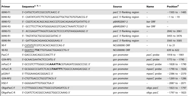
Table 1.Oligonucleotides used in this study.
Primer Sequencea, b, c Source Name Positiond
0090-F1 5’-CATGCTCATCCGCCGTCAACC-3’ pacC5’-flanking region – 21505 to21485 0090-R2 5’- CAATATCATCTTCTGTCGACGGTTGCTGCTGTGTGACCG-3’ pacC5’-flanking region – 21 to219 0090-F2 5’- CGGTCACACAGCAGCAACCGTCGACAGAAGATGATATTG-3’ pBARGEM7-2 barORF – 0090-R3 5’- GCCTTCCTTACATGGGGAGCGTCGACCTAAATCTCGGT-3’ pBARGEM7-2 barORF –
0090-F3 5’- ACCGAGATTTAGGTCGACGCTCCCCATGTAAGGAAGGC-3’ pacC3’-flanking region – 2042 to 2061 0090-R1 5’- TAGTATGCTGCGCGGCGATAC-3’ pacC3’-flanking region – 3455 to 3476 0090-R4 5’- GATTGGACAGAAGCAGGGAAG-3’ pacC3’-flanking region – 3565 to 3585
90-F 5’-CATATGTCGTCCACACCAGCCCAG-3’ NCU00090 ORF – 1 to 21
90-R2 5’-GGATCCTTACTTGTGAACTGGAGCCTG-3’ NCU00090 ORF – 639 to 622
PacC-F 5’-GACCCAACAGCCCAACTT-3’ gsnpromoter pacCprobe 1918 to21901 GSN-RP3 5’-GCAACGAATACTCCCATG-3’ gsnpromoter pacCprobe 1773 to21790 mPacC-F 5’-GCCGTCTTTGGGCCAGAAATTACGTGAGATCGGGCCCGC-3’ gsnpromoter mpacCprobe 21828 to21790 mPacC-R 5’-GCGGGCCCGATCTCACGTAATTTCTGGCCCAAAGACGGC-3’ gsnpromoter mpacCprobe 21790 to21826 pGSN-F 5’-TTGGAAAGACGGGACC-3’ gsnpromoter mpacCprobe 22386 to22370 GSN-RP2 5’-CTGTTGACCCTGCGTTACG-3’ gsnpromoter mpacCprobe 21269 to21286
HSE-F 5’-GGGGAATTTGTGGCTGA-3’ gsnpromoter – 22087 to22071
OligoPacC-F 5’-CTTTGGGCCAGCTTGGCCGTGAGATCG-3’ gsnpromoter oligopacC 21823 to21797 OligoPacC-R 5’-CGATCTCACGGCCAAGCTGGCCCAAAG-3’ gsnpromoter oligopacC 21797 to21823
aTheNdeI andBamHI restriction sites are underlined in the 90-F and 90-R2 sequences, respectively. bThe TAA stop codon inserted in the ORF NCU00090 sequence is represented in bold italics. cThe nucleotides mutated in the PacC motif are represented in bold.
dPrimer positions are in relation to the ATG start codon.
doi:10.1371/journal.pone.0044258.t001
ChIP-PCR Analysis
ChIP assays were performed according to Tamaruet al. [49], with modifications. Briefly, the wild-type strain was grown in liquid VM medium at 30uC, 250 rpm, for 24 h, and subjected to pH stress as previously described. Mycelial samples (125 mL aliquots) were subsequently transferred into 500 mL flasks and the chromatin was fixed by adding formaldehyde to 1% final concentration followed by incubation for 30 min at 30uC and 250 rpm. Formaldehyde was quenched by adding 125 mM glycine to each sample and then incubating at 30uC, 250 rpm, for 10 min. Chromatin prepared from each sample was pre-cleared with normal rabbit IgG (Upstate) and protein A agarose (Sigma) 50% slurry pre-blocked with sonicated salmon-sperm DNA and then immunoprecipitated with polyclonal anti-PACC antibody and protein A-agarose. As a negative control, a reaction without anti-PACC antibody was done. The 146 bp DNA fragment associated with the PACC protein was amplified by
PCR using the oligonucleotides PacC-F and GSN-RP3 and the reaction products were analyzed on a 2% agarose gel. A plasmid containing the entire sequence of thegsngene, including its 59- and 39-flanking regions, was used as a positive control for PCR.
Acknowledgments
We thank Stela Virgilio for helping with the morphological analysis and Antonio Tarcisio Delfino for technical assistance. We wish to thank Dr. Michael Freitag from Oregon State University, Corvallis, Oregon, USA for help with ChIP-PCR analysis.
Author Contributions
Conceived and designed the experiments: FBC FZF RMP MCB. Performed the experiments: FBC FZF. Analyzed the data: FBC FZF MCB. Contributed reagents/materials/analysis tools: MCB. Wrote the paper: FBC FZF MCB.
References
1. Galagan JE, Calvo SE, Borkovich KA, Selker EU, Read ND, et al. (2003) The genome sequence of the filamentous fungusNeurospora crassa. Nature 422: 859– 868.
2. de Paula RM, Wilson WA, Terenzi HF, Roach PJ, Bertolini MC (2005) GNN is a self-glucosylating protein involved in the initiation step of glycogen biosynthesis inNeurospora crassa. Arch Biochem Biophys. 435: 112–124.
3. Rothman-Denes LB, Cabib E (1971) Glucose-6-phosphate dependent and independent forms of yeast glycogen synthetase. Their properties and interconversions. Biochemistry 10: 1236–1242.
4. Noventa-Jorda˜o MA, Polizeli MLTM, Bonini BM, Jorge JA, Terenzi HF (1996) Effects of temperature shifts on the activities ofNeurospora crassa glycogen synthase, glycogen phosphorylase and trehalose-6-phosphate synthase. FEBS Lett 378: 32–36.
5. de Paula R, de Pinho CA, Terenzi HF, Bertolini MC (2002) Molecular and biochemical characterization of theNeurospora crassaglycogen synthase encoded by thegsncDNA. Mol Genet Genomics 267: 241–253.
6. Freitas FZ, Bertolini MC (2004) Genomic organization of theNeurospora crassa gsn gene. Possible involvement of the STRE and HSE elements in the modulation of gene transcription during heat shock. Mol Genet Genomics 272: 550–561. 7. Freitas FZ, de Paula RM, Barbosa LCB, Terenzi HF, Bertolini MC (2010)
cAMP signaling pathway controls glycogen metabolism inNeurospora crassaby regulating the glycogen synthase gene expression and phosphorylation. Fungal Genet Biol 47: 43–52.
8. Gonc¸alves RD, Cupertino FB, Freitas FZ, Luchessi AD, Bertolini MC (2011) A genome-wide screen forNeurospora crassatranscription factors regulating glycogen metabolism. Mol Cell Proteomics 10: DOI 10.1074/mcp.M111.007963-2. 9. Tilburn J, Sarkar S, Widdick DA, Espeso EA, Orejas M, et al. (1995) The
AspergillusPacC zinc finger transcription factor mediates regulation of both acid-and alkaline-expressed genes by ambient pH. EMBO J 14: 779–790. 10. Dı´ez E, Alvaro J, Espeso EA, Rainbow L, Sua´rez T, et al. (2002) Activation of
theAspergillusPacC zinc finger transcription factor requires two proteolytic steps. EMBO J 21: 1350–1359.
11. Herva´s-Aguilar A, Rodrı´guez JM, Tilburn J, Arst HN Jr, Pen˜alva MA (2007) Evidence for the direct involvement of the proteasome in the proteolytic processing of theAspergillus nidulanszinc finger transcription factor PacC. J Biol Chem 282: 34735–34747.
12. Arst HN Jr, Pen˜alva MA (2003a) pH regulation inAspergillusand parallels with higher eukaryotic regulatory systems. Trends Genet 19: 224–231.
13. Pen˜alva MA, Tilburn J, Bignell E, Arst HN Jr (2008) Ambient pH gene regulation in fungi: making connections. Trends Microbiol 16: 291–300. 14. Su SS, Mitchell AP (1993) Identification of functionally related genes that
stimulate early meiotic gene expression in yeast. Genetics 133: 67–77. 15. Lamb TM, Mitchell AP (2003) The transcription factor Rim101p governs ion
tolerance and cell differentiation by direct repression of the regulatory genes NRG1 and SMP1 inSaccharomyces cerevisiae. Mol Cell Biol 23: 677–686. 16. Li W, Mitchell AP (1997) Proteolytic activation of Rim1p, a positive regulator of
yeast sporulation and invasive growth. Genetics 145: 63–73.
17. Arin˜o J (2010) Integrative responses to high pH stress inSaccharomyces cerevisiae. OMICS 14: 517–523.
18. Davis D (2003) Adaptation to environmental pH inCandida albicans and its relation to pathogenesis. Curr Genet 44: 1–7.
19. Nozawa SR, Ferreira-Nozawa MS, Martinez-Rossi NM, Rossi A (2003) The pH-induced glycosylation of secreted phosphatases is mediated inAspergillus nidulansby the regulatory genepacc-dependent pathway. Fungal Genet Biol 39: 286–295.
20. Squina FM, Leal J, Cipriano VT, Martinez-Rossi NM, Rossi A (2010) Transcription of theNeurospora crassa70-kDa class heat shock protein genes is
modulated in response to extracellular pH changes. Cell Stress Chaperones 15: 225–231.
21. Ninomiya Y, Suzuki K, Ishii C, Inoue H (2004) Highly efficient gene replacements in Neurospora strains deficient for nonhomologous end-joining. Proc Natl Acad Sci USA 101: 12248–12253.
22. Colot HV, Park G, Turner GE, Ringelberg C, Crew CM, et al. (2006) A high-throughput gene knockout procedure forNeurosporareveals functions for multiple transcription factors. Proc Natl Acad Sci USA 103: 10352–10357.
23. Pen˜alva MA, Arst HN Jr (2004) Recent advances in the characterization of ambient pH regulation of gene expression in filamentous fungi and yeasts. Annu Rev Microbiol 58: 425–451.
24. Serrano R, Ruiz A, Bernal D, Chambers JR, Arin˜o J (2002) The transcriptional response to alkaline pH inSaccharomyces cerevisiae: evidence for calcium-mediated signalling. Mol Microbiol 46: 1319–1333.
25. Viladevall L, Serrano R, Ruiz A, Domenech G, Giraldo J, et al. (2004) Characterization of the calcium-mediated response to alkaline stress in Saccharomyces cerevisiae. J Biol Chem 279: 43614–43624.
26. Platara M, Ruiz A, Serrano R, Palomino A, Moreno F, et al. (2006) The transcriptional response of the yeast Na+
-ATPaseENA1gene to alkaline stress involves three main signaling pathways. J Biol Chem 281: 36632–36642. 27. Espeso EA, Arst HN Jr (2000) On the mechanism by which alkaline pH prevents
expression of an acid-expressed gene. Mol Cell Biol 20: 3355–3363. 28. Arst HN Jr, Pen˜alva MA (2003b) Recognizing gene regulation by ambient pH.
Fungal Genet Biol 40: 1–3.
29. Flaherty JE, Pirttila¨ AM, Bluhm BH, Woloshuk CP (2003) PAC1, a pH-regulatory gene fromFusarium verticillioides. Appl Environ Microbiol 69: 5222– 5227.
30. Meyer V, Spielvogel A, Funk L, Tilburn J, Arst HN Jr, Stahl U (2005) Alkaline pH-induced up-regulation of the afp gene encoding the antifungal protein (AFP) ofAspergillus giganteusis not mediated by the transcription factor PacC: possible involvement of calcineurin. Mol Genet Genomics 274: 295–306.
31. Merhej J, Richard-Forget F, Barreau C (2011) The pH regulatory factor Pac1 regulates Tri gene expression and trichothecene production in Fusarium graminearum. Fungal Genet Biol 48: 275–284.
32. Martinez-Pastor M, Marchler G, Schuller C, Marchler BA, Ruis H, et al. (1996) TheSaccharomyces cerevisiaezinc finger proteins Msn2p and Msn4p are required for transcriptional induction through the stress response element (STRE). EMBO J 15: 2227–2235.
33. Freitas FZ, Chapeaurouge A, Perales J, Bertolini MC (2008) A systematic approach to identify STRE-binding proteins of thegsnglycogen synthase gene promoter inNeurospora crassa.Proteomics 8: 2052–2061.
34. Gorner W, Durchschlag E, Martinez-Pastor MT, Estruch F, Ammerer G, et al. (1998) Nuclear localization of the C2H2zinc finger protein Msn2p is regulated
by stress and protein kinase A activity. Genes Dev 12: 586–597.
35. Casado C, Gonza´lez A, Platara M, Ruiz A, Arin˜o J (2011) The role of the protein kinase A pathway in the response to alkaline pH stress in yeast. Biochem J 438: 523–533.
36. Ramo´n AM, Fonzi WA (2003) Diverged binding specificity of Rim101p, the Candida albicansortholog of PacC. Eukaryot Cell 2: 718–728.
37. Pen˜as MM, Herva´s-Aguilar A, Mu´nera-Huertas T, Reoyo E, Pen˜alva MA, et al. (2007) Further characterization of the signaling proteolysis step in theAspergillus nidulanspH signal transduction pathway. Eukaryot Cell 6: 960–970. 38. Herranz S, Rodrı´guez JM, Bussink HJ, Sa´nchez-Ferrero JC, Arst HN Jr, et al.
(2005) Arrestin-related proteins mediate pH signaling in fungi. Proc Natl Acad Sci USA 102: 12141–12146.
39. Herva´s-Aguilar A, Galindo A, Pen˜alva MA (2010) Receptor-independent ambient pH signaling by ubiquitin attachment to fungal arrestin-like PalF. J Biol Chem 285: 18095–18102.
40. Kullas AL, Martin SJ, Davis D (2007) Adaptation to environmental pH: integrating the Rim101 and calcineurin signal transduction pathways. Mol Microbiol 66: 858–871.
41. McCluskey K (2003) The Fungal Genetics Stock Center: from molds to molecules. Adv Appl Microbiol 52: 245–262.
42. Vogel HJ (1956) A convenient growth medium forNeurospora crassa(medium N). Microbiol Genet Bull 13: 42–43.
43. Brunelli JP, Pall ML (1993) A series of yeast/Escherichia colik expression vectors designed for directional cloning of cDNAs and cre/lox-mediated plasmid excision. Yeast 9: 1309–1318.
44. Laemmli UK (1970) Cleavage of structural proteins during the assembly of the head of bacteriophage T4. Nature 227: 680–685.
45. Hartree EF (1972) Determination of protein: a modification of the Lowry method that gives a linear photometric response. Anal Biochem 48: 422–427. 46. Sokolovsky V, Kaldenhoff R, Ricci M, Russo VEA (1990) Fast and reliable
mini-prep RNA extraction fromNeurospora crassa. Fungal Genet Newslett 37: 41–43. 47. Sambrook J, Russell DW (2001) Molecular Cloning. A Laboratory Manual (3rd
ed.), Cold Spring Harbour Laboratory Press, Cold Spring Harbour. 48. Ausubel F, Brent R, Kingston RE, Moore DD, Seidman JG, et al. (1996)
Current Protocols in Molecular Biology, John Wiley and Sons, Inc., New York. 49. Tamaru H, Zhang X, McMillen D, Singh PB, Nakayama J, et al. (2003) Trimethylated lysine 9 of histone H3 is a mark for DNA methylation in Neurospora crassa. Nat Genet 34: 75–79.