Post-treatment Vascular Leakage and
Inflammatory Responses around Brain Cysts
in Porcine Neurocysticercosis
Siddhartha Mahanty1,2*, Miguel Angel Orrego2, Holger Mayta3, Miguel Marzal2, Carla Cangalaya2, Adriana Paredes2, Eloy Gonzales-Gustavson4, Gianfranco Arroyo4,
Armando E. Gonzalez4, Cristina Guerra-Giraldez2,3, Hector H. García2,3,5, Theodore E. Nash1,2, the Cysticercosis Working Group in Peru
1Laboratory of Parasitic Diseases, National Institutes of Allergy and Infectious Diseases, National Institutes of Health, Bethesda, Maryland, United States of America,2Laboratory of Immunopathology, Universidad Peruana Cayetano Heredia, Lima, Peru,3Facultad de Ciencias y Filosofía, Universidad Peruana Cayetano Heredia, Lima, Peru,4Faculty of Veterinary Sciences, Universidad Nacional Mayor de San Marcos, Lima, Peru,5Cysticercosis Unit, Instituto Nacional de Ciencias Neurologicas, Lima, Peru
¶ Membership of the Cysticercosis Working Group in Peru is provided in the Acknowledgments. *smahanty@niaid.nih.gov
Abstract
Cysticidal treatment of neurocysticercosis, an infection of humans and pig brains with Tae-nia solium, results in an early inflammatory response directed to cysts causing seizures and
focal neurological manifestations. Treatment-induced pericystic inflammation and its asso-ciation with blood brain barrier (BBB) dysfunction, as determined by Evans blue (EB) extrav-asation, was studied in infected untreated and anthelmintic-treated pigs. We compared the magnitude and extent of the pericystic inflammation, presence of EB-stained capsules, the level of damage to the parasite, expression of genes for proinflammatory and regulatory cy-tokines, chemokines, and tissue remodeling by quantitative PCR assays between treated and untreated infected pigs and between EB-stained (blue) and non stained (clear) cysts. Inflammatory scores were higher in pericystic tissues from EB-stained cysts compared to clear cysts from untreated pigs and also from anthelmintic-treated pigs 48 hr and 120 hr after treatment. The degree of inflammation correlated with the severity of cyst wall damage and both increased significantly at 120 hours. Expression levels of the proinflammatory genes for IL-6, IFN-γ, TNF-αwere higher in EB-stained cysts compared to clear cysts and
unaffect-ed brain tissues, and were generally highest at 120 hr. Additionally, expression of some markers of immunoregulatory activity (IL-10, IL-2Rα) were decreased in EB-stained capsules.
An increase in other markers for regulatory T cells (CTLA4, FoxP3) was found, as well as significant increases in expression of two metalloproteases, MMP1 and MMP2 at 48 hr and 120 hr post-treatment. We conclude that the increase in severity of the inflammation caused by treatment is accompanied by both a proinflammatory and a complex regulatory re-sponse, largely limited to pericystic tissues with compromised vascular integrity. Because treatment induced inflammation occurs in porcine NCC similar to that in human cases, this model can be used to investigate mechanisms involved in host damaging inflammatory re-sponses and agents or modalities that may control damaging post treatment inflammation.
OPEN ACCESS
Citation:Mahanty S, Orrego MA, Mayta H, Marzal
M, Cangalaya C, Paredes A, et al. (2015) Post-treatment Vascular Leakage and Inflammatory Responses around Brain Cysts in Porcine Neurocysticercosis. PLoS Negl Trop Dis 9(3): e0003577. doi:10.1371/journal.pntd.0003577
Academic Editor:Malcolm K. Jones, University of
Queensland, AUSTRALIA
Received:October 31, 2014
Accepted:January 29, 2015
Published:March 16, 2015
Copyright:This is an open access article, free of all
copyright, and may be freely reproduced, distributed, transmitted, modified, built upon, or otherwise used by anyone for any lawful purpose. The work is made available under theCreative Commons CC0public domain dedication.
Data Availability Statement:All relevant data are
within the paper and its Supporting Information files.
Funding:This research was supported in part by the
Intramural Research Program of the National Institute of Allergy and Infectious Diseases, NIH. Partial support from Fogarty International Center—NIH
Author Summary
Neurocysticercosis is caused by infection of the brain with the larval (cyst) stage of the tape wormTaenia soliumin humans and pigs. Antiparasitic drug treatment is
compro-mised by worsening of neurological symptoms during therapy due to reactive inflamma-tion triggered by the dying parasite. The immune mechanisms that cause this
inflammation are poorly understood. In this study, we investigated the nature of inflam-mation after treatment in pigs naturally infected withT.soliumcysts. Evans blue dye
in-jected into infected pigs marks areas in the brain where the normally impermeable capillaries have become more permeable, allowing damaging cells and molecules to leak out into the brain. By microscopy and measurement of gene expression for inflammation-inducing immune mediators, we show that inflammation in the brain tissues around cysts is more severe with increased vessel leakage. Furthermore, the levels of these mediators in-creased after antiparasitic drug treatment. A significant implication of these findings is that it may be possible to inhibit the inflammation around parasites using drugs or biolog-ics that inhibit these inflammatory pathways and, thereby, reduce local brain damage dur-ing treatment. These observations may also be applicable to other inflammatory
conditions that affect the brain.
Introduction
Neurocysticercosis (NCC), an infection with the larval stage of the tapeworm,Taenia solium, is
the most common cause of adult onset epilepsy worldwide. Infection is acquired from adult tapeworm carriers who excrete infectious ova either within proglottids or free in the feces. Fol-lowing ingestion of feces by free roaming pigs or accidental ingestion of ova by humans, the ova release oncospheres that burrow into the intestine, enter blood vessels and disseminate to the tissues, where they develop into viable larval cysts mostly in the muscles, subcutaneous tis-sue and brain. The life cycle is completed when tapeworms develop in the small intestine after humans ingest raw pork containing cysts. Symptomatic disease is the consequence of brain and spinal cord infection and its severity and course are determined by the location, number and degree of inflammation directed to the cysts [1,2].
Viable cysts invoke little host inflammatory response. For reasons that are unclear, degener-ating cysts or those damaged due to anthelmintic treatment provoke pathologic host inflamma-tory responses [1–3]. Inflammation around degenerating cysts in the brain parenchyma usually give rise to seizures while those in the subarachnoid spaces cause diffuse and/or focal arachnoiditis resulting in hydrocephalus, infarcts and nerve entrapments. Cysts in the ventri-cles commonly cause hydrocephalus due to mechanical obstruction of CSF outflow or as a re-sult of ventriculitis and scarring [2].
The detrimental inflammatory response induced by cysticidal drugs complicates treatment; in practice, corticosteroids are almost always employed to suppress inflammation and control symptoms. The optimal regimen for the safe and effective use of corticosteroids or other anti-inflammatory agents have not been studied in multicystic or complicated NCC. Therefore the dose, duration and type of corticosteroid selected are based upon the experience of the individ-ual practitioner [4]. A better understanding of the acute inflammatory responses induced by treatment is necessary to devise simple, safe and more effective treatment measures.
Competing Interests:The authors have declared
The pig is the natural intermediate host ofT.soliumand the only other animal readily
in-fected with cysts ofT.solium. Although used intermittently over the years for a variety of
stud-ies [5–9], pigs have not been utilized for the development of an animal model for cysticercosis or neurocysticercosis. Since the pig harbors cysts in both the brain and muscles, it has the po-tential to serve as an excellent model of human infections, and provides opportunities to study the pathogenesis of inflammation as well as immunoregulation in the vicinity of live and dying cysts. A limited number of studies investigating the pathology and underlying mechanisms of inflammation around cysts in naturally infected pigs have shown that the host reaction to the cyst results in a progressive local inflammatory response that develops into a fibrotic reaction with collagen deposition and granuloma formation [5,8]. The granuloma formation is charac-terized by infiltration with mononuclear cells and eosinophils [5,6,8,10], accompanied by apo-ptosis of infiltrating lymphocytes in early degenerating cysts and neuronal degeneration as well as astrogliosis [11,12], in the brain tissue adjacent to granulomas [11]. A comparison of histo-pathology with magnetic resonance imaging (MRI) confirmed a correlation of MRI findings with viable, early and late degenerating cysts [13,14].
Extravasation of vascular fluids around cysts, often revealed by contrast enhancement and/ or edema in radiological imaging (computed tomography [CT] or MRI), appears to be a crucial element of the pathology around degenerating cysts and a feature of post-treatment pericystic inflammation [10,15]. Using extravasation of intravenously delivered Evans blue (EB) as a marker for vascular leakage in and around cysts [16], we investigated the relationship between vascular permeability around cysts and the induction of inflammatory pathways following treatment with anthelmintic drugs. Gene expression analysis of the pericystic tissues of a subset of brain cysts revealed that increased permeability following anthelmintic treatment is accom-panied by an upregulation of proinflammatory genes and a corresponding down regulation of some regulatory (anti-inflammatory) genes. These data reveal a close relationship between vas-cular leakage and pericystic inflammation in neurocysticercosis and confirm the utility of a pig
“model”of infection to study inflammatory mechanisms involved in this disease.
Materials and Methods
Study animals
Thirteen outbred pigs naturally infected withTaenia solium, as determined by a positive
ton-gue examination, were obtained in Huancayo, Peru [17]. Pigs were anesthetized with ketamine (10 mg/K intramuscular injection, Agrovetmarket SA, Peru) and xylazine (2 mg/Kg, Agrovet-market SA, Peru), for intravascular infusion and euthanized with sodium pentobarbital (100–
125 mg/Kg, intravenous injection, Agrovetmarket SA, Peru). The study protocol and proce-dures were reviewed and approved by the Ethics Committee of the Veterinary School of San Marcos University in Lima, Peru. Pigs received one of three treatments prior to EB infusions: 1) no anthelmintic treatment (infected, n = 5); 2) praziquantel (100 mg/kg po, once) two days earlier (infected, n = 4,) or, 3) praziquantel (100 mg/kg po, once) five days earlier (infected, n = 4).
Ethics statement
Animal experiments were performed at the Facultad de Medicina Veterinaria, Universidad Nacional Mayor de San Marcos, Lima, Peru, under the protocol“Evaluación de la permeabili-dad vascular en cerebro y músculo de cerdos naturalmente infectados conTaenia solium”, with
Treatment conditions and EB infusion
In two separate experiments infected pigs were treated with a single dose of praziquantel (100 mg/Kg, Merck, Darmstadt, Germany) and sacrificed 48h (n = 4) and 120h later (n = 4). Five untreated infected pigs were used as controls. Two hours before euthanasia, pigs were anesthetized and infused with 2% Evans blue (EB, 80 mg/Kg; Sigma-Aldrich, St. Louis, MO) in normal saline (NaCl 0.85%, Laboratorios Baxter, Colombia, Baxter del Peru) by intravenous injection via the carotid artery. Just after euthanasia, the pigs were perfused with chilled saline solution with heparin (NaCl 0.85% with 10 UI of heparin/mL) and the brain was immediately removed at necropsy.
Collection and selection of specimens for qPCR and histology
Brains were cut into ~1 cm slices on dry ice. The presence of blue and clear stained tissue around cysts was documented by gross examination. Of a total of 371 cysts harvested for this study, 30 (8%) of capsules were randomly selected for qPCR analysis. The samples selected, in-cluding cysts with both blue and clear pericystic capsular tissues, were harvested and placed in chaotropic buffer (RNALater, Qiagen/Life Technologies, Gaithersburg, MD) for further pro-cessing. For histopathological studies, the remainder of the brain cysts including the pericapsu-lar tissue were fixed in 10% neutral buffered formalin. Samples from unaffected brain
parenchymal tissue clearly distant from pericystic tissues were used as reference tissues for qPCR analysis.
Histological examination
Paraffin sections were processed using standard procedures and stained with hematoxylin and eosin (HE) and Masson’s Trichrome stain (MT). Only complete cysts (151/371, or 41%) were used because the generation of the inflammatory and cyst damage scores required inclusion of the circumference. Quantitative inflammatory responses were assessed histologically in cysts with complete capsules and/or cyst walls in bright field microscopic images (Primo Start, Zeiss, Germany) of each cyst using a calibrated camera (AxioCam ICc1, Zeiss, Germany) with Axio-Vision v4.6 software (Zeiss, Germany).
Initially, an image of each complete cyst including the surrounding host reaction (capsule) was examined to determine the range of inflammatory changes present around each cyst and to calculate the proportion of the cyst circumference showing each of the designated inflamma-tory scores (IS) 0 to IS 4 (seeS1 TableandS1 Fig.). The classification of the inflammatory stages broadly followed the schema described by Álvarezet al[5] and Londoñoet al[8] with
additional semiquantitative assessment of the extent and severity of the inflammation around each cyst, based on the average number of cells per high power field and the area of the pericys-tic tissue that contained the inflammatory infiltrates (SeeS1 Fig.). Using these measurements, IS 1 to 4 represented increasing extent and severity of inflammatory reactions (S1 Fig.). The presence of typical granules and nuclear morphology allowed us to identify eosinophils on HE stains, but for determination of semi-quantitative inflammatory and cyst wall damage scores we modified the approach used by Alvarezet al. [5], using an assessment of the location and
density of total cellular infiltrate without identifying constituent cell types.
Cyst wall damage was categorized into five stages of damage corresponding to damage scores (DS)0 to DS4, based on the degree of tissue disruption in the cyst walls, as outlined by Londoñoet al. [8], defined inS1 Tableand illustrated inS1 Fig.
composite = Sum (IS Score x % of capsular circumference)DS-composite = Sum (DS Score x % of cyst circumference)For the purpose of this calculation, the percentage of the circumference was rounded off to increments of 20% (i.e., 0, 20, 40, 60, 80 or 100%). As an example, a cyst-capsule that had 20% of the cyst-capsule with IS 2, 40% with IS 3 and 40% with IS 4 would have the composite IS score of 320 (2x20+3x40+4x40 = 320). Similar calculations were used for com-posite DS scores. Using these formulae the theoretical maximum value for the IS- and DS-com-posite scores for a given cyst is 400 (4 x 100%).
Generation of cDNA and real time PCR (qPCR) assays for gene
expression studies
Total RNA was isolated from 50–100 mg of tissue samples using TRIzol reagent (Invitrogen, Carlsbad, CA). RNA concentrations were determined using a NanoDrop (NanoDrop Products, Wilmington, DE). cDNA was generated from one microgram of extracted total RNA using multiscribe RT polymerase (Applied Biosystems, Inc, Bedford, MA) in 100-ul reactions by in-cubation for 10 min at 25°C followed by 60 min at 37°C, 5 min at 95°C on a thermocycler (MJ Research PTRC-200, BioRad-MJ Research, Hercules, CA). Real-time PCR was performed in 20μl reactions using TaqMan Gene Expression Assays (Applied Biosystems, Foster City, CA)
and primer probe pairs for each gene of interest. PCR reactions, run in triplicate, used the fol-lowing cycling parameters: 40 cycles of 20 sec at 95°C, 1 sec at 95°C and 1 min at 60°C, on an AB StepOnePlus cycler, normalized against 18S ribosomal RNA as a control gene. Primers and probes used were off-the-shelf reagents purchased from the vendor (Life Technologies, Grand Island, NY). We used 18S rRNA (Life Technologies code 4319413E) as a control gene to confirm RNA integrity and primer probe pairs for porcine IL-1β(Ss03393804_m1), IL-6 (Ss03384604_u1), IL-13 (Ss03392353_m1), IFN-γ(Ss03391053_g1), TNF-α(Ss03391318_g1), IL-10 (Ss03391318_g1), CTLA4 (Ss03213761_m1), IL-2RA (CD25; Ss03381754_u1), FoxP3 (Ss03376695_u1), matrix metalloprotease (MMP) 1 (Ss03374796_u1), MMP9 (Ss03392097_ g1), tissue inhibitor of metalloprotease (TIMP) 1 (Ss03381944_u1) and TIMP2 (Ss03375440_ u1) genes. PCR reactions, run in triplicate, used the following cycling parameters: 40 cycles of 20 sec at 95°C, 1 sec at 95°C and 1 min at 60°C, on an AB StepPlusOne cycler (Life Technolo-gies, Grand Island, NY). Gene expression was analyzed as anti-log2 transformed threshold cycle number corrected for the housekeeping gene (2-ΔΔCT), representing a fold increase (or
decrease) over the control gene.
Statistical analysis
Non-parametric statistics, Mann-Whitney U test for two groups and Kruskal-Wallis test for multiple groups, were calculated using Prism software (Graphpad, San Diego, CA) for compar-isons of the histological and gene expression parameters mentioned above between uninfected and infected pigs and between clear capsules and those with EB staining. Differences with p-values of<0.05 were considered statistically significant.
Results
Study population and treatment subgroups
11 pigs were examined histopathologically (using haematoxylin-eosin staining) for evidence of inflammation and damage to the cyst wall. An available sample set of thirty capsules (20 blue and 10 clear) from the brains of 6 pigs were selected randomly among cysts with complete capsules for assessment of gene expression of pro-inflammatory and regulatory pathway genes by qPCR. Different capsule samples were used for gene expression and for histopathological analyses.
Assessment of Evans Blue extravasation
The presence and number of both cysts with clear and blue capsules was confirmed in 1-cm coronal sections of both cerebral hemispheres in all pigs that received EB infusions (Fig. 1) as previously reported [16]. The proportion of cysts with capsules that demonstrated EB staining was significantly higher in PZQ treated than in untreated infected pigs [16]. The increase in EB stained capsules is consistent with the increase in enhancement reported in treated patients within the first week after the start of cysticidal agents [2,4]. Among these 371 total cyst sam-ples, 33 of 47 (70%) cysts from untreated and 89 of 104 (86%) cysts from treated infected pigs demonstrated EB extravasation.
Relationship between inflammation, cyst damage and Evans Blue
extravasation
Semiquantitative histologic inflammatory and cyst damage scores were determined in 151 cysts with an evaluable capsule and cyst. Irrespective of anthelmintic treatment (i.e., in all pigs, untreated, and at 48 hr and 120 hr post-treatment), blue capsules had significantly higher in-flammatory scores compared to clear capsules (Fig. 2). In addition, the inflammatory scores of
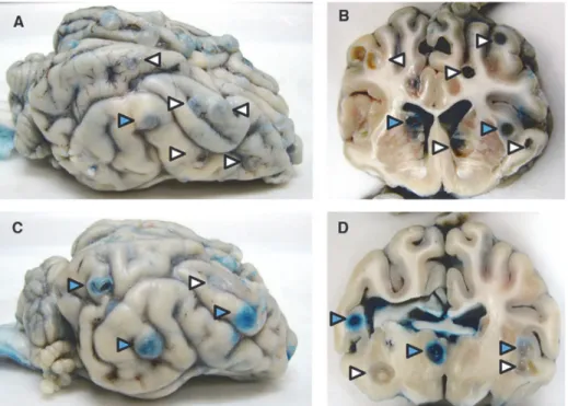
Fig 1. Clear and blue-stained capsules (EB capsules) on the surface and cut surfaces of brains from infected pigs.Photographs of the surface and coronal sections of representative brains from an untreated pig (A and B) and a pig treated with praziquantel 48 h previously (C and D), showing cysts with clear (white arrowheads) and blue capsules (blue arrowheads) visible within the parenchyma and on the surface.
blue capsules increased between 48 hr and 120 hr of treatment but the difference in inflamma-tory or damage scores between clear and blue capsules only reached significance at 120 hr. In contrast to capsules with EB staining, no significant changes in either inflammation or damage were found between clear capsules in treated pigs at all time points.
We analyzed cyst damage scores for both the effect of treatment with PZQ and the effect of BBB disruption (EB stained capsules). We found that PZQ treatment alone did not significant-ly increase damage scores, and that increased scores were seen onsignificant-ly in cysts with EB stained capsules in PZQ treated pigs not only when compared with untreated cysts but also when com-pared to treated clear cysts (Fig. 2B). In contrast, damage scores for clear capsules were not sig-nificantly increased in pigs treated with PZQ when compared to untreated pigs (Fig. 2B). As shown inFig. 2B, damage scores of cysts with EB-stained capsules at 120 hr post-treatment were significantly increased above both the corresponding cysts with EB stained capsules at 48 hr as well as cysts with either EB-stained or clear capsules in untreated pigs (Fig. 2B). The
Fig 2. EB capsules show increased inflammation and cyst wall damage compared to clear capsules.Cysts were harvested from the brains of infected untreated pigs (n = 3; seeMethods) or pigs treated with PZQ 48h (n = 4) or 120h (n = 4) earlier. For each cyst, scores reflecting the degree and extent of the inflammatory cell infiltrates (A) and cyst wall damage (B) at 48h (PZQ 48h) and 120h (PZQ 120h) after PZQ treatment (seeMethodsandS1 Fig.) are shown (open squares refer to clear capsules and closed squares, to EB capsules, bar = mean). Statistical significance in comparison between groups is represented by asterisks (*: p<0.05;**: p<0.01;***: p<0.005).
increase in the proportion of EB capsules over time as well as the increase in inflammatory and cyst damage scores at 120 hr indicates that treatment had induced or exacerbated pericystic in-flammation and cyst damage following praziquantel treatment, an expected result.
Upregulation of pro-inflammatory and regulatory genes in cysts with
Evans Blue leakage
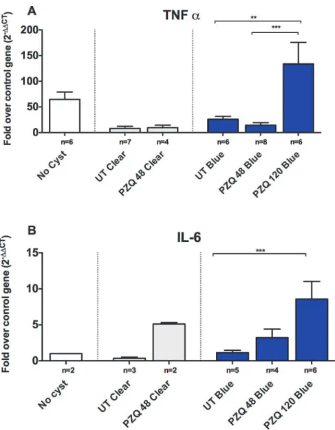
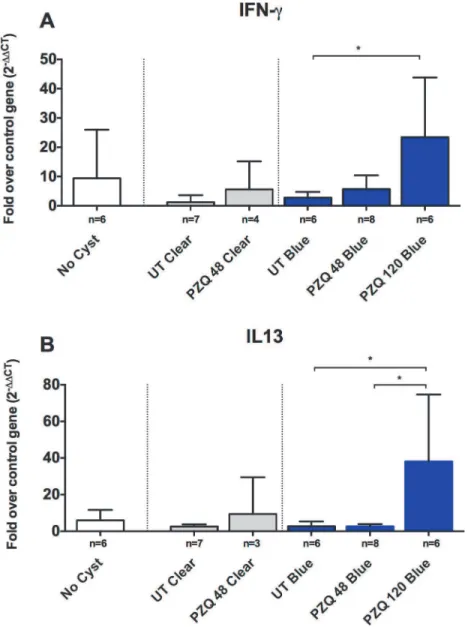
Given that there was a significant increase in the inflammatory reaction around the blue cysts after PZQ treatment, we investigated a number of pro-inflammatory and immunoregulatory pathways to identify molecules involved in regulating these responses in vivo. As was observed with damage scores, clear capsules did not show significant changes in the levels of gene ex-pression for inflammatory markers (Figs.3–5) at 48h post treatment, however, it should be noted that no clear capsules were analyzed for gene expression analysis at 120h because all cysts harvested for RNA extraction were found to have EB stained capsules. In contrast, as shown in Figs.3,4and5, there was a relative increase in expression levels of all the proinflam-matory gene tested (TNF-α, IL-6 and interferon (IFN)-γ) in EB-stained capsules compared to clear capsules from untreated pigs and brain tissues from infected pigs distant from the cyst location. Interestingly, IL-13, a gene associated with fibrosis and Th2 responses in other hel-minth infections, also increased in EB-stained capsules, but not in clear capsules, at 120 hr post treatment (Fig. 4). Differences between blue and clear capsule-associated cysts at 120 hr and at 48 hr did not achieve statistical significance, likely due to the small number of clear cysts avail-able for analysis.
Unlike the proinflammatory markers, a variable pattern of change in gene expression was observed in the regulatory genes evaluated. IL-10 and CD25 decreased transiently after treat-ment by 48 hr post treattreat-ment in the EB capsules, whereas, CTLA4 and FoxP3 showed tran-siently reduced expression levels in EB-stained cysts at 48 hr post treatment, which rebounded to higher levels at 120 hr (Fig. 5). These data indicate that higher expression of proinflamma-tory genes is accompanied by a transient reduction in the levels of mRNA for several regulaproinflamma-tory genes. However, all the significant changes were seen in EB-stained cysts; clear cysts did not demonstrate an increase in pro-inflammatory or regulatory genes (See Figs.3–5). Of note, there were no clear capsules in the brains of animals studied for gene expression 120 hr post treatment with PZQ, because the few cysts with clear capsules were used for histopathologic analysis.
To determine if expression of genes associated with tissue remodeling and possibly in gran-uloma formation were increased, as reported in other animal models of NCC [18], we com-pared gene expression levels of matrix metalloproteases (MMP) 1 and 9 and tissue inhibitors of MMPs (TIMPs) 1 and 2 (Fig. 6) in capsules with and without EB staining. There were no sig-nificant differences in expression of these genes between EB-stained and clear capsules or unin-fected tissues (Fig. 6). In contrast, expression of the genes for of MMP1 and MMP9, as well as the TIMP1 and TIMP2 increased significantly in EB-stained (blue) capsules at 48h and 120 hr after PZQ treatment. Interestingly, the increase in expression of MMP1 and MMP9 peaked at different times (48h for MMP9 and 120h for MMP1).
Discussion
treatment. The use of EB injection allowed anatomic identification and analysis of cysts with BBB leakage, which is equivalent to gadolinium extravasation around cysts during MRI. By performing semi-quantitative analysis of the infiltrating cyst-associated inflammatory cells, the expression of genes for cytokine and other regulatory molecules and the extent of cyst damage over time in relation to BBB leakage, we were able to determine the dynamics and relationship of treatment-induced changes to the pathology.
Fig 3. Upregulation of proinflammatory genes in EB capsules compared to clear capsules.Expression levels of RNA in six types of brain tissue samples, uninfected brain tissue distant from cysts (no cysts), clear pericystic brain tissue in untreated pigs (UT Clear) and at 48h post treatment (PZQ 48 Clear) and EB capsules in untreated pigs (UT Blue) and at 48h (PZQ 48 h Blue) and 120h post treatment (PZQ 120 h Blue) were quantified using real-time reverse transcriptase polymerase chain reaction (RT-PCR) assays. Gene expression levels are depicted as fold increase of studied RNA over reference (housekeeping) RNA (18S rRNA), TNF-α(A; number [n] of samples for the indicated types of capsules are shown below each bar) and
IL-6 (B). The bars represent means and CI95for the group. Statistically significant differences in levels of gene expression are indicated by asterisks (p<0.05, Mann-Whitney U test). Asterisks represent level of
significance:*: p<0.05;**: p<0.01;***: p<0.005.
Our most significant finding was that treatment with praziquantel led to the induction of in-flammation and blood-brain barrier disruption 48 hr and 120 hr post treatment similar to the increase in pericystic enhancement and perilesional edema observed by MRI in treated humans [19–21]. Using EB injection we found a positive correlation between the increase in inflamma-tory pathology and BBB disruption over time as shown by the increase in EB stained capsules. In comparison, cysts without BBB disruption, identified by a lack of EB staining, for the most part showed little inflammatory cell reaction or changes in cytokine gene expression. The cysts with acute inflammatory responses and BBB leakage and were associated with elevated expres-sion of proinflammatory cytokines, but somewhat unexpectedly, also demonstrated heightened immunoregulatory responses, notably, of IL-13 and CD25 (IL-2 receptor). The correlation of
Fig 4. Upregulation of both Th1 and Th2 markers, IFN-γand IL-13, in blue capsules following PZQ
treatment.Expression levels of RNA in brain tissues and EB-stained and clear capsules for IFN-γ(A;
number [n] of samples for the indicated types of capsules are shown below each bar), and IL-13 (B). In each graph, the bars represent means and CI95for the group. Statistically significant differences in levels of gene expression are indicated by asterisks (p<0.05, Mann-Whitney U test). Asterisks represent level of
significance:*: p<0.05;**: p<0.01;***: p<0.005.
the BBB leakage with inflammation is logical and expected since the pericystic inflammatory response primarily consists of white blood cells from outside the brain, which require enhanced access to the cyst by way of blood vessels located within the capsule.
In these experiments the inflammatory response directed to the treated, degenerating cysts and damage to the cysts apparently occurred at the same time. The magnitude of the inflamma-tory response was also found to be proportional to the degree of cyst wall damage. In addition, we found that the damage occurred predominantly in cysts that showed EB leakage in their capsules, best appreciated at 5 days post treatment (Fig. 2A). These findings are consistent with the notion that the inflammatory reaction and BBB disruption are likely important determi-nants of the degree of cyst damage. Our data are also consistent with previously reported in-flammatory pathology in rodent models [22–25] and experimental pig infections [6,8–10]. In this study, we did not determine the types of inflammatory cells at the sites of EB leakage and cyst wall damage but focused on the intensity of inflammatory infiltrates by assessing the extent of infiltrate (described inS1 Tableand depicted inS1 Fig.). However, eosinophils were notable and distinguishable by morphology and the presence of granules on HE staining (S1 Fig.).
Fig 5. Increased expression of CD-25 and IL-10 in EB capsules following PZQ treatment.Expression levels of RNA in brain tissue samples from EB-stained and clear capsules were quantified (SeeMethods). Shown are expression levels, depicted as fold increase over the RNA levels for the reference (housekeeping) gene (18S rRNA), encoding four representative regulatory T cells (Treg) phenotyping and functional markers: CD25 (A; number [n] of samples for the indicated types of capsules are shown below each bar), FoxP3 (B), IL-10 (C) and CTLA4 (D). Statistically significant differences in levels of gene expression are indicated by asterisks (p<0.05, Mann-Whitney U test). Asterisks represent level of significance:*: p<0.05;**: p<0.01;***: p<0.005.
The evidence for activation of inflammatory responses comes from increased expression of the pro-inflammatory mediators TNF-α, IL-6 and IFN-γafter treatment with PZQ (Figs3
and4A) and decreased levels of IL-10 in treated cysts (Fig. 5). However, the increased expres-sion of inflammatory mediators (and conversely, lower levels of the regulatory molecule, IL-10), were seen around some, but not all, cyst types; the differences between untreated and PZQ-treated cysts were significant only in cysts that demonstrated BBB disruption (Figs.4and
5). These findings show that pre- and post-treatment degeneration of cysts and inflammation are closely tied to increased BBB disruption. The presence of gadolinium enhancement in human NCC is commonly used as a surrogate for the presence and degree of inflammation. These studies confirm the association.
An interesting observation was the post-treatment downregulation of IL-10, a major anti-inflammatory cytokine [9,18,19,26] and a marker of regulatory T cells (CD25) in the EB-stained capsules but not in unEB-stained capsules (Fig. 5C). High levels of expression of mRNA for IL-10 have been reported in infected (but untreated) pigs previously [27]. Other markers in-dicative of modulation of inflammation, including FoxP3 (a marker for Treg cell populations) and CTLA4 (an inhibitory co-factor that functionally modulates Th and Treg cells and has been associated with T cell hyporesponsiveness in other chronic infectious diseases such as
Fig 6. Increased expression of fibrosis/granuloma promoting genes in EB and clear capsules.Methods and tissues examined are identical to those analyzed in Figs.3–5. Shown are four genes representative of fibrosis and granuloma promoting molecules: MMP1 (A; number [n] of samples for the indicated types of capsules are shown below each bar; n = 7, 6, 3, 6, 6, respectively), MMP9 (B), TIMP1 (C) and TIMP2 (D). Statistically significant differences in levels of gene expression are indicated by asterisks (p<0.05, Mann-Whitney U test). Asterisks represent level of significance:*: p<0.05;
**: p<0.01;***: p<0.005.
filarial infections [28] and in tuberculosis [29]), were upregulated in the same capsules that had lower levels of IL-10 and CD25 (Fig. 5). The high levels of IL-10 around untreated cysts or those without disruption of the BBB suggest that they are in a relatively hyporesponsive state and that treatment may be“releasing”or exposing the parasite to an unimpeded proinflamma-tory responses after the parasite is damaged or killed. In the proinflammaproinflamma-tory environment, an increase in the expression of regulatory mediators, notably IL-13, may also represent a com-pensatory or homeostatic release of regulatory responses aimed at limiting potentially pathological inflammation.
Helminth infections are frequently associated with modulation of the host immune system [30]. The immunomodulatory state is a consequence of a dominant T helper (Th) type 2 adap-tive response induced or associated with these infections, and is characterized by high levels of IL-4, IL-13, IL-5, IgE and IgG subtypes IgG1 and IgG4 specific for the parasite antigens [31], recruitment of immunomodulatory populations such as regulatory T cells (Tregs: CD4+CD25+FoxP3+cells) [30,32] and inhibitory
“alternatively activated”macrophages, or M2 macrophages [33,34]. Our results that show a relative lack of inflammation around untreated cysts (similar to unaffected, distant brain tissues) are consistent with an immunomodulatory state.
Although a regulatory environment appears to dominate in helminth infections [30,35] as well as in humans and pigs infected withT.solium[27,36] and in rodent models of NCC
[25,34,37,38], a more inflammatory process with edema and inflammation around degenerat-ing cysts has long been recognized as an important feature of NCC [2,39]. Symptomatic NCC is associated with higher levels of circulating pro-inflammatory mediators, including TNF-α, IFN-γ, IL-1βand IL-6 [40–43]. A good model of human infection should, ideally, reflect this mixed picture of inflammatory and regulatory immune activation. Indeed, this is what was ob-served in the cysts from pigs after PZQ treatment. The unexpectedly complex pattern of ex-pression of different counterregulatory markers (IL-10 vs. CTL4 and FoxP3;Fig. 5) may be influenced by the anatomical location of the response. Since infection by this parasite occurs in the brain, in contrast to peripheral tissues, the immune response may differ from peripheral host responses, which are more reflective of naïve T and B cells, and monocytic cell populations than the tissue-derived immune cells found in the brain. The similarities in histopathology and evolution of the cysts in this pig model and the ability to study the parasite and host response directly in the brain make this a promising model for research in human NCC.
These in vivo studies are difficult, labor intensive and limited by the number of infected ani-mals available. Changes earlier than 48 hr were not analyzed but may be important. In addition unlike MRI studies that can be performed sequentially and can demonstrate changes of the same cysts over time, in the present model the status of individual cysts before treatment can-not be determined. Instead, we show changes related to untreated animals and changes in pigs studied at earlier and later time points. In previous studies that focused on pathologic changes following anthelmintic treatment, we and others noted an increase in the eosinophilic migra-tion into the pericystic region [9,10,52]. In a previous report we showed that eosinophils were at times numerous and associated with focal regions of cyst wall damage and infiltration, which increased with cysticidal treatment [10]. These regions were enriched in eosinophils, which had also ingested EB-bound albumin. The eosinophilic infiltrates may important if not essential for cyst degeneration but may also be responsible for much of the inflammatory side effects of anthelmintic treatment.
Although pigs, like humans, are natural hosts forT.soliumand histopathological features
and immune responses observed in pigs are similar to what has been surmised from human studies [19,39,53–55], there are important differences that advise caution in interpreting obser-vations from the pig as a model for human disease. An obvious difference is the parasite bur-den, which is generally significantly higher in pigs than in humans [6,56]. The longevity of the infection is also shorter in pigs, and consequently there are no calcified cysts in the pigs used in our experiments. The heavy burden of extracerebral cysts in pigs is also at variance with human infections at the time of presentation. The heavy burden of parasite antigens that results may have a dampening effect on inflammatory responses, as has been seen in other helminth infections [31]. These factors have unknown influences on the nature of the immune response, and warrant further investigation in experimental infections with better defined infection pa-rameters (such as parasite burden, duration of infection, etc).
In summary, our results indicate that BBB disruption is accompanied by both pro-inflam-matory and regulatory host responses, and that the percentage of cysts with these characteris-tics increases with treatment. We have demonstrated BBB leakage directly by using EB, which allowed us to perform characterization of specific cysts presumably at different stages of dam-age or degeneration. We also found evidence of increased fibrosis in the pericystic capsular re-gions at both gene expression and histopathological levels in comparison to cysts that did not show BBB leakage. This model has many of the characteristics and features of human infec-tions and therefore can be used to investigate the mechanisms involved in the genesis of in-flammation, treatment-induced immunopathology and host parasite interactions. This will assist in identifying therapeutic agents with a narrower but more rational spectrum of targets to suppress inflammation in the CNS. Further studies in this model, using natural and experi-mental infections can be used for selection of better drugs and biological agents to suppress damaging post treatment inflammatory responses.
Supporting Information
S1 Table. Histological criteria used for staging of pericystic inflammatory reaction and cyst wall damage.Histological features of cysts capsules and cysts that define inflammatory stages (IS) 1–4 and cyst wall damage scores (DS) 1–4, respectively.
(DOCX)
damage scores (DS): DS0 (E), DS1 (F), DS2 (G) and DS3 (H). All tissue sections were stained with Masson´s trichromic stain. Magnification 400x in all panels. Key: collagen (co), cyst wall (cw), cellular infiltrate (ci), eosinophils (eos), tegument (t, hollow arrowhead), subtegument (st, gray arrowhead) and internal region (ir, black arrow head).
(TIF)
Acknowledgments
We thank the staff of the animal facilities at Faculty of Veterinary Sciences at the Universidad Nacional Mayor de San Marcos, Lima, Peru, for invaluable assistance in care and treatment of animals used in this work.
The Cysticercosis Working Group in Peru: Hector H. Garcia, MD, PhD (Universidad Per-uana Cayetano Heredia, Instituto Nacional de Ciencias Neurológicas, Lima, Peru, lead contact,
hgarcia@jhsph.edu), Robert. H. Gilman, MD, DTMH (Johns Hopkins Bloomberg School of Public Health, Baltimore, MD); Armando E. Gonzalez, DVM, PhD; Gianfranco Arroyo, DVM; Eloy Gonzales-Gustavson, DVM, MSc (School of Veterinary Medicine, Universidad Nacional Mayor de San Marcos, Lima, Peru); Victor C. W. Tsang (Georgia State University, Atlanta, GA); Manuela Verastegui, PhD; Mirko Zimic, PhD; Holger Mayta, PhD, Miguel Angel Orrego, BSc; Patricia Saenz, TM (Universidad Peruana Cayetano Heredia, Lima, Peru´); Isidro Gonza-les, MD; Herbert Saavedra, MD (Instituto Nacional de Ciencias Neurológicas, Lima, Peru).
Author Contributions
Conceived and designed the experiments: SM CGG HHG TEN AP. Performed the experi-ments: MAO HM MM CC EGG AEG CGG GA. Analyzed the data: SM MAO CC CGG HHG TEN. Contributed reagents/materials/analysis tools: AEG SM TEN. Wrote the paper: SM TEN. Acquired the data: AP.
References
1. Garcia HH, Gonzalez AE, Gilman RH (2011) Cysticercosis of the central nervous system: how should it be managed? Curr Opin Infect Dis 24: 423–427. doi:10.1097/QCO.0b013e32834a1b20PMID: 21788891
2. Nash TE, Garcia HH (2011) Diagnosis and treatment of neurocysticercosis. Nat Rev Neurol 7: 584– 594. doi:10.1038/nrneurol.2011.135PMID:21912406
3. Nash TE (2003) Human case management and treatment of cysticercosis. Acta Trop 87: 61–69. PMID:12781379
4. Nash TE, Mahanty S, Garcia HH, Cysticercosis Group in P (2011) Corticosteroid use in neurocysticer-cosis. Expert Rev Neurother 11: 1175–1183. doi:10.1586/ern.11.86PMID:21797658
5. Alvarez JI, Londono DP, Alvarez AL, Trujillo J, Jaramillo MM, et al. (2002) Granuloma formation and parasite disintegration in porcine cysticercosis: comparison with human neurocysticercosis. J Comp Pathol 127: 186–193. PMID:12354530
6. de Aluja A, Vargas G (1988) The histopathology of porcine cysticercosis. Vet Parasitol 28: 65–77. PMID:3388737
7. Garrido GS, de Aluja AS, Casas FC (2007) Early stages of development of the Taenia solium metaces-tode in pigs. J Parasitol 93: 238–241. PMID:17539403
8. Londono DP, Alvarez JI, Trujillo J, Jaramillo MM, Restrepo BI (2002) The inflammatory cell infiltrates in porcine cysticercosis: immunohistochemical analysis during various stages of infection. Vet Parasitol 109: 249–259. PMID:12423936
10. Marzal M, Guerra-Giraldez C, Paredes A, Cangalaya C, Rivera A, et al. (2014) Evans blue staining re-veals vascular leakage associated with focal areas of host-parasite interaction in brains of pigs infected with Taenia solium. PLoS One 9: e97321. doi:10.1371/journal.pone.0097321PMID:24915533
11. Sikasunge CS, Phiri IK, Johansen MV, Willingham AL 3rd, Leifsson PS (2008) Host-cell apoptosis in Taenia solium-induced brain granulomas in naturally infected pigs. Parasitology 135: 1237–1242. doi: 10.1017/S0031182008004678PMID:18620623
12. Terrazas LI, Montero D, Terrazas CA, Reyes JL, Rodriguez-Sosa M (2005) Role of the programmed Death-1 pathway in the suppressive activity of alternatively activated macrophages in experimental cysticercosis. Int J Parasitol 35: 1349–1358. PMID:16126211
13. Chawla S, Gupta RK, Husain N, Garg M, Kumar R, et al. (2004) Prediction of viability of porcine neuro-cysticercosis with proton magnetic resonance spectroscopy: correlation with histopathology. Life Sci 74: 1081–1092. PMID:14687649
14. Chawla S, Husain N, Kumar S, Pal L, Tripathi M, et al. (2004) Correlative MR imaging and histopatholo-gy in porcine neurocysticercosis. J Magn Reson Imaging 20: 208–215. PMID:15269945
15. de Aluja AS, Gonzalez D, Rodriguez Carbajal J, Flisser A (1989) Histological description of tomograph-ic images of Taenia solium cysttomograph-icerci in pig brains. Clin Imaging 13: 292–298. PMID:2598111
16. Guerra-Giraldez C, Marzal M, Cangalaya C, Balboa D, Orrego MA, et al. (2013) Disruption of the blood-brain barrier in pigs naturally infected with Taenia solium, untreated and after anthelmintic treatment. Exp Parasitol 134: 443–446. doi:10.1016/j.exppara.2013.05.005PMID:23684909
17. Gonzalez AE, Cama V, Gilman RH, Tsang VC, Pilcher JB, et al. (1990) Prevalence and comparison of serologic assays, necropsy, and tongue examination for the diagnosis of porcine cysticercosis in Peru. Am J Trop Med Hyg 43: 194–199. PMID:2389823
18. Wang IC, Fan PC, Lu SC, Fan CK, Su KE (2008) Suppression of host Th1-type granulomatous inflam-mation by Taenia solium metacestodes is related to down-regulation of osteopontin gene expression. Int J Parasitol 38: 239–248. PMID:17765901
19. Mahanty S, Garcia HH, Cysticercosis Working Group in P (2010) Cysticercosis and neurocysticercosis as pathogens affecting the nervous system. Prog Neurobiol 91: 172–184. doi:10.1016/j.pneurobio. 2009.12.008PMID:20035822
20. Nash TE, Singh G, White AC, Rajshekhar V, Loeb JA, et al. (2006) Treatment of neurocysticercosis: current status and future research needs. Neurology 67: 1120–1127. PMID:17030744
21. Sikasunge CS, Johansen MV, Willingham AL 3rd, Leifsson PS, Phiri IK (2008) Taenia solium porcine cysticercosis: viability of cysticerci and persistency of antibodies and cysticercal antigens after treat-ment with oxfendazole. Vet Parasitol 158: 57–66. doi:10.1016/j.vetpar.2008.08.014PMID:18834668
22. Alonso-Trujillo J, Rivera-Montoya I, Rodriguez-Sosa M, Terrazas LI (2007) Nitric oxide contributes to host resistance against experimental Taenia crassiceps cysticercosis. Parasitol Res 100: 1341–1350. PMID:17206501
23. Ortiz-Flores AM, Ledesma-Soto Y, Calleja EA, Rodriguez-Sosa M, Juarez I, et al. (2013) Taenia crassi-ceps infection does not influence the development of experimental rheumatoid arthritis. Biomed Res Int 2013: 316980. doi:10.1155/2013/316980PMID:23509709
24. Peon AN, Espinoza-Jimenez A, Terrazas LI (2013) Immunoregulation by Taenia crassiceps and its an-tigens. Biomed Res Int 2013: 498583. doi:10.1155/2013/498583PMID:23484125
25. Terrazas LI (2008) The complex role of pro- and anti-inflammatory cytokines in cysticercosis: immuno-logical lessons from experimental and natural hosts. Curr Top Med Chem 8: 383–392. PMID: 18393901
26. Sciutto E, Cardenas G, Adalid-Peralta L, Fragoso G, Larralde C, et al. (2013) Human neurocysticerco-sis: immunological features involved in the host's susceptibility to become infected and to develop dis-ease. Microbes Infect 15: 524–530. doi:10.1016/j.micinf.2013.03.007PMID:23542217
27. Singh AK, Prasad KN, Prasad A, Tripathi M, Gupta RK, et al. (2013) Immune responses to viable and degenerative metacestodes of Taenia solium in naturally infected swine. Int J Parasitol 43: 1101– 1107. doi:10.1016/j.ijpara.2013.07.009PMID:24184156
28. Babu S, Blauvelt CP, Kumaraswami V, Nutman TB (2006) Regulatory networks induced by live para-sites impair both Th1 and Th2 pathways in patent lymphatic filariasis: implications for parasite persis-tence. J Immunol 176: 3248–3256. PMID:16493086
29. Babu S, Bhat SQ, Kumar NP, Jayantasri S, Rukmani S, et al. (2009) Human type 1 and 17 responses in latent tuberculosis are modulated by coincident filarial infection through cytotoxic T lymphocyte anti-gen-4 and programmed death-1. J Infect Dis 200: 288–298. doi:10.1086/599797PMID:19505258
31. McSorley HJ, Maizels RM (2012) Helminth infections and host immune regulation. Clin Microbiol Rev 25: 585–608. doi:10.1128/CMR.05040-11PMID:23034321
32. McSorley HJ, Harcus YM, Murray J, Taylor MD, Maizels RM (2008) Expansion of Foxp3+ regulatory T cells in mice infected with the filarial parasite Brugia malayi. J Immunol 181: 6456–6466. PMID: 18941236
33. Reyes JL, Terrazas CA, Alonso-Trujillo J, van Rooijen N, Satoskar AR, et al. (2010) Early removal of al-ternatively activated macrophages leads to Taenia crassiceps cysticercosis clearance in vivo. Int J Parasitol 40: 731–742. doi:10.1016/j.ijpara.2009.11.014PMID:20045000
34. Reyes JL, Terrazas LI (2007) The divergent roles of alternatively activated macrophages in helminthic infections. Parasite Immunol 29: 609–619. PMID:18042168
35. Maizels RM, McSorley HJ, Smyth DJ (2014) Helminths in the hygiene hypothesis: sooner or later? Clin Exp Immunol 177: 38–46. doi:10.1111/cei.12353PMID:24749722
36. Chavarria A, Fleury A, Bobes RJ, Morales J, Fragoso G, et al. (2006) A depressed peripheral cellular immune response is related to symptomatic neurocysticercosis. Microbes Infect 8: 1082–1089. PMID: 16520076
37. Terrazas CA, Sanchez-Munoz F, Mejia-Dominguez AM, Amezcua-Guerra LM, Terrazas LI, et al. (2011) Cestode antigens induce a tolerogenic-like phenotype and inhibit LPS inflammatory responses in human dendritic cells. Int J Biol Sci 7: 1391–1400. PMID:22110390
38. Rodriguez-Sosa M, David JR, Bojalil R, Satoskar AR, Terrazas LI (2002) Cutting edge: susceptibility to the larval stage of the helminth parasite Taenia crassiceps is mediated by Th2 response induced via STAT6 signaling. J Immunol 168: 3135–3139. PMID:11907063
39. White AC Jr., Robinson P, Kuhn R (1997) Taenia solium cysticercosis: host-parasite interactions and the immune response. Chem Immunol 66: 209–230. PMID:9103671
40. Verma A, Prasad KN, Cheekatla SS, Nyati KK, Paliwal VK, et al. (2011) Immune response in symptom-atic and asymptomsymptom-atic neurocysticercosis. Med Microbiol Immunol 200: 255–261. doi:10.1007/ s00430-011-0198-xPMID:21533784
41. Saenz B, Fleury A, Chavarria A, Hernandez M, Crispin JC, et al. (2012) Neurocysticercosis: local and systemic immune-inflammatory features related to severity. Med Microbiol Immunol 201: 73–80. doi: 10.1007/s00430-011-0207-0PMID:21698478
42. Kashyap B, Das S, Jain S, Agarwal A, Kaushik JS, et al. (2012) Correlation between the clinico radio-logical heterogeneity and the immune-inflammatory profiles in pediatric patients with neurocysticerco-sis from a tertiary referral centre. J Trop Pediatr 58: 320–323. doi:10.1093/tropej/fmr093PMID: 22123942
43. Chavarria A, Fleury A, Garcia E, Marquez C, Fragoso G, et al. (2005) Relationship between the clinical heterogeneity of neurocysticercosis and the immune-inflammatory profiles. Clin Immunol 116: 271– 278. PMID:15935735
44. Ooi WW, Wijemanne S, Thomas CB, Quezado M, Brown CR, et al. (2011) Short report: A calcified Tae-nia solium granuloma associated with recurrent perilesional edema causing refractory seizures: histo-pathological features. Am J Trop Med Hyg 85: 460–463. doi:10.4269/ajtmh.2011.11-0221PMID: 21896805
45. Alvarez JI, Krishnamurthy J, Teale JM (2009) Doxycycline treatment decreases morbidity and mortality of murine neurocysticercosis: evidence for reduction of apoptosis and matrix metalloproteinase activity. Am J Pathol 175: 685–695. doi:10.2353/ajpath.2009.081073PMID:19574432
46. Antoniou GA, Georgiadis GS, Antoniou SA, Murray D, Smyth JV, et al. (2013) Plasma matrix metallo-proteinase 9 levels may predict endoleaks after endovascular aortic aneurysm repair. Angiology 64: 49–56. doi:10.1177/0003319712458537PMID:22969161
47. Moore CS, Crocker SJ (2012) An alternate perspective on the roles of TIMPs and MMPs in pathology. Am J Pathol 180: 12–16. doi:10.1016/j.ajpath.2011.09.008PMID:22033229
48. Verma A, Prasad KN, Nyati KK, Singh SK, Singh AK, et al. (2011) Association of MMP-2 and MMP-9 with clinical outcome of neurocysticercosis. Parasitology 138: 1423–1428. doi:10.1017/
S0031182011001259PMID:21813044
49. Rascon AA, Jr., McKerrow JH (2013) Synthetic and Natural Protease Inhibitors Provide Insights into Parasite Development, Virulence, and Pathogenesis. Curr Med Chem.
50. Nash TE, Del Brutto OH, Butman JA, Corona T, Delgado-Escueta A, et al. (2004) Calcific neurocysti-cercosis and epileptogenesis. Neurology 62: 1934–1938. PMID:15184592
52. Torres A, Plancarte A, Villalobos AN, de Aluja AS, Navarro R, et al. (1992) Praziquantel treatment of porcine brain and muscle Taenia solium cysticercosis. 3. Effect of 1-day treatment. Parasitol Res 78: 161–164. PMID:1557328
53. Fleury A, Escobar A, Fragoso G, Sciutto E, Larralde C (2010) Clinical heterogeneity of human neuro-cysticercosis results from complex interactions among parasite, host and environmental factors. Trans R Soc Trop Med Hyg 104: 243–250. doi:10.1016/j.trstmh.2010.01.005PMID:20116079
54. Keiser PB, Nash TE (2003) Prolonged perilesional edema after treatment of parenchymal neurocysti-cercosis: methotrexate as a corticosteroid-sparing agent. Clin Infect Dis 36: e122–126. PMID: 12746791
55. Perez-Torres A, Ustarroz M, Constantino F, Villalobos N, de AA (2002) Taenia solium cysticercosis: lymphocytes in the inflammatory reaction in naturally infected pigs. Parasitol Res 88: 150–152. PMID: 11936504