Copyright © 2012 The Korean Society of Plastic and Reconstructive Surgeons
This is an Open Access article distributed under the terms of the Creative Commons Attribution Non-Commercial License (http://creativecommons.org/
licenses/by-nc/3.0/) which permits unrestricted non-commercial use, distribution, and reproduction in any medium, provided the original work is properly cited. www.e-aps.org
INTRODUCTION
Fat grafts for restoring soft tissue defects have been used for many decades [1,2], and various methods are currently in use. In particular, the results of studies on restoring fat, cartilage, bone, muscle, and nerve tissue using adipose tissue-derived stem cells (ASC) have been reported [3-7]. However, the advantages and disadvantages of fat grafts are still under debate, and an ideal method has not yet been established. Furthermore, in spite of the various studies suggesting methods for enhancing the
sur-vival rate of grated adipose tissues, no method has been recog-nized to be ideal.
In a previous study, we reported the long-term follow-up results of facial fat grats performed using the Coleman procedure, and we conirmed fat grating to be a reliable procedure [2]. We also conirmed that autologous stromal vascular fraction (SVF) could be efectively used for treating phalangeal bone defects [6].
Based on our previous study results, the efects of SVF on the generation and long-term survival rate of adipose tissue were investigated in the present study by comparing grats of adipose
Facial Sot Tissue Augmentation using Autologous
Fat Mixed with Stromal Vascular Fraction
Sang Kyun Lee, Deok-Woo Kim, Eun-Sang Dhong, Seung-Ha Park, Eul-Sik Yoon
Department of Plastic Surgery, Korea University Anam Hospital, Korea University College of Medicine, Seoul, KoreaCorrespondence: Eul-Sik Yoon Department of Plastic Surgery, Korea University Anam Hospital, Korea University College of Medicine, 73 Inchon-ro, Seongbuk-gu, Seoul 136-705, Korea
Tel: +82-2-920-5368 Fax: +82-2-922-7437 E-mail: yesanam2@korea.ac.kr
Background Autologous fat grafting evolved over the twentieth century to become a quick, safe, and reliable method for restoring volume. However, autologous fat grafts have some problems including uncertain viability of the grafted fat and a low rate of graft survival. To overcome the problems associated with autologous fat grafts, we used uncultured adipose tissue-derived stromal cell (stromal vascular fraction, SVF) assisted autologous fat grafting. Thus, the purpose of this study was to evaluate the effect of SVF in a clinical trial.
Methods SVF cells were freshly isolated from half of the aspirated fat and were used in combination with the other half of the aspirated fat during the procedure. Between March 2007 and February 2008, a total of 9 SVF-assisted fat grafts were performed in 9 patients. The patients were followed for 12 weeks after treatment. Data collected at each follow-up visit included clinical examination of the graft site(s), photographs for historical comparison, and information from a patient questionnaire that measured the outcomes from the patient perspective. The photographs were evaluated by medical professionals.
Results Scores of the left facial area grafted with adipose tissue mixed with SVF cells were signiicantly higher compared with those of the right facial area grafted with adipose tissue without SVF cells. There was no signiicant adverse effect.
Conclusions The subjective patient satisfaction survey and surgeon survey showed that SVF-assisted fat grafting was a surgical procedure with superior results.
Keywords Cell transplants / Tissue grafts / Mesenchymal stem cells / Adipose tissue
Received: 25 May 2012 • Revised: 3 Aug 2012 • Accepted: 3 Aug 2012
pISSN: 2234-6163 • eISSN: 2234-6171 • http://dx.doi.org/10.5999/aps.2012.39.5.534 • Arch Plast Surg 2012;39:534-539
No potential conlict of interest relevant to this article was reported.
tissue mixed with SVF obtained from adipose tissue and grats of adipose tissue not mixed with SVF to the facial area of patients.
METHODS
Patients
All patients provided writen informed consent. Out of the 35 patients who wanted a fat graft during face-lift and facial con-touring surgeries in the period from March 2007 to February 2008, 9 patients that could be followed up were selected for the present study. Follow-up was conducted every 2 weeks up to the postoperative 12th week in order to evaluate the results of the fat grats, and then every month ater that up to 11 months post-operatively according to the condition of the patient in order to evaluate complications. he age of the patients ranged from 29 to 68 years with a mean of 43.3 years. he numbers of male and female patients were 3 and 6, respectively (Table 1).
Procedures
Taking into consideration the individual features of each patient, donor sites were selected mainly from the lower abdomen, hip, and thigh before developing the appropriate design. Sleep an-esthesia was induced through intravenous injection of 1.0-2.0 mg/kg of ketamine (Huons Co., Seoul, Korea), 0.1-0.2 mg/kg of midazolam (Bukwang Pharm. Co., Seoul, Korea), and 1.5-2.5 mg/kg of propofol (Dongkook Co., Seoul, Korea); the heart rate and oxygen saturation were then monitored. A tumescent solu-tion consisting of a mixture of Hartmann’s solusolu-tion (JW Pharm. Co., Seoul, Korea), 0.1% lidocaine (Huons Co.), and 1:100,000 epinephrine (Daihan Pharm. Co., Seoul, Korea) was injected us-ing a 10 mL syrus-inge into the fat donor sites.
Ater waiting for 10 minutes until the tumescent solution pen-etrated into the tissue, a cannula with a round tip was connected to a 10 mL Luer-Lok (Becton, Dickinson and Co., Franklin Lakes, NJ, USA) syringe. Using this device, a negative pressure of 1-2 mL vacuum was then applied to aspirate adipose tissue [8].
In order to minimize destruction of adipose tissue, the vacuum space in the 10 mL syringe was limited to less than 2 mL. he syringe loaded with the aspirated fat tissue was capped so that the syringe was air-tight and kept upright. he adipose tissue was centrifuged at 3,000 rpm for 3 minutes to separate the adipose tissue in the supernatant fluid from the blood cells, tumescent solution, and water in the lower layers, which were subsequently removed [2].
Half of the aspirated adipose tissue was kept in a refrigerator while the remaining half was used for the process of extracting SVF cells. Extracted adipose tissue was rinsed with phosphate-bufered saline to remove blood cells, tumescent solution, and water before treatment with 0.075% collagenase at 37°C for 30 minutes. After that, the collagenase was inactivated using Dul-becco’s modiied Eagle medium (DMEM, Gibco, Carlsbad, CA, USA), which was added in the same amount as the collagenase. The treated cells were centrifuged at 1,500 rpm for 5 minutes. Supernatant fluid was removed after centrifugation, and only the SVF cells containing ASCs remained. After resuspending the SVF cells in 10% fetal bovine serum, centrifugation was conducted again before removing the remnants using 100 µm nylon ilters [9]. SVF cells obtained from the above process were mixed with half of the aspirated adipose tissue before injection, and the remaining adipose tissue was also used for injection.
Adipose tissue with SVF cells, and adipose tissue without SVF cells were loaded into separate 1 mL syringes, and the syringes were connected with an injection cannula. While moving the cannula back and forth beneath the skin, 0.3 to 0.5 mL of fat was grafted each time the cannula was moved back. The recipient site was filled with fat from the bottom up during fat grafting. In order to reduce tissue damage, and prevent inlow of fat into the blood vessels, only a Coleman type cannula (Byron Medical Inc., Tucson, AZ, USA), which has a round tip, was used for the fat grat [2].
Fat was grafted multiple times, in a minimum amount each time, to form the facial contour. Tape was atached to the grat sites to ix the grated fat in position. Adipose tissue mixed with SVF cells was grated into the let facial area, while adipose tissue without SVF cells was grated into the right facial area for com-parison. The volume of the graft was decided by the surgeon who took into consideration the individual conditions of the pa-tients, but the amount of fat grated into the right and let side of the face of the same patient were about the same. Fat was grated into the malar eminence, infraorbital region, and nasolabial fold. No additional grat was conducted ater the initial grat.
Results evaluation
Follow-up and progress evaluation were conducted during
out-Table 1. Age distribution of the patients
Patient number Age Sex
1 62 F
2 40 F
3 37 M
4 43 M
5 29 F
6 53 F
7 26 M
8 32 F
9 68 F
patient visits through clinical observation of the transplantation sites, photographs, patient satisfaction assessment reports, and a questionnaire answered by a plastic surgeon who did not per-form the procedure. Evaluation of the areas of the grat including the malar eminence, infraorbital region, and nasolabial fold was performed using a patient satisfaction assessment and a ques-tionnaire illed out by a plastic surgeon. he 5 categories of as-sessment included in the questionnaire were volume consistency, sotness, irregularity, naturality, and overall satisfaction [2]. Each category had an evaluation score between 1 and 10, with a higher score indicating a beter outcome (Fig. 1). Evaluation was con-ducted using photos of the patient to compare the images before surgery and 12 weeks after surgery and noting the scores. The evaluation score of the let facial area grated with adipose tissue mixed with SVF cells was compared with that of the right facial area grated with adipose tissue without SVF cells for statistical analysis.
Statistical analysis
All results were expressed as a median (range, min-max), and the Wilcoxon signed-rank test was used for the statistical processing of the results. Statistical significance was recognized when the P-value was 0.05 or less. he data were analyzed using SPSS ver. 12.0 (SPSS Inc., Chicago, IL, USA) sotware.
RESULTS
Results of the patient and plastic surgeon evaluations were ana-lyzed separately. According to the patient evaluation, the evalu-ation score of the malar eminence of the let facial area grated with adipose tissue mixed with SVF cells was signiicantly higher than that of the malar eminence of the right facial area grated with adipose tissue without SVF cells in all 5 categories of vol-ume consistency, softness, irregularity, naturality, and overall satisfaction. In addition, evaluation scores of the let infraorbital region and nasolabial fold were also significantly higher than
those of the right facial area (Tables 2-4).
Moreover, according to the evaluation conducted by the plas-tic surgeons, scores of the malar eminence, infraorbital region, and nasolabial fold of the left facial area grafted with adipose tissue mixed with SVF cells were signiicantly higher compared with those of the right facial area grafted with adipose tissue without SVF cells (Tables 5-7).
In both the patient evaluations and surgeon evaluations, the highest evaluation score was given to the nasolabial fold followed by the malar eminence and infraorbital region (Tables 8, 9). Dif-ferences in scores between the patient evaluations and surgeon
Fig. 1. Questionnaires for the results evaluation
Table 2. Surgical outcome on malar eminence as evaluated by the patients
Subjective category
Mean patient rating (malar eminence, left)
Mean patient rating (malar eminence, right)
P-valuea)
Volume consistency 7 (7-8) 6 (5-7) 0.007
Softness 7 (6-9) 6 (4-8) 0.047
Irregularity 7 (6-8) 6 (5-7) 0.026
Naturality 8 (6-9) 6 (5-7) 0.024
Overall satisfaction 7 (6-8) 6 (5-8) 0.043
Total 36 (34-42) 30 (27-33) 0.008
a)Wilcoxon signed-rank test was used for the statistical processing of the results.
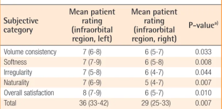
Table 3. Surgical outcome on infraorbital region as evaluated by the patients
Subjective category
Mean patient rating (infraorbital region, left)
Mean patient rating (infraorbital region, right)
P-valuea)
Volume consistency 7 (6-8) 6 (5-7) 0.033
Softness 7 (7-9) 6 (5-8) 0.008
Irregularity 7 (5-8) 6 (4-7) 0.044
Naturality 7 (6-9) 5 (4-7) 0.007
Overall satisfaction 8 (7-9) 6 (5-7) 0.010
Total 36 (33-42) 29 (25-33) 0.007
a)Wilcoxon signed-rank test was used for the statistical processing of the results.
Table 4. Surgical outcome on nasolabial fold as evaluated by the patients
Subjective category
Mean patient rating (nasolabial fold,
left)
Mean patient rating (nasolabial fold,
right)
P-valuea)
Volume consistency 8 (6-9) 7 (5-8) 0.011
Softness 8 (7-9) 7 (5-8) 0.016
Irregularity 8 (6-8) 6 (4-7) 0.007
Naturality 7 (7-8) 6 (5-7) 0.016
Overall satisfaction 8 (7-9) 7 (5-8) 0.011
Total 39 (36-42) 30 (27-35) 0.008
a)Wilcoxon signed-rank test was used for the statistical processing of the results.
evaluations were negligible and statistically insigniicant. In all 5 categories of volume consistency, sotness, irregularity, natural-ity, and overall satisfaction, the area grated with adipose tissue mixed with SVF cells showed significantly better results com-pared with those of areas grafted with adipose tissue without cells.
Evaluation was conducted 12 weeks postoperatively. Ater the initial overcorrection, the appearance of the grated areas became more natural during the follow-up period, and all of the patients were satisied with their appearance (Figs. 2, 3).
DISCUSSION
Since fat grat was irst introduced in 1893 by Neuber, various methods have been suggested [10]. Adipose tissue is easily and repeatedly obtained, and fat grat is a simple and low-risk proce-dure. Accordingly, fat grat is now one of the most common and widely used procedures in plastic surgery. Because the fat tissue used in these procedures is autologous, no side efects caused by immune responses have been reported, and any part of the body can be a recipient site. However, grated fat can be absorbed into the body, and the level of absorption is diicult to anticipate. In addition, results can be significantly different from surgeon to surgeon and accordingly, quantitative analysis and evaluation of fat grats are not easy.
Studies on embryonic stem cells and adult stem cells are active in the ield of biotechnology [5]. Embryonic stem cells are ideal stem cells for tissue reconstruction because they can diferenti-ate into various tissues. However, diferentiation of embryonic stem cells is diicult to control, and the risk of malignant degen-eration is present. Ethical issues are another factor restricting their clinical applications [6]. In contrast, adult stem cells are
ob-Table 5. Surgical outcome on malar eminence as evaluated by a separate surgeon
Subjective category
Mean patient rating (malar eminence, left)
Mean patient rating (malar eminence, right)
P-valuea)
Volume consistency 7 (6-8) 6 (5-7) 0.015
Softness 7 (5-8) 5 (4-7) 0.044
Irregularity 7 (6-8) 5 (5-7) 0.019
Naturality 7 (6-9) 6 (4-7) 0.011
Overall satisfaction 7 (7-8) 6 (5-7) 0.014
Total 36 (32-38) 29 (25-31) 0.008
a)Wilcoxon signed-rank test was used for the statistical processing of the results.
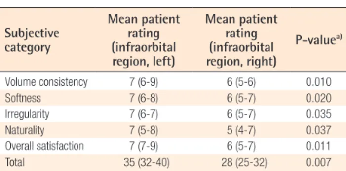
Table 6. Surgical outcome on infraorbital region as evalu-ated by a separate surgeon
Subjective category
Mean patient rating (infraorbital region, left)
Mean patient rating (infraorbital region, right)
P-valuea)
Volume consistency 7 (6-9) 6 (5-6) 0.010
Softness 7 (6-8) 6 (5-7) 0.020
Irregularity 7 (6-7) 6 (5-7) 0.035
Naturality 7 (5-8) 5 (4-7) 0.037
Overall satisfaction 7 (7-9) 6 (5-7) 0.011
Total 35 (32-40) 28 (25-32) 0.007
a)Wilcoxon signed-rank test was used for the statistical processing of the results.
Table 7. Surgical outcome on nasolabial fold as evaluated by a separate surgeon
Subjective category
Mean patient rating (nasolabial fold,
left)
Mean patient rating (nasolabial fold,
right)
P-valuea)
Volume consistency 8 (7-9) 6 (5-8) 0.017
Softness 8 (6-9) 6 (4-8) 0.014
Irregularity 7 (6-8) 6 (5-6) 0.011
Naturality 7 (6-8) 5 (4-6) 0.027
Overall satisfaction 8 (7-9) 6 (5-7) 0.011
Total 37 (36-41) 30 (24-34) 0.008
a)Wilcoxon signed-rank test was used for the statistical processing of the results.
Table 8. Surgical outcome on nasolabial fold versus malar eminence and infraorbital region as evaluated by the patients
Mean patient rating (nasolabial
fold, left)
Mean patient rating (malar eminence, left)
P-valuea) rating (nasolabial Mean patient
fold, left)
Mean patient rating (infraorbital
region, left)
P-valuea)
Total 39 (36-42) 36 (34-42) 0.041 39 (36-42) 36 (33-42) 0.048
a)Wilcoxon signed-rank test was used for the statistical processing of the results.
Table 9. Surgical outcome on nasolabial fold versus malar eminence and infraorbital region as evaluated by a separate surgeon
Mean patient rating (nasolabial
fold, left)
Mean patient rating (malar eminence, left)
P-valuea) rating (nasolabial Mean patient
fold, left)
Mean patient rating (infraorbital
region, left)
P-valuea)
Total 37 (36-41) 36 (32-38) 0.042 37 (36-41) 35 (32-40) 0.043
tained from already diferentiated tissues, and are free from ethi-cal controversy. Among adult stem cells, adipose stem cells are actively studied because they are easily obtainable. According to an in vitro experiment conducted by Zuk et al. [3], adipose stem cells can diferentiate into cells of fat, cartilage, muscle, and bone. In practice, the clinical application of adipose stem cells in a grat is not easy because cell culture facilities that gain the approval of the Korea Food and Drug Administration need to be used. Due to these limitations, recent studies have focused on SVF cells that are obtained when adipose tissue is centrifuged without the use of cultivation. Leblanc et al. [11] reported that SVF cells ob-tained from adipose tissue increased blood low in the coronary artery ater the development of myocardial infarction. Lendeckel et al. [12] reported cases of successful bone reconstruction using
fibrin glue and SVF cells obtained from adipose tissue, which were grated into the area of the cranial defect.
In the present study, we tried to delay or prevent absorption of fat ater grating by mixing SVF cells with adipose tissue. At post-operative 12 weeks, the patient and doctor surveys confirmed that the results of grats with adipose tissue mixed with SVF cells were beter than those grated with adipose tissue without SVF cells in all 5 categories of volume consistency, sotness, irregular-ity, naturalirregular-ity, and overall satisfaction. As suggested by multiple studies [11,12], SVF cells have been considered to facilitate the growth of surrounding tissues. In the present study, this function of SVF cells was observed. However, the mechanism of SVF cells facilitating the growth of surrounding fat cells is not clearly understood. Additional studies investigating this issue are ex-pected in the future.
According to the present study, the highest evaluation score ater a fat grat mixed with SVF cells was assigned to the nasola-bial fold followed by the malar eminence and infraorbital region (Tables 8, 9). In our previous study [2], the lowest satisfaction score ater grat was assigned to the nasolabial fold area. he rea-son for the diferent results between the previous and the pres-ent study is considered to be due to the SVF cells used in the present study having a remarkable efect in the area of nasolabial fold, which usually has low satisfaction scores due to the deep-ness of the fold.
Quantitative analysis or measurement of the adipose tissue that remains ater a fat grat is diicult to perform in practice. here-fore, the efects of the fat grats were evaluated through surveys using the clinical photos. However, this method is limited by the subjective nature of the evaluation carried out by the patients or surgeons. In the future, an objective and quantitative method of
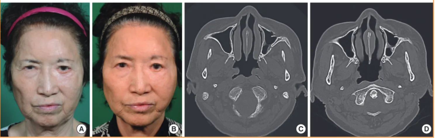
Fig. 2. Fat graft with SVF cells (case 1)
(A, B) Improved nasolabial fold, malar eminence, and infraorbital region after a fat graft with SVF cells (left face) or a fat graft without SVF cells (right face). The left face showed the better result. (A) Preoperative view. (B) Postoperative view at 12 weeks. (C, D) Facial bone computed tomography. Improved nasolabial fold region after a fat graft with SVF cells (left face) or a fat graft without SVF cells (right face). (C) Preoperative view. (D) Postoperative view at 12 weeks. SVF, stromal vascular fraction.
B C D
A
Fig. 3. Fat graft with SVF cells (case 2)
Improved nasolabial fold, malar eminence, and infraorbital region after a fat graft with SVF cells (left face) or a fat graft without SVF cells (right face). The left face showed the better result. (A) Preoperative view. (B) Postoperative view at 12 weeks. SVF, stromal vascular fraction.
measuring the fat amount should be developed to calculate the survival rate of SVF cells that are mixed with adipose tissue.
In order to establish a control group, the right side of the face of the patients was grated with adipose tissue without SVF cells. Consequently, bias was minimized in the present study. Even though this method was used with the permission of the patients, there was a risk of causing an imbalance in the appearance of the right and let side of the face in the patients. For this reason, only 9 patients were enrolled in the present study. A larger number of patients, and a more accurately established control group than the present study may be necessary to obtain more reliable data on the efects of SVF cells on the survival rate of fat cells.
Moreover, there were some limitations to the present study. First, facial bone computed tomography was conducted only at 12 weeks postoperatively. However, it was not conducted immediately ater the fat grats were performed. If we conduct facial bone computed tomography immediately ater the opera-tion and at 12 weeks postoperatively, it may measure the absorp-tion rate of fat cells. Second, only 9 patients were enrolled in this study and the follow-up period for the evaluation results was 12 weeks postoperatively. If the number of patients and the follow-up period were increased, it could verify the efects of SVF cells on the survival rate of fat cells.
The positive effects of SVF cells on the generation and long-term survival rate of adipose tissue were conirmed in the present study. In particular, the efects of SVF cells were more evident in the area of deep wrinkles in the face than in other areas. In con-clusion, fat grats using SVF cells may enhance the survival rate of fat cells and the efects of fat grats.
REFERENCES
1. Illouz YG. he fat cell “grat”: a new technique to ill depres-sions. Plast Reconstr Surg 1986;78:122-3.
2. Kim SB, Kim DW, Yoon ES. Facial fat injection: long-term
follow-up results. J Korean Soc Aesthetic Plast Surg 2010; 16:35-40.
3. Zuk PA, Zhu M, Mizuno H, et al. Multilineage cells from human adipose tissue: implications for cell-based therapies. Tissue Eng 2001;7:211-28.
4. Dhar S, Yoon ES, Kachgal S, et al. Long-term maintenance of neuronally differentiated human adipose tissue-derived stem cells. Tissue Eng 2007;13:2625-32.
5. Yoon E, Dhar S, Chun DE, et al. In vivo osteogenic potential of human adipose-derived stem cells/poly lactide-co-glycol-ic acid constructs for bone regeneration in a rat critlactide-co-glycol-ical-sized calvarial defect model. Tissue Eng 2007;13:619-27.
6. Jeong T, Ji YH, Kim DW, et al. Treatment of phalageal bone defect using autologous stromal vascular fraction from li-poaspirated tissue. J Korean Soc Plast Reconstr Surg 2011; 38:438-44.
7. Poloni A, Maurizi G, Leoni P, et al. Human dediferentiated adipocytes show similar properties to bone marrow-derived mesenchymal stem cells. Stem Cells 2012;30:965-74. 8. Coleman SR. Hand rejuvenation with structural fat grating.
Plast Reconstr Surg 2002;110:1731-44.
9. Tabit CJ, Slack GC, Fan K, et al. Fat grating versus adipose-derived stem cell therapy: distinguishing indications, tech-niques, and outcomes. Aesthetic Plast Surg 2012;36:704-13. 10. Ersek A, Chang P, Salisbury MA. Lipo layering of autolo-gous fat: an improved technique with promising results. Plast Reconstr Surg 1998;101:820-6.
11. Leblanc AJ, Touroo JS, Hoying JB, et al. Adipose stromal vascular fraction cell construct sustains coronary microvas-cular function ater acute myocardial infarction. Am J Physiol Heart Circ Physiol 2012;302:H973-82.