Int J Anat Res 2014, 2(2):296-04. ISSN 2321-4287 Original Article
SURGICAL ANATOM Y OF DORSAL ROOT ENTRY ZONE OF
CERVICAL SPINAL NERVES : CADAVERIC STUDY
A.Arun Kumar *
1, Sudha Seshayyan
2, V.Tamilalagan
3, M .Sindou
4.
ABSTRACT
Address for Correspondence: Dr.A.Arunkumar, Depart ment of Anat omy, Sri Lakshmi Narayana Inst it ute of M edical Sciences, Osudu, Agaram Village, Villianur Commune, Kudupakkam Post , Puducherry-605502, India. M obile no: +91 9962972243. E-M ail: [email protected]
Access this Article online
Quick Response code Web site:
* 1 Depart ment of Anat omy, Sri Lakshmi Naryana Inst it ute of M edical Sciences, Puducherry, India.
2 Inst it ute of Anat omy, M adras M edical College, Chennai, India.
3 Depart ment of Anat omy, Nat ional Inst it ute of Siddha, Chennai, India.
4 Depart ment of Neurosurgery, Neurological Hospital P.Wert heimer, Universit y of Lyon, France.
Background:The m ain pur pose of t his st udy is t o det erm ine t he detai led m orphom et r ic data of Dorsal Root
Ent ry Zone (DREZ) of cervical spinal nerves. This know ledge is necessary for diagnosis, t reat m ent and surgical m anagem ent of pain due t o m any conditions like brachial plexus avulsion injury, post-herpet ic neuralgia, phantom pain and cancer pain involved in cervical myelo-radiculopat hy. There are few er st udies report ed in t his field of DREZ.
M aterials and M ethods:Tw ent y f ive adult form alin fixed cadavers are taken for t his st udy. Convent ional Spinal
cord dissect ion is follow ed as per Cunningham ’s Dissect ion M annual.
Findings: The paramet ers included are Num ber of dorsal root let s, Longit udinal Lengt h of DREZ, Distance betw een t w o successive DREZ, Lengt h of dorsal root let s, Distance bet w een r ight and left DREZ, Dist ance bet w een DREZ and Ligam ent um dent iculat um , Cranial angles of Superior & inferior root let s.
Results:Result s w ere not ed for all t he param et ers and are com pared w it h t he previous st udies. The significant
observat ions are obtained.
Conclusion:Surgical anat omy of Dorsal Root Ent ry Zone (DREZ) of cer vical spinal nerves w ill be useful for t he
neurosurgeons doing Drezot omy procedure, in w hich t he nocicept ive fibres alone are specifically severed w it h preservat ion of ot her sensat ions.
KEYW ORDS:DREZ, Dorsal Root Ent r y Zone, Cervical Spinal Nerves, Pain.
INTRODUCTION
Int ernat ional Journal of Anatomy and Research, Int J Anat Res 2014, Vol 2(2):296-04. ISSN 2321- 4287
Received: 21 M arch 2014
Peer Review : 21 M arch 2014 Published (O):30 April 2014 Accepted: 08 April 2014 Published (P):30 June 2014 Internat ional Journal of Anat omy and Research
ISSN 2321-4287 w w w.ijmhr.org/ ijar.ht m
Dorsal Root Ent ry Zone (DREZ) is t he region on t he posterolateral sulcus of spinal cord, w here t he dorsal root let s carrying sensory fibers enter int o t he spinal cord. It is t he junct ion bet w een spinal cord (CNS) and spinal nerves (PNS).
Through t hese dorsal root let s, all t he sensat ions are carried t o t he spinal cord. Number of dorsal root let s is not constant for all t he spinal nerves. Region bet w een ent ry point s of first t o last root let of one spinal nerve is called as Dorsal
Root Ent ry Zone of t hat nerve.
Chronic pat ient s w it h cancer, brachial plexus avulsion injuries [1,2,3,4], paraplegia[5,6], spinal cord lesions[ 2,7] , peripheral ner ve lesions, p han t om l im b pai n [ 1,8] , p ost her p et i c neuralgia[2,9,10]etc., suffer severe int olerable pain. W hen conser vat ive t reat m ent s fai l t o abolish pain, surgeons prefer any one of t he follow ing.
1. Rhizot omy[11], 2. Sympat hect omy,
3. Cordot omy, 4. Neurost imulat ion, 5. Ganglionect omy[12,13]
6. DREZot omy[14,15,16,17,18,19,20,21,22] Of t hese, DREZot omy is considered t o be t he recent advanced procedure w hich select ively dest roys nocicept ive fibres alone and preserve ot her sensat ions.
At t he root ent ry zone each root let present s m edial and lat eral divisions. M edial division consists of thickly myelinated fibers. These fibers carry t ouch, pressure and vibrat ion sensat ions.
Lateral division consist of t hinly myelinated and unmyelinated fibers. These fibers conveys pain and temperat ure sensat ions.
In lat eral division, t he t em perat ure carrying fibers are dorsal and pain carrying fibers are vent ral in posit ion. M aking an incision along t he lat eral aspect of t he DREZ w ill cut t he pain carr ying f ibres alone. Ot her sensat ions li ke t ouch, pressure, vibrat ion and posit ion senses carried by medial division are preserved.
DREZotomy PROCEDURE:
It is do ne by creat ing incision and bipolar coagulat ions on t he vent ro-lateral aspect of t he Dorsal Root Ent ry Zone. Incision is done at t he lateral part of DREZ and medial part of Tract of Lissauer. The incision is about 2 mm dept h at an angle of 45º . The incision cut s select ively t he nocicept ive fibers grouped in lat eral division, excitat ory medial fibers of t ract of Lissauer and hyperact ive neurons of dorsal horn.
It is t he schemat ic representat ion of DREZ and t arget of m icro-DREZot omy[ 22] . (t hanks t o Co-aut hor M .Sindou)
Upper part of t he schem at ic represent at ion show s pial ring w hich divides t he root let int o a
peripheral and a cent ral segment . The t ransit ion bet w een t he t w o segment s is at t he pial ring (PR), w hich is locat ed approxim at ely 1m m out side t he penet rat ion of t he root let .
As they approach PR, the fine fibers (nociceptive) move t oward t he root let surfaces. In t he cent ral segment , they group in the ventrolateral port ion of t he DREZ, t o ent er t he dorsal hor n (DH) t hrough t he t ract of Lissauer (TL). The large myotat ic fibers (myot ) are sit uated in t he middle of t he DREZ, w hereas t he large lemniscal fibers are located dorsomedially t o reach t he dorsal column (DC).
Low er part of schemat ic representat ion show s Dorsal Horn (DH) circuit ry.
Rexed’s laminae are marked from I t o VII. M DT (arrow head) cut s most of t he fine and myotat ic fibers and enters t he medial (excitat ory) port ion of TL and t he apex of t he dorsal horn. This procedure preserves most lemniscal presynapt ic fibers, t he lateral (inhibit ory) port ion of TL, and most of t he DH.
AIM
Aim of t his st udy is t o determine t he cadaveric morphomet ric data of Dorsal Root Ent ry Zone (DREZ) o f cer v ical spi nal ner v es. Det ai l ed anat omical know ledge of DREZ is necessary for t he d iagn osi s, t r eat m en t an d su r gical management of many diseases. M orphomet ric data of DREZ has been reported in many previous st udies. Previous st udies w ere done in Turkey [23], Georgia [24], China [25], France [16, 17, 18, 19, 20, 21, 22] etc. As for as Indian populat ion are concerned t here are no st udies available in t he field of DREZ. Hence t his st udy is intended t o record t he basic anat omic data of DREZ of Sout h Indian populat ion.
Know ledge of t he parameters ment ioned below w ill be useful for t he surgeons in planning t he surgeries and m anagement of post-operat ive complicat ions. They are.,
1. Number of dorsal root let s. 2. Longit udinal Lengt h of DREZ.
3. Distance bet w een t w o successive DREZ. 4. Lengt h of dorsal root let s.
5. Distance bet w een right and left DREZ. 6. Dist ance bet w een DREZ and Ligament um dent iculat um.
Int J Anat Res 2014, 2(2):296-04. ISSN 2321-4287 M ATERIALS AND M ETHODS
M ATERIALS:
Twent y five formalin fixed adult human cadavers of Sout h Indian origin are taken for t his st udy from t he Inst it ute of Anat omy, M adras M edical College, Chennai. The mean age of t his group is 55. St udy sample included bot h male and female cadavers w it h no hist ory of diseases.
Required materials for dissect ion are scalpel, BP hand le and blades, Bone cut t er, Spine saw, spat ula, artery forceps, t oot hed forceps, blunt forceps, chisel , hammer, scissors, hand lens, loupes, gloves, cot t on, gauze, t oken, t hread, formalin container,
Required materials for m easurement are inch tape, Digital Vernier Caliper – 15 cm, prot ract or, Digital Camera – used in t his st udy is- Sony (DSC-WX- 150, 18.1 M egapixels).
M ETHOD:
Dissect i o n m et h o d i s fo l l ow ed as t h e Convent ional dissect ion met hod given in t he Cunningham ’s M anual of Pract ical Anat omy, Volume Three - Head, Neck and Brain, fifteent h edit ion, p.no 192 t o 202. The spinal cords rem oved f rom t he cadaver are st ored in a formalin filled container. Unique t oken number for each spinal cord is given. The parameters taken for t his st udy are measured and noted.
RESULTS AND DISCUSSION
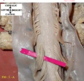
1. NUM BER OF DORSAL ROOTLETS:
In t his parameter t he t otal number of root let s present in dorsal root of each cervical spinal
nerve is counted. These root let s are t he cent ral process of dorsal root ganglion. They enter t he spinal cord t hrough t he postero lateral sulcus for each spinal nerve. Num bers of root let s are counted on bot h sides.
Fig : 1 – A , show s t he dorsal surface of t he spinal cord at cervical level. Number of dorsal root let s in cervical level is counted on bot h sides. The root let s are variable in t hickness.
Fig. 1:Do rsal View, sho ws dorsal root let s at cervical level.
Table (1a): Obser ved Result s - Num ber of Dorsal
Root let s (NODR).
Table (1b):Discussion - Num ber Of Dorsal Root let s
(NODR).
AUTHOR YEAR CADAVER COUNT
SEGM ENT TAKEN
FOR STUDY
OBSERVED RESULT (Num bers)
KUBA Y[ 26] 1994 18 C5 – T1 5 to 16
TANAKA[27] 2000 18 C5 – C8 8 to 12
A.KARATAS[23 ] 2004 15 C1 – T1 2 to 13
JIAN-PING
XIANG,M .D[25 ] 2007 20 C5 – T1 7.76 NUM BER OF DORSAL ROOTLETS (NODR)
SINDOU[22] 1974 - C4-T1
Present study 2013 25 C1 – T1
C2,C3,C4 = 4 C5,C6,C7,C8 = 6
C6 = 8.7 T1= 6.7
CERVICAL SEGM ENTS :
Maxi mum: C5 (10.16 ± 1.595)
M i ni mum: C8 (5.52 ± 0.646)
CARGILL
H.ALLEYNE,Jr[24] 1998 10 C3- T1
Previous st udies w ere m ainly done for t he diagnosis and t reat m ent of brachial plexus injuries. When compared w it h t he present study t he obser vat ions for t he sam e level, show correlat ion w it h t he range 3 -10 num ber of dorsal root let s in t he st udies of CARGILL H.
Sl. No SEGM ENT NODR-LEFT
(M EAN) (Nos.)
NODR-RIGHT (M EAN) (Nos.)
1 C 1 7.6 7.56
2 C 2 8.56 8.92
3 C 3 6.28 6.88
4 C 4 9.12 9.44
5 C 5 9.96 10.36
6 C 6 7.24 7.4
7 C 7 6.52 6.68
ALLEYNE, Jr[24] et al, TANAKA[27] et al, KUBA Y[26] et al and JIAN-PING XIANG,M .D[25].,et al.
2. LONGITUDINAL LENGTH OF DREZ :
In t his parameter, lengt h bet w een superior and inferior root let of each dorsal root is measured using a digital vernier caliper and it is noted in m illim et ers. It is t he point of ent ry of dorsal ro ot let s i nt o t he sub st an ce of spinal co rd t hrough postero lateral sulcus. It is measured on bot h sides.
Fig : 2 – A , show s t he dorsal surface of t he spinal cord at cervical level. Longitudinal lengt h of DREZ in cervical level is measured on Right side.
When compared w it h t he previous st udies, t he
p resent st u d y sh o w s cor r elat i on f or t hi s parameter.
Fig. 2-A:Dorsal V iew, longit udinal lengt h of DREZ at
Cer vical level.
Table (2-A): Observed result s of longit udinal Lengt h
of DREZ(LLODREZ).
1 C 1 5.64 5.75
2 C 2 7.45 7.93
3 C 3 11.62 11.45
4 C 4 12.47 12.54
5 C 5 13.12 12.91
6 C 6 10.1 9.98
7 C 7 10.88 10.3
8 C 8 10.03 10.1
LLODREZ-LEFT M EAN - mm
Sl. No SEGM ENT LLODREZ-RIGHT
M EAN - mm
Table (2b): Discussion of Longit udinal Lengt h of Drez
(LLODREZ).
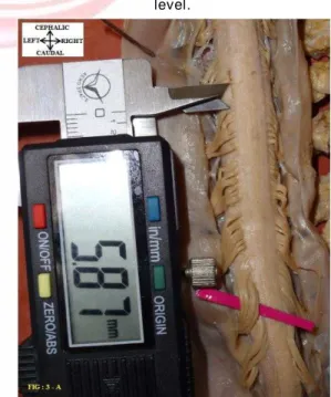
3. DISTANCE BETW EEN SUCCESSIVE DREZ:
In t h i s p aram et er t h e di st an ce b et w een successive spinal segment s is measured. This measurement is done by measuring t he distance bet w een inferior root let of spinal nerve above t o the superior rootlet of spinal nerve below. This m easu r em en t i s d o n e o n b o t h si des. M easurem ent is t aken using digit al vernier caliper and it is noted in millimeters.
Fig: 3 – A, show s t he dorsal surface of t he spinal cor d at cer v i cal lev el. Dist ance bet w een successive DREZ in cervical level is measured on left side.
Fig. 3: Distance bet w een successive DREZ at Cervical level.
The present st udy show ed, distance bet w een successive DREZ is maximum at t he interval C2 & C3 (2.42 ± 1.272) and minimum at t he interval C7 & C8 (0.56 ± 1.010). This param et er is reported for t he first t ime. There are no previous st udies for discussion.
AUTHOR YEAR CADAVER COUNT
SEGM EN T TAKEN FOR
STUDY
OBSERVED RESULT(m m )
18 C5 – T1
Present study 2 013 25 C1 – T1
LONGITUDINAL LEN GTH OF DREZ (LLODREZ)
6 – 14 mm, Lower cer vi cal segment – Decr eases
C7 = 10 .7 mm (r an ge 8-1 6) C4= 12 .7 mm (r ange 10-1 6)
4.3 – 17.7 LONGEST – C5 CERVICAL SEGM ENTS :
M axi mum: C5 (13.01 ± 1.38 0) M i ni mum : C1
(5.70 ± 0.78 1)
CARGILL
H.ALLEYN E.,Jr[ 24] 1 998 10 C3- T1
A.KARATAS[ 2 3] 2 004 15 C1 – T1
Int J Anat Res 2014, 2(2):296-04. ISSN 2321-4287
Table (3a): Observed result s of Dist ance Bet w een
Successive DREZ (DBSDREZ).
1 C1&C2 1.92 1.6
2 C2&C3 3.24 1.61
3 C3&C4 2.03 2.42
4 C4&C5 1.81 1.67
5 C5&C6 1.29 1.24
6 C6&C7 1.45 1.84
7 C7&C8 0.54 0.59
8 C8&T1 0.4 0.69
Sl. No SEGM ENT DBSDREZ-LEFT M EAN - mm
DBSDREZ-RIGHT
M EAN – mm
4. DISTANCE BETW EEN LEFT AND RIGHT DREZ:
In t his parameter t he distance bet w een left and r ight DREZ of sam e segm ent is m easured. M easured using digit al vernier calipers and noted in millimeters.
Fig : 4 – A , show s t he dorsal surface of t he spinal cord at cervical level. Distance bet w een left and right DREZ at cervical level is measured.
Fig. 4:Dorsal V iew, Distance bet w een Lef t & Right DREZ at Cervical level.
When compared w it h t he previous st udies, t he
present st udy show s correlat ion for t his param-eter. In t his present st udy, distance bet ween left
& right DREZ show ed maximum at C6 segment ,
minimum in C8.
Table (4a): Observed Result s of Dist ance Bet w een Left
And Right DREZ (DBL& RDREZ).
Sl. No SEGM ENT DBL& RDREZ (M EAN) (mm)
1 C 1 6.44
2 C 2 6.85
3 C 3 6.83
4 C 4 6.34
5 C 5 6.82
6 C 6 7.15
7 C 7 6.2
8 C 8 4.86
Table (4b): Discussion of Distance Bet w een Left And
Right DREZ (DBL& RDREZ).
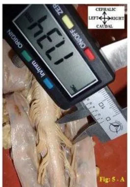
5. LENGTH OF DORSAL ROOTLETS :
In t his parameter t he lengt h of dorsal root let s from postero-lateral sulcus t o t he point w here it pierces t he duram at er is m easured. This measurem ent is t aken for t he middle root let using a digit al vernier caliper and not ed in m i l l i m et ers. Len gt h o f d o rsal r o o t l et s i s measured on bot h sides.
Fig : 5 – A , show s t he dorsal surface of t he spinal cord at cervical level. This pict ure show s lengt h of dorsal root let s at cervical level.
Fig. 5:Dorsal View, show s lengt h of dorsal root let s at
cer vical level.
AUTHOR YEAR CADAVER COUNT
SEGM ENT TAKEN
FOR STUDY
OBSERVED RESULT(m m )
Pre se nt St udy 2 01 3 2 5 C1 – T1
5 to 9 mm LONGEST = C6 2.2 to 9.4 mm LONGEST - C2 & C3
LOW ER CERVICAL SEGM ENT-DECREASES
5.90 mm C5 = 3.54 mm T1 = 2.23 mm CERVICAL SEGM ENTS : M axi mum: C6(7.15 ± 0.494) M i ni mum: C8(4.86 ± 0.628)
KARATAS[ 2 3 ] 2004 15 C1 – T1
JIAN -PIN G
XIANG,M .D[ 2 5] 2007 20 C5 – T1
DISTANCE BETW EEN LEFT & RIGHT DREZ (DBL& RDREZ)
Table (5a): Observed result s of Lengt h of Dorsal Root let s (LODR).
Sl. No SEGM ENT LODR-LEFT
(M EAN)(mm)
LODR-RIGHT (M EAN) (mm)
1 C 1 7.1 6.92
2 C 2 9.08 9.04
3 C 3 11.1 10.94
4 C 4 10.61 10.46
5 C 5 12.93 12.82
6 C 6 15.17 14.56
7 C 7 17.88 16.8
8 C 8 18.69 17.94
Table (5b): Discussion of Lengt h of Dorsal Root let s
(LODR).
W hen com pared w it h KUBA Y[ 26] et al and A.KARATAS[23] et al st udies w it h respect t o t he lengt h of dorsal root let s, longest lengt h alone correlates w it h t he present st udy.
The st udies done by CARGILL H.ALLEYNE.,Jr[24] et al and TANAKA[27] et al show s correlat ion w it h present st udy.
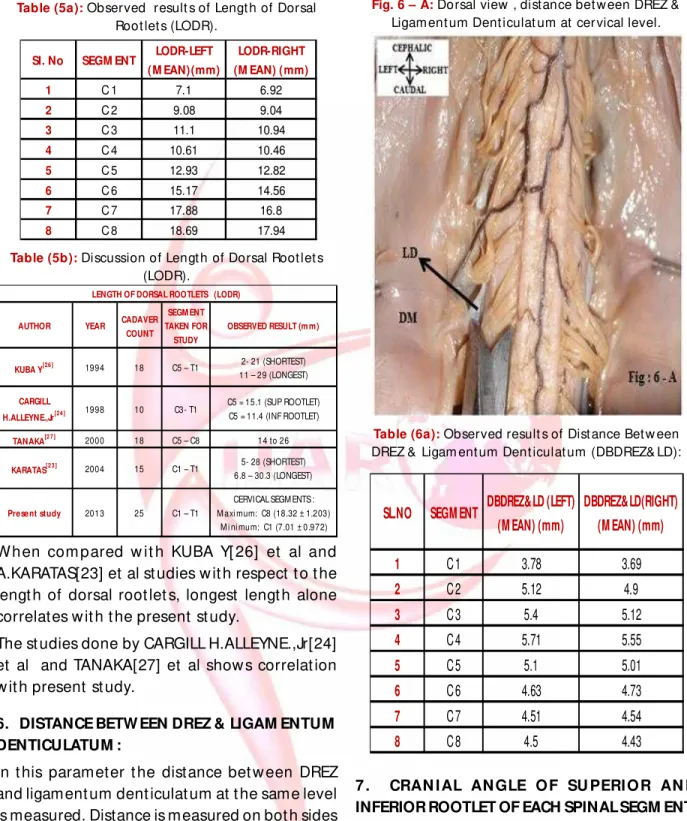
6. DISTANCE BETW EEN DREZ & LIGAM ENTUM DENTICULATUM :
In t his parameter t he distance bet w een DREZ and ligament um dent iculat um at t he same level is measured. Distance is measured on bot h sides u si n g d igi t al ver ni er cal i per an d n o t ed i n millimeters.
Fig : 6 – A , show s t he dorsal surface of t he spinal cord at cervical level. Distance bet w een DREZ and ligament um dent iculat um at cervical level is measured on left side.
The present st udy show s maximum DBDREZ & LD is at C4 (5.63 ± 0.691) and minimum at C1 (3.73 ± 0.508). This parameter is reported for t he first t ime. There are no previous st udies for dis-cussion.
Fig. 6 – A: Dorsal view , distance bet w een DREZ & Ligam ent um Dent iculat um at cer vical level.
Table (6a): Observed result s of Dist ance Bet w een
DREZ & Ligam ent um Dent iculat um (DBDREZ& LD):
1 C 1 3.78 3.69
2 C 2 5.12 4.9
3 C 3 5.4 5.12
4 C 4 5.71 5.55
5 C 5 5.1 5.01
6 C 6 4.63 4.73
7 C 7 4.51 4.54
8 C 8 4.5 4.43
SL.NO SEGM ENT DBDREZ&LD(RIGHT) (M EAN) (mm) DBDREZ&LD (LEFT)
(M EAN) (mm)
7 . CRAN I AL AN GLE O F SU PERI O R AN D INFERIOR ROOTLET OF EACH SPINAL SEGM ENT:
A. CRANIAL ANGLE OF SUPERIOR ROOTLET:
In this parameter t he angle formed in t he cranial aspect at t he point w here t he superior or inferior rootlet enters postero-lateral sulcus is measured. Cranial angle is measured using prot ract or on bot h sides and noted in degrees.
Fig : 7 – A , show s t he dorsal surface of t he spinal cord at cervical level. Angle form ed in bet w een superior root let and post erolat eral su lcu s i n cr an ial asp ect i s n o t ed u si n g prot ract or.
AUTHOR YEAR CADAVER COUNT
SEGM ENT TAKEN FOR
STUDY
OBSERVED RESULT (m m)
TANAKA[2 7 ] 2000 18 C5 – C8 14 to 26
5- 28 (SHORTEST) 6.8 – 30.3 (LONGEST) C5 = 15.1 (SUP ROOTLET)
C5 = 11.4 (INF ROOTLET) 2- 21 (SHORTEST) 11 – 29 (LONGEST)
Present study 2013 25 C1 – T1
CERVICAL SEGM ENTS : M axi mum: C8 (18.32 ± 1.203) M i ni mum: C1 (7.01 ± 0.972) CARGILL
H.ALLEYNE.,Jr[24 ] 1998 10 C3- T1
KARATAS[2 3 ] 2004 15 C1 – T1 LENGTH OF DORSAL ROOTLETS (LODR)
Int J Anat Res 2014, 2(2):296-04. ISSN 2321-4287
Fig. 7-A: Dorsal V iew, yellow arrow – Superior Root let ,
show s cranial angle of superior root let w it h post ero-lat eral sulcus of spinal cord.
Table (7a): Observed result s of Cranial Angle of
Superior And Inferior Root let of Each Spinal Segm ent (CAOSR).
SL. NO SEGM ENT CAOSR-LEFT
(MEAN) (˚)
CAOSR-RIGHT
(MEAN) (˚)
1 C 1 111.6 108.8
2 C 2 121.2 119.2
3 C 3 138 135.2
4 C 4 143.6 141.2
5 C 5 121.8 120
6 C 6 146.8 146
7 C 7 144.4 144
8 C 8 144 143.6
Table (7b): Discussion of Cranial Angle of Superior and
Inferior Root let of Each Spinal Segm ent (CAOSR).
AUTHOR YEAR CADAVER
COUNT SEGM ENT TAKEN FOR
STUDY
OBSERVED RESULT (in degrees) CRANIAL ANGLE OF SUPERIOR ROOTLET (CAOSR)
CARGILL
H.ALLEYNE.,Jr[24]
1998 10 C3- T1 C5 = 116˚ Smallest T1 = 156˚ Largest
Present study 2013 25 C1 – T1
CERVICAL SEGM ENTS : M aximum : C5 (144.20 ± 5.379) M i ni mum : C1 (110.20 ± 8.204) According t o CARGILL H.ALLEYNE.,Jr [24] et al cranial angle of superior root let at C5 (1160) is minimum. In t he present st udy, w hen compared among t he cervical segment s cranial angle of superior root let at C1 (1100) is minimum. The present st udy show s CAOSR is maximum at C5 and minimum at C1 segment .(Ref : Fig 7-A).
B. CRANIAL ANGLE OF INFERIOR ROOTLET:
Fig : 7 – B , show s t he dorsal surface of the spinal cord at cervical level. Angle formed in bet w een inferior root let and post erolat eral sulcus in cranial aspect is noted using prot ract or.
Fig. 7 – B:Dorsal V iew, yellow arrow – Infer ior Root let , show s cranial angle of inferior root let w it h
post erolat eral sulcus of spinal cord.
Table (8a): Observed result s of Cranial Angle of
Inferior Root let of Each Spinal Segm ent (CAOIR).
SL.NO SEGM ENT CAOIR-LEFT
(MEAN) (˚)
CAOIR-RIGHT
(MEAN) (˚)
1 C 1 91.2 92.8
2 C 2 100 101.6
3 C 3 115.2 115.6
4 C 4 116.8 117.6
5 C 5 129.2 123.2
6 C 6 130 129.6
7 C 7 130.4 125.2
8 C 8 135.2 126
Table (8b): Discussion of Cranial Angle of Infer ior
Root let of Each Spinal Segm ent (CAOIR).
The present st udy show s CAOIR is maximum at
C8 segm en t . Then it d ecr eases i n cr an ial
direct ion, minimum in C1 (Ref : Fig 7-B).
JIAN-PING
XIANG,M .D[25]
2007 20 C5 – T1 65.6 to 19.8
CERVICAL SEGMENTS : Maximum : C8(130.60 ± 8.184) M i nimum : C1 (92.00 ± 8.081)
OBSERVED RESULT (in degrees)
C6 = Smal l est C5 = next smal l est CARGILL
H.ALLEYNE.,Jr[24] 1998 10 C3- T1
Present study 2013 25 C1 – T1
CRANIAL ANGLE OF INFERIOR ROOTLET (CAOIR)
AUTHOR YEAR CADAVER
COUNT
CONCLUSION
Surgical anat omy of Dorsal Root Ent r y Zone (DREZ) of cervical spinal nerves w ill be useful for t he neurosurgeons doing Drezot omy procedure, i n w h ich t he n o ci cept i ve f i br es al on e ar e specifically severed w it h preservat ion of ot her sensat ions. The cadaveric dat a also helps t he n eu r o su r geo n s t o av o id i nj u r y t o n eu ral st ruct ures as w ell as t o avoid post-operat ive complicat ions. The highest and low est root let num ber m ay be a useful int ra-dural surgical landmark.
These cadaver ic dat a w i ll be usef ul in t he u n der st an d in g an d t r eat m en t o f l esio n s compressing t he int ra dural nerve root s. It w ill also help in other int ra dural procedures like intra dural nerve st imulat ion, endoscopic navigat ion [28] etc.
The cadaveric dat a m ay also help in clinical n eu r o phy sio l o gy t o co r r el at e w i t h any discrepancies in t he clinical sympt oms and signs or elect rophysiological findings w it h t he site of pat hology.
The cadaveric data will also be useful for imaging specialist s in t he interpretat ion of CT and M RI of spinal cord.
When compared t o ot her surgical procedures, DREZotomy procedure preserves most lemniscal presynapt ic fibers, inhibit ory port ion of t ract of Lissauer (lat eral port ion) and m ost of dorsal horn. The pain relief from t his procedure is long last ing and permanent .
ABBREVIATIONS:
DREZ - Dorsal Root Ent ry Zone C - Cervical
T - Thoracic
LD - Ligam ent um Dent iculat um DR - Dorsal Root let s
DM - Duram at er
SPR - Select ive Post er ior Rhizot omy M DT - M icrosurgical Drezot omy TL - Tract of Lissauer
DH - Dorsal Horn DC - Dorsal Colum n PR - Pial Ring M N - M ot or Neuron IN - Int er Neuron M YOT - M yotact ic fibres
NODR - Num ber of Dorsal Root let s LODR - Lengt h of Dorsal Root let s LLODREZ - Longit udinal Lengt h of DREZ
DBSDREZ - Distance bet w een successive DREZ DBL& RDREZ - Dist ance bet w een Left & Right DREZ DBDREZ& LD - Dist ance bet w een DREZ & Ligam ent um Dent iculat um
CAOSR - Cranial Angle of Superior Root let CAOIR - Cranial Angle of Inferior Root let
Acknow ledgement
I d ed i cat e t h i s w o r k t o m y b e l o v ed Fat h e r -V.Arivalagan.
I co nv ey m y h e ar t f e l t t h an k s t o Dr. Su gan t h i Balasubram anium , Dr.J.Sreevidya, M D, Dr.A.Balaji, K.Vedi, A.Rajeswari, R.Keert hi, A.Ashok kum ar and Abinaya Palanisamy.
Conflicts of Interests: None
REFERENCES
[1]. Cam pbell. JM . The Hopkins experience w it h lesions of dorsal hor n for pain from avulsion of brachial plexus. Appl. Neurophysiol 1988;51:170-174. [2]. Friedm an AH, Bullit .E. Dorsal root ent ry zone lesions
in t he t reat m ent of pain follow ing brachial plexus avulsion, spinal cord injury and herpes zost er. Appl. Neurophysiol 1988;51:164-169.
[3] . Friedm an AH, Nashold Jr BS, Bronec P. Dorsal root ent ry zone lesions for t he t reat m ent of brachial p l exu s av u l si o n i n j u r i es, a f o l l o w - u p st u d y : Neurosurgery 1988;22:369– 373.
[ 4] . Tho m as DG. Dor sal r oot ent r y zone l esions in b r ach i al p l exu s av u l si o n : N e u r o su r ger y 1984;15:966–968.
[ 5] . Bo t t e r el l . EH. Pai n i n p ar ap l e gi a : cl i n i cal m anagem ent and surgical t reat m ent : Proc R Soc M ed 1954;47:281-288.
[6]. Nashold Jr BS, Bullit t E. Dorsal root ent ry zone lesi on s t o co nt rol cen t ral pain in p ar apl egics. J.Neurosurg 1981;55:414–419.
[7] . Friedm an AH, Nashold Jr BS. DREZ lesion for relief of pain relat ed t o spinal cord injury: J Neurosurg 1986;65:465-469.
[8]. Saris SC, Iacono.RP, Nashold BS Jr. Dorsal root ent ry zone lesions for post am putat ion pain: J Neurosurg 1985;62:72-76.
[9] . Fr iedm an AH. Dorsal root ent ry zone lesions for t h e t r e at m e n t o f p o st h e r p e t i c n e u r al gi a: Neurosurg 1984;15:969-970.
[ 10] .Fr iedm an AH, Nashold Jr BS, Ovelm an-Levit t J. Dorsal root ent r y zone lesions for t he t reat m ent of POST HERPETIC NEURALGIA : J Neurosurg 1984; 60:1258-1262.
[11]. Seoville. W.B. Ext radural spinal sensor y rhizot omy. J.Neurosurg 1966;25:94-95.
[ 12 ] . Osgo o d .C. P, Du j o v ny. M , Fai l l e. R, Ab assy.M . M i cr osugical ganglio nect om y f or chro ni c pain syndrom es. J.Neurosurg 1976;45:113-115. [13]. Sm it h.F.P. Trans-spinal ganglionect omy for relief of
int ercostals pain. J.Neurosurg 1970;32:574-577. [14]. Charles E, Raw lings III. The DREZ procedure : An
Int J Anat Res 2014, 2(2):296-04. ISSN 2321-4287
[ 15] .Nasho l d Jr BS. The DREZ o p er at i o n : m o d er n t echniques in surgery : Neurosurger y 1984;35:1– 17.
[16].Sindou.M , Fischer.G, Gout elle.A, Schott .B, M ansuy.L. La radicellot om ie post erieure dans le t rait em ent des spast icit es.Rev.neurol. 1974 b;130:201-215. [ 17].Sindou.M , Fischer.G, M ansuy.L. Post er ior spinal
r hizot omy and select ive post erior r hizidiot omy. Prog. Neurol.surg. 1976;7:201-205.Basel;karger [ 18] .Sin d ou .M , Keravel ,Y. Anal gesie p ar l a m et ho d
d’elect ro st im ulat ion t ranscutanee. Neurochirurgie 1980;26:153–157.
[ 19] .Si n do u .M , Fi sch er.G, Go u t el l e.A, Al legr e.G.E. M icrosurgical select ive post er ior r hizot om y (69 cases). Pain 1981;suppl.1, abst ract no.354,289. [20]. Sindou.M , Lapras, C. Neurosurgical t reat m ent of
pain in t he Pancoast -Tobias syndrom e: select ive post erior rhizot om y and open ant erolat eral C2-cor do t o m y.In; Advan ces i n Pain Research an d Therapy1982;.4:199-209.
[ 21 ] . Si n d o u . M An d A.Go t e l l e. Su r gi cal p o st er i o r rhizot om ies for t he t reat m ent of pain: Advances and Technical St randards in Neurosurgery Vol.10. Spr inger-Ver lag W ien New York: 193;147-185. [22]. Sindou.M . M icrosurgical DREZot omy(M DT) for pain,
spast i ci t y an d hyp eract i ve bl ad d er : a 20-year experience: Acta Neurochir (w ien) 1995;137:1-5. [23]. Karatas.A, Caglar.S, Savas.A, Elhan.A And Erdogan.A.
Anat om ical Research: M icrosurgical anat omy of t he dorsal root ent ry zones : Springer-Verlag 2004.
[24]. Cargill.H Alleyne, Jr., M .D., C.M ichael Caw ley,M .D., Danieal L.Barrow, M .D, And Gar y D.Bonner,M .B.A. M icrosurgical Anat omy of t he dorsal cervical nerve root s and t he cervical dorsal root ganglion / vent ral root com plexes: Elsevier Science Inc, Surg Neurol 1998;50:213-218.
[25]. Jian-Ping Xiang,M .D, Xiao-Ling Liu,M .D, Yang-Bing Xu,M .D, Jian-Yun Wang,M .D, And Jun Hu, M .D. M icrosurgical anat omy of dorsal root ent ry zone of brachial plexus : W iley-Liss,Inc. M icrosurger y 2008;28:17–20.
[ 26] . Kuba. Y, Waga.S, Kojim a.T, M at subara.T, Kuga.Y, Nakagaw a.Y. M icrosurical anat omy of t he low er cer v i cal sp i n e an d co r d : N eu r o su r ger y 1994;34:895–890:discussion 901-902.
[27] . Tanaka.N, Fujim ot o.Y, AN HS, Ikut a.Y, Yasuda.M . The an at o m i c r e l at i o n am o n g t h e n e r v e r o o t s , int erver t ebral foram ina and int er vert ebral discs of t h e cer v i cal sp i n e : Sp i n e ( Ph i l a Pa 1 976 ) 2000;25:286-291.
[ 28]. Takuya Fujim ot oa. V isualizat ion of sacral nerve root s via percut aneous int raspinal navigat ion (PIN) : Spine 2005.
[ 29] .Cu n n i n gh am ’s M an u al Of Pr act i cal An at o m y: Fif t eent h ed it ion, Volum e -3, Head, Neck and Brain., p.no-192-202.
[30]. Datt a.A.K (3rd edit ion): Essent ials of Neuroanat omy,
Cur rent Books Int ernat ional, Kolkatta. p.no: 209-234.