Abst ract
Submitted: March 8, 2016 0RGL¿FDWLRQ$XJXVW Accepted: August 11, 2016
Root lengt h and alveolar bone level of
im pact ed canines and adj acent t eet h
aft er ort hodont ic t ract ion: a long- t erm
evaluat ion
Obj ect ive: The aim of t his ret rospect ive st udy was t o evaluat e t he
long-t erm effeclong-t s of orlong-t hodonlong-t ic long-t raclong-t ion on roolong-t lenglong-t h and alveolar bone level in
im pact ed canines and adj acent t eet h. Mat erial and Met hods: Sam ple consist ed
of 16 pat ient s ( nine m ales and seven fem ales) , m ean init ial age 11 years and
8 m ont hs pr esent ing w it h unilat erally m ax illar y im pact ed canines, palat ally
displaced, t reat ed w it h t he sam e surgical and ort hodont ic approach. Teet h from
t he im pact ed- canine side w ere assigned as Group I ( GI ) , and cont ralat eral t eet h
as cont rol, Group I I ( GI I ) . The m ean age of pat ient s at t he end of ort hodont ic
t r eat m ent was 14 year s and 2 m ont hs and t he m ean post - t r eat m ent t im e
was 5 years and 11 m ont hs. Bot h cont ralat eral erupt ed m axillary canines and
adj acent t eet h served as cont rol. Root lengt h and alveolar bone level ( buccal and
palat al) w ere evaluat ed on cone- beam com put ed t om ography ( CBCT) im ages.
The com parison of root lengt h and alveolar bone level changes bet w een groups
ZHUHDVVHVVHGE\DSSO\LQJSDLUHGWWHVWDWDVLJQL¿FDQFHOHYHORIS 5HVXOWV7KHUHZHUHQRVWDWLVWLFDOO\VLJQL¿FDQWGLIIHUHQFHVLQURRWOHQJWKDQG
buccal and palat al bone levels of canines and adj acent t eet h am ong groups.
Conclusions: I m pact ed canine t reat m ent by closed- erupt ion t echnique associat ed
w it h canine crow n perforat ion, has a m inim al effect on root lengt h and buccal
and palat al alveolar bone level in bot h canine and adj acent t eet h, dem onst rat ing
t hat t his t reat m ent prot ocol has a good long- t erm prognosis.
Ke yw or ds: I m pact ed t oot h. Root resorpt ion. Correct ive ort hodont ics. Cone-beam com put ed t om ography.
Aldir Cordeiro da SILVA1
Anderson CAPISTRANO1
Renata Rodrigues de ALMEIDA-PEDRIN1
Maurício de Almeida CARDOSO1
Ana Cláudia de Castro Ferreira CONTI1
Leopoldino CAPELOZZA FILHO1
http://dx.doi.org/10.1590/1678-77572016-0133
1Universidade do Sagrado Coração, Depart am ent o de Ort odont ia, Bauru, Brasil.
I nt roduct ion
Dent al abnorm alit ies are oft en found during t he
d iag n osis of or t h od on t ic p at ien t s, esp ecially t h e
ect opic erupt ions30. Several st udies have associat ed
t he canine im pact ion wit h ot her anom alies1,18,27,29, such
as agenesis, m icrodont ia and dent al t ransposit ions,
point ing t o t he hypot hesis t hat t hese event s have t he
sam e genet ic origin1. Disregarding t he t hird m olars,
m axillary canines present t he great est prevalence of
ect opic erupt ion, ranging from 1% t o 3% depending
on t he st udied populat ion group5- 7,13DQGVSHFL¿FDOO\
t he palat al displacem ent is m ore frequent t han t he
buccal one2.
I t is im por t ant t o highlight t hat failur e in ear ly
diagnosing and t reat ing t he im pact ed t oot h can result
in serious dam ages, such as ext ernal resorpt ion of
adj acen t t eet h est h et ic pr oblem s, r edu ced den t al
arches, and increased follicular cyst form at ion, t hat
m ay ev en t u ally cau se t oot h loss an d per iodon t al
involvem ent9,12.
The m ain side effect of ort hodont ic t ract ion w hen
m anaging ect opic canines is root resorpt ion, w hich
can affect not only canines but also adj acent t eet h20,25.
I n a st udy using periapical radiographs t o evaluat e
pat ient s present ing palat ally displaced canines t reat ed
by m eans of open sur gical ex posur e and lev eling
approach, t he root s of im pact ed canines and lat eral
incisors w ere sm aller t han t hose of cont ralat eral t eet h
used as cont rol28.
Fact ors, such as t he init ial posit ioning of t he t eet h,
t he size of t he follicle and t he proxim it y of im pact ed
FDQLQHWRWKHDGMDFHQWWHHWKKDYHEHHQLGHQWL¿HGDV
responsible for root resorpt ion of t he involved t eet h.
Ericson and Kurol14 ( 1988) concluded t hat t he size
of t he follicle or t he posit ioning of t he lat eral incisor
showed no correlat ion wit h root resorpt ion. However, it
VHHPVWKDWXQHUXSWHGFDQLQHVLQFUHDVHWKHULVNRIURRW
resorpt ion in t he adj acent t eet h especially because
of t he physical proxim it y ( < 1 m m ) bet w een t hem32.
Anot her im port ant sequelae relat ed t o ort hodont ic
t ract ion of im pact ed canines is t he alveolar bone loss
around t he canine and t he adj acent t eet h as well as t he
¿QDOSHULRGRQWDOVWDWXV11,18,31. The diagnosis of t hese
com plicat ions and specially it s ext ension can be crit ical
in deciding t he t reat m ent plan t o be adopt ed and t he
prognosis of t he t oot h im pact ion. I n t his regard, t he
advent of cone- beam com put ed t om ography ( CBCT)
was ext rem ely im port ant , because it enabled m inor
changes t o be det ect ed w it h great er accuracy24. Thus,
r oot r esor pt ion and alv eolar bone loss of suppor t
t issues sur r ounding each t oot h can now be m or e
accurat e and precisely diagnosed24.Ericson and Kurol15
( 2000) dem onst rat ed t hat t he use of CBCT increased
t he det ect ion of root resorpt ion in approxim at ely 50%
com pared w it h convent ional x- ray exam s.
Therefore, t he aim of t his st udy was t o evaluat e
t he long- t erm effect s of ort hodont ic t ract ion on root
lengt h and alveolar bone insert ion in im pact ed canines
and adj acent t eet h.
Mat erial and m et hods
Th is r et r ospect iv e st u dy w as appr ov ed by t h e
Resea r ch Et h i cs Co m m i t t ee o f Un i v er si d a d e d o
Sagrado Coração, under prot ocol num ber 541- 211.
To perform sam ple size calculat ion, t he root lengt h
m easurem ent in t he upper canines, lat eral incisors and
¿UVWSUHPRODUVZDVFRQGXFWHGLQDSLORWVWXG\ZLWKVL[
subj ect s. I t was det erm ined t hat t he largest st andard
deviat ion of t he difference bet w een t he t oot h and it s
cont ralat eral occurred in t he buccal root m easurem ent
RIWKH¿UVWSUHPRODUPP7KXVDGRSWLQJDQ ĮRIDQGDSRZHURIZLWKDPLQLPXPPHDQ
difference t o be det ect ed of 10% in root lengt h ( 1.47
m m ) , t he sam ple size calculat ion show ed t hat 10
subj ect s w ere necessary t o achieve reliable result s.
I nit ially, 28 subj ect s present ing w it h unilat erally
im pact ed m axillary canine, palat al displaced, t reat ed
w it h t he sam e sur gical and or t hodont ic t echnique,
w er e con secu t iv ely select ed f r om an or t h od on t ic
JUDGXDWH SURJUDP DQG D SULYDWH SUDFWLFH 7KH ¿QDO
t ot al sam ple com prised 16 pat ient s ( nine m ales and
seven fem ales) , m ean init ial age of 11 years and 8
PRQWKV ZKR KDG &%&7V DV ¿QDO UHFRUGV EHFDXVH WKH\ZHUHWDNHQIRUWKLUGPRODUVGLDJQRVLVSXUSRVHV
At t he end of t he ort hodont ic t reat m ent , all pat ient s
present ed a m ean age of 14 years and 2 m ont hs and
w ere observed for a m ean post - t reat m ent period of 5
years and 11 m ont hs, varying from 1 t o 12 years. As
an inclusion crit erion, t he follow- up should be done at
least 1 year aft er t reat m ent .
Teet h from t he im pact ed- canine side were assigned
as Group I ( GI ) , and cont ralat eral t eet h as cont rol,
Group I I ( GI I ) . Pat ient s w ere t reat ed w it h t he sam e
t r act ion pr ot ocol or ien t ed by on ly on e su per v isor
( LCF) . The sam e professional perform ed t he surgery
t hat , a sm all per forat ion w it h a spher ical Car bide
b u r ( 1 / 4 ) w as d on e in or d er t o p ier ce a 0 . 0 1 2 ”
PHWDOOLFZLUHOLJDWXUH'HQWDXUXP*PE+ &R.* ,VSULQJHQ%DGHQ:UWWHPEHUJ*HUPDQ\DOORZLQJ
t he ort hodont ic t ract ion. The advant ages of t he crow n
SHUIRUDWLRQ DUH WKH ORZHU ULVN RI D VHFRQG VXUJLFDO SURFHGXUH VLQFH LW LV FRPPRQ ZKHQ D EUDFNHW LV
bonded t o a t oot h under surgical condit ions; t he ot her
advant age is relat ed t o t he applicat ion of force in t he
long axis of t he t oot h t hat suffered t ract ion in order t o
bet t er cont rol t he direct ion of t he t ract ion procedure.
I n addit ion , less t issu e m an ipu lat ion an d sh or t er
surgery t im e are also observed10. Aft er t his procedure, WKH ÀDS ZDV UHSRVLWLRQHG DQG FDQLQH WUDFWLRQ ZDV
perform ed w it h segm ent ed arch m echanics by using
0.019x0.025” TMA w ires exert ing a cont inuous force
GHÀHFWLRQ RI J $ VWDLQOHVV VWHHO SDVVLYH ´
t ranspalat al ar ch w as used as anchorage and t he
im pact ed canines w ere ort hodont ically guided t o it s
correct arch posit ion.
I n o r d e r t o co m p a r e , i n a l o n g t e r m - b a si s,
r o o t l en g t h an d al v eo l ar b o n e l ev el i n can i n es,
lat er al in cisor s an d f ir st p r em olar s ( b ot h sid es) ,
m easurem ent s were perform ed in t om ographic scans.
7KH&%&7VFDQVZHUHWDNHQLQDSRVWWUHDWPHQWSHULRG
of 5 years and 11 m ont hs ( m ean) , w it h t he follow ing
m achines and acquisit ion set t ings: Prexion3D ( PreXion
,QF6DQ0DWHR&D86$N9P$H[SRVXUHWLPH RIVHFRQGVFPGLDPHWHU¿HOGRIYLHZDQG
m m voxel size; i- CAT ( I m aging Sciences I nt ernat ional,
+DW¿HOG3$86$N9P$H[SRVXUHWLPHRI VHFRQGVFPGLDPHWHU¿HOGRIYLHZDQGDPP
voxel size.
The acquired im ages w ere convert ed int o DI COM
f o r m a t ( D i g i t a l I m a g i n g a n d Co m m u n i ca t i o n i n
Medicine) and m easurem ent s w ere m ade using t he
Prexion 3D View er soft ware ( PreXion I nc., San Mat eo,
Ca, USA) . The reconst ruct ed im ages w ere analyzed
ZLWKDPPWKLFNQHVVSDUDPHWHU,QWKLVVWXG\WKH PHDVXUHPHQW PHWKRG SURSRVHG E\ .LP 3DUN DQG .RRN21 ( 2009) w as adapt ed for Pr ex ion 3D View er
soft ware. To det erm ine root lengt h ( RL) , t he dist ance
from t he cem ent oenam el j unct ion ( CEJ) t o t he apex
w as per for m ed. I n or der t o det er m ine t he buccal
alveolar bone levels ( BABL) and palat al alveolar bone
levels ( PABL) , dist ances from CEJ t o buccal and palat al
alveolar crest w ere m easured, respect ively. Canines
and lat eral incisors m easurem ent s were perform ed on
VDJLWWDOVHFWLRQV&RURQDOVHFWLRQVZHUHXVHGWR¿UVW
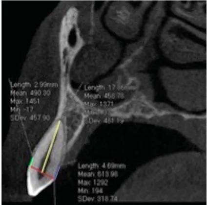
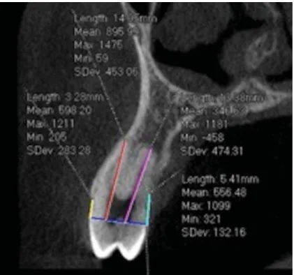
prem olars m easurem ent s ( Figures 1 and 2) .
Figure 1- 6DJLWWDOYLHZDVVHVVLQJURRWOHQJWK5/EXFFDODOYHRODUERQHOHYHO%$%/DQGSDODWDODOYHRODUERQHOHYHO3$%/PHDVXUHPHQWV <HOORZOLQH5/IURPWKHFHPHQWRHQDPHOMXQFWLRQ&(-WRURRWDSH[*UHHQOLQH%$%/IURPWKHFHPHQWRHQDPHOMXQFWLRQ&(-WR EXFFDOFUHVW%OXHOLQH3$%/IURPWKHFHPHQWRHQDPHOMXQFWLRQ&(-WRSDODWDOFUHVW5HGOLQH/LQHFRQQHFWLQJWKHFHPHQWRHQDPHO
St at ist ical analysis
I nt ra- exam iner syst em at ic errors w ere assessed
by apply ing pair ed t - t est and random er r or s w er e
analyzed wit h Dahlberg’s form ula. To verify t he norm al
dist ribut ion of t he variables, Kolm ogorov- Sm irnov t est
was used. Dat a present ed a norm al dist ribut ion. The
com par ison of r oot lengt h and alveolar bone level
changes bet w een groups was assessed by applying
SDLUHGWWHVWDWDVLJQL¿FDQFHOHYHORIS
All st at ist ical analyses w ere perform ed w it h St at ist ica
soft ware ( St at ist ica for Window s 5.0; St at soft , Tulsa,
OK, USA) .
Result s
Root lengt h
Regarding root length, results showed no statistically
VLJQL¿FDQWGLIIHUHQFHEHWZHHQJURXSV7DEOH7KH
highest st andard deviat ion of t he difference bet w een
t he m eans in t he t w o gr oups was found in canine
m easur em ent s ( 0 . 9 5 m m ) , and t he low est in t he
SDODWDOURRWRIWKH¿UVWSUHPRODUVPP:KHQ
com paring t he t w o groups, GI show ed decreased root
lengt h in 67.18% ( 0.63 m m ) of t he sam ple, w it h t he
largest reduct ion equivalent t o 26% of t he root lengt h
of t he cont ralat eral t oot h.
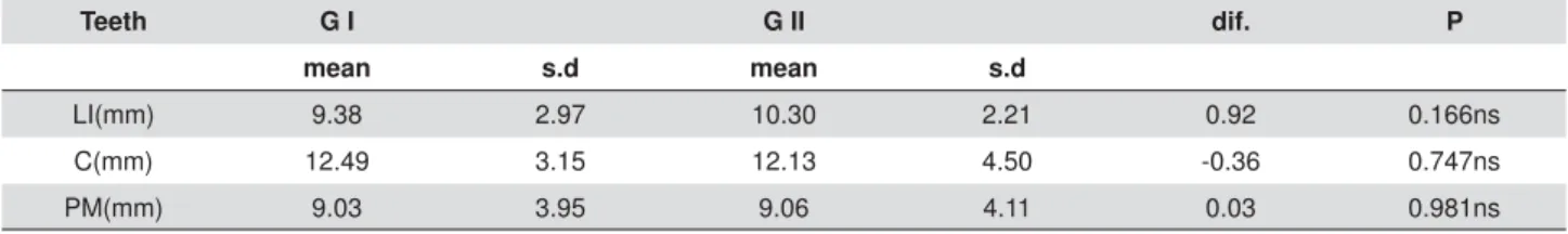
Alveolar bone level
St at ist ical an aly sis of t h e r esu lt s sh ow ed n o
VLJQL¿FDQW GLIIHUHQFH EHWZHHQ JURXSV 7KH KLJKHVW
Teeth G I G II dif. P
mean s.d mean s.d
LI(mm) 13.01 1.58 13.57 2.16 0.56 0.095ns
C(mm) 15.83 2.13 16.78 2.81 0.95 0.105ns
PM - B(mm) 13.77 1.62 14.41 1.77 0.64 0.083ns PM - P(mm) 13.65 1.57 14.05 1.85 0.40 0.322ns
QVQRQVLJQL¿FDQW&FDQLQH/,ODWHUDOLQFLVRU30%EXFFDOSUHPRODU303SDODWDOSUHPRODU Table 1- Intergroup comparison regarding root length measurements (paired t-test)
SDODWDOFUHVW3XUSOHOLQH/LQHFRQQHFWLQJWKHFHPHQWRHQDPHOMXQFWLRQ&(-st an dar d dev iat ion of t h e differ en ce bet w een t h e
m eans regarding t he buccal alveolar bone level ( BABL)
in t he t wo groups was 0.03 m m found in t he prem olars
( Table 2) and regarding t he palat al alveolar bone level
( PABL) was 0.39 m m , found in lat eral incisors ( Table
3) . When com paring GI and GI I , 56% of GI show ed
a decrease in BABL m easurem ent and 58.3% in t he
PABL m easurem ent . I n t hree prem olars, one in GI
( im pact ed) and t wo in GI I ( cont rol group) , t he absence
of buccal alveolar bone was not ed.
Discussion
Th e n eed of sig n if ican t t oot h m ov em en t an d
t he lengt hy or t hodont ic t r eat m ent associat ed w it h
forced erupt ion of ect opic canines m ay increase t he
suscept ibilit y t o root resorpt ion and alveolar bone level
changes in such pat ient s.
Consider ing t he sor t of sequelae involved in
ort hodont ic t ract ion of im pact ed canines, t he decision
of using CBCT im ages in t his st udy was based upon t he
higher accuracy and precision24 of t his m et hod w hen
com par ed w it h 2- D im ages. Fur t her m or e, int raoral
UDGLRJUDSKVKDYHGLVDGYDQWDJHVVXFKDVWKHGLI¿FXOW\
of st an d ar d izat ion an d t h e im ag e d ist or t ion1 9 , 2 6.
Moreover, it w ould not be possible t o visualize buccal
DQGSDODWDODOYHRODUERQHOHYHOVZKHQSHULDSLFDO¿OPV DUHWDNHQGXHWRWKHVXSHULPSRVLQJRILPDJHVDQG
in lat eral cephalogram s, due t o t he t eet h posit ions in
t he dent al arch. I n addit ion, such im ages do not allow
t he det ect ion of fenest rat ions.
To perform root lengt h m easurem ent , eit her an
ordinal scale ( 0- 4) could be em ployed or t he direct root
m easurem ent . Regarding t he periodont al condit ions,
HLWKHUWKHSURELQJSRFNHWGHSWK11 could be perform ed
or t he m easuring of t he alveolar crest using periapical
radiographs19. The m et hod chosen in t his st udy, t o
obt ain t he root lengt h and alveolar palat al bone and
level m easurem ent s, has been previously described by
.LP3DUNDQG.RRN21PRGL¿HGE\+DQGHOPDQ17 DQG%HFNPDQQHWDO4 ( 1998) , w hich used t he
cem ent- enam el j unct ion as reference. The accuracy of
t his m et hod has already been proven earlier and t he
r esult s of syst em at ic er r or assessm ent s, evaluat ed
E\ SDLUHG WWHVW VKRZHG QR VWDWLVWLFDOO\ VLJQL¿FDQW
difference, w it h except ion of t he buccal bone level of
t he lat eral incisors, and t he random error, assessed
by applying Dahlberg’s form ula was from 0.02 of t he
buccal alveolar bone level of t he canines t o 0.21 of
t he root lengt h from lat eral incisors.
Som e im port ant advant ages of t he closed- erupt ion
t echnique associat ed w it h a canine crow n perforat ion
SHUIRUPHGLQWKLVVWXG\DUHKLJKOLJKWHGUHGXFHGULVN
of a new surgical procedure; less t issue m anipulat ion,
HVSHFLDOO\WKHGHQWDOIROOLFOHLPSRUWDQWIUDPHZRUNIRU
t oot h erupt ion) and a m echanical advant age of allowing
t he applicat ion of force in t he long axis of t he t eet h. A
segm ent ed arch and t he use of t ranspalat al arch as an
anchorage device was t he select ed m echanics for t he
ort hodont ic t ract ion of im pact ed canines, as proposed
by Lindauer and I ssacson23 ( 1995) . The canines w ere
guided t o erupt on t he palat e avoiding t he cont act wit h
WKHURRWVRIDGMDFHQWWHHWKDQGWKXVUHGXFLQJWKHULVN
of root resorpt ion.
Teeth G I G II dif. P
mean s.d mean s.d
LI(mm) 9.38 2.97 10.30 2.21 0.92 0.166ns
C(mm) 12.49 3.15 12.13 4.50 -0.36 0.747ns
PM(mm) 9.03 3.95 9.06 4.11 0.03 0.981ns
QVQRQVLJQL¿FDQW&FDQLQH/,ODWHUDOLQFLVRU30SUHPRODU
Table 2- Intergroup comparison regarding buccal bone level measurements (paired t-test)
Teeth G I G II dif. P
mean s.d mean s.d
LI(mm) 11.15 1.29 11.54 1.99 0.39 0.280ns
C(mm) 13.43 2.43 14.26 3.13 0.83 0.163ns
PM(mm) 10.86 2.02 10.14 2.77 -0.72 0.135ns
QVQRQVLJQL¿FDQW&FDQLQH/,ODWHUDOLQFLVRU30SUHPRODU
Despit e t he result s of t his st udy had point ed out t o
a m inim al decrease in t he root lengt h m easurem ent s
of canines and adj acent t eet h from GI com pared w it h
*,,QRVWDWLVWLFDOVLJQL¿FDQWGLIIHUHQFHZDVIRXQG7KLV VOLJKWGLIIHUHQFHEHVLGHVQRWVWDWLVWLFDOO\VLJQL¿FDQW KDVQRFOLQLFDOVLJQL¿FDQFHVLQFHWKHPD[LPXPYDOXH
of t he com parison bet ween sides was 0.95 m m . These
UHVXOWVFRXOGEHLQÀXHQFHGE\OLJKWIRUFHVDSSOLFDWLRQ
during t he t ract ion m echanics.
Th ese f in d in g s r ein f or ce t h e r esu lt s f ou n d b y
Brusveen, et al.8 ( 2012) and Lem pesi, et al.22 ( 2014) WKDW WKHUH LV QR VWDWLVWLFDOO\ VLJQL¿FDQW GLIIHUHQFH
am ong t he groups. These aut hors, however, evaluat ed
only t he root lengt h of t he incisors. Woloshyn, et al.31 DQG 6FKPLGW DQG .RNLFK28 ( 2007) evaluat ed
t h r o u g h p er i a p i ca l r a d i o g r a p h s t h e r o o t l en g t h
RI WKH LQFLVRUV FDQLQHV DQG ¿UVW SUHPRODUV 7KH
only difference bet w een t hese t w o st udies was t he
VXUJLFDODSSURDFKLQWKH¿UVWVWXG\WKH\DGRSWHGWKH
closed- erupt ion t echnique and ort hodont ic t ract ion,
sim ilar ly t o our st udy, and in t he second one, an
DSLFDOO\SRVLWLRQHGÀDSZLWKRXWRUWKRGRQWLFWUDFWLRQ
Co r r o b o r at i n g o u r r esu l t s, t h e au t h o r s f o u n d a
sm all decrease in root lengt h, but w it hout st at ist ical
VLJQL¿FDQFH+RZHYHUWKHURRWOHQJWKRISUHPRODUVLQ WKHVWXG\RI6FKPLGWDQG.RNLFK28 ( 2007) , present ed
sim ilar result s am ong groups. A lim it at ion in t hese
st udies was t he use of periapical radiographs, w hich
FRXOGLQÀXHQFHWKHUHVXOWVEHFDXVHRIWKHRYHUODSSLQJ
of buccal and palat al root s. Anot her difference bet ween
RXUV DQG 6FKPLGW DQG .RNLFK¶V VWXG\28 ( 2007) was WKH DSLFDOO\ SRVLWLRQHG ÀDS WHFKQLTXH HPSOR\HG RQ
t hat research. We underst and t hat not all canines can
EH WUHDWHG E\ DSLFDO SRVLWLRQLQJ RI WKH ÀDS ZLWKRXW
ort hodont ic t ract ion, part icularly t hose m ost ect opic
posit ioned, in which ort hodont ic t ract ion is a challenge
IRUWKHRUWKRGRQWLVWDQGPD\DOVRLQÀXHQFHRQWKHURRW
resorpt ion induced by ort hodont ic t reat m ent22.
Regar ding t he buccal and palat al alveolar bone
level, alt hough our result s show ed t hat com paring GI
and GI I , 56% of GI show ed a decrease in BABL, t his
GLIIHUHQFHZDVQRWFRQVLGHUHGVWDWLVWLFDOO\VLJQL¿FDQW
These result s are sim ilar t o t hose report ed by Schm idt
DQG.RNLFK28 ( 2007) w hen evaluat ing t he m esial and
dist al bone level using per iapical radiographs. The
VDPH UHVXOWV UHJDUGLQJ SHULRGRQWDO ¿QDO FRQGLWLRQ
( probing dept h) w ere found by Caprioglio, Vanni and
Bolam per t i1 1 ( 2 0 1 3 ) evaluat ing palat ally im pact ed
canines t hat suffer ed t ract ion. On t he ot her hand,
%HFNHU DQG &KDXVKX3 ( 2 0 0 5 ) an d Ev r en , et al.1 6
( 2014) , assessing t he m esial and dist al bone level
in canines t hat suffer ed t ract ion, also in per iapical
UDGLRJUDSKVIRXQGDVWDWLVWLFDOO\VLJQL¿FDQWERQHORVV
am ong groups. On t he lat est , not only palat ally, but
buccally displaced canines present ed reduced bone
levels com pared w it h t heir cont ralat erals. I t should
be highlight ed t hat palat ally displaced canines, w hen
suffering t ract ion, m ay not com prom ise t he periodont al
st at us as t hose buccally displaced.
6RPHFRQÀLFWLQJ¿QGLQJVFRXOGEHDFFRXQWHGE\WKH
m et hod of evaluat ion, eit her CBCT scans or periapical
radiograph, w hich could st and for such differ ence.
Also, t he t ract ion pr ot ocol and t he init ial posit ion
RI WKH FDQLQHV FRXOG LQÀXHQFH WKHVH UHVXOWV VLQFH
t he role of adequat e oral hygiene during appliance
WKHUDS\PD\EHVLJQL¿FDQW6,16,18. More reliable result s
sh ou ld be ach iev ed in per f or m in g m easu r em en t s
in CBCT scans, but in t w o different periods, before
and aft er t reat m ent . The lim it at ions of t his st udy are
WKDWRQO\WKH¿QDO&%&7LPDJHVZHUHDYDLODEOHDQG
it s r et r ospect iv e design. But ex posing pat ient s t o
unnecessary radiat ion should also be avoided, even
considering only t he m axillary area. Ot her lim it at ion
of our st udy is t hat t he sam ple size seem ed sm all ( 16
subj ect s) , but t he sam ple size calculat ion showed t hat
10 subj ect s were necessary t o achieve reliable result s.
I t is im port ant t o em phasize t hat an early diagnosis
is always bet t er t o prevent irreversible dam ages t o t he
involved and adj acent t eet h9,12. Even aft er an early
diagnosis, in som e cases it is necessary t o perform
t oot h t ract ion. Besides t hat , according t o t his research
we can st at e t hat t he t ract ion prot ocol associat ed wit h
t he ort hodont ic correct ive t reat m ent did not negat ively
affect t he periodont al st at us and t he root lengt h of t he
im pact ed canines and adj acent t eet h.
Conclusion
The t reat m ent of im pact ed canines had m inim al
effect on root lengt h and buccal and palat al alveolar
bon e lev els, n ot on ly in or t h odon t ic can in es t h at
suffered t ract ion, but also in adj acent t eet h ( lat eral
LQFLVRUDQG¿UVWSUHPRODUGHPRQVWUDWLQJDJRRGORQJ
References
1- Baccet t i T. A cont rolled st udy as associat ed dent al anom alies. Angle Ort hod. 1998; 68( 3) : 267- 7.
2- Bayram M, Ozer M, Sener I . Maxillary canine im pact ions relat ed t o im pact ed cent ral incisors: t w o case report s. J Cont em p Dent Pract . 2007; 8( 6) : 72- 81.
%HFNHU$&KDXVKX6/RQJWHUPIROORZXSRIVHYHUHO\UHVRUEHG
m axillary incisors aft er resolut ion of an et iologically associat ed im pact ed canine. Am J Ort hod Dent ofacial Ort hop. 2005; 127: 650- 4.
%HFNPDQQ6+.XLWHUW5%3UDKO$QGHUVHQ%6HJQHU'7KH53 7XLQ]LQJ'%$OYHRODUDQGVNHOHWDOGLPHQVLRQVDVVRFLDWHGZLWKORZHU
face height . Am J Ort hod Dent ofacial Ort hop. 1998; 113: 498- 506. 5- Bishara SE. I m pact ed m axillary canines: a review. Am J Ort hod Dent ofacial Ort hop. 1992; 101: 159- 71.
6- Bishara SE. Clinical m anagem ent of im pact ed m axillary canines. Sem in Ort hod. 1998; 4: 87- 98.
%URZQ /+ %HUNPDQ 6 &RKHQ ' .DSODQ $/ 5RVHQEHUJ 0 $
radiological st udy of t he frequency and dist ribut ion of im pact ed t eet h. J Dent Assoc S Afr. 1982; 37: 627- 30.
%UXVYHHQ(0%UXGYLN3%¡H2(0DYUDJDQL0$SLFDOURRWUHVRUSWLRQ
of incisors aft er ort hodont ic t reat m ent of im pact ed m axillary canines: a radiographic st udy. Am J Ort hod Dent ofacial Ort hop. 2012; 141: 427- 35. 9- Cam pbell CM, DiBiase A, Flem ing PS. Concom it ant dilacerat ion, t ransposit ion, and int raosseous m igrat ion: report of a pat ient t reat ed w it h m ax illar y can in e- cen t r al in cisor su b st it u t ion . Am J Or t h od Dent ofacial Ort hop. 2014; 146: 514- 21.
10- Capelozza Filho L, Consolaro A, Cardoso MA, Siqueira DF. Enam el dr illing for canine t ract ion: advant ages, disadvant ages, descr ipt ion of su r gical t ech n iqu e an d biom ech an ics. Den t al Pr ess J Or t h od. 2011; 16: 172- 205.
11- Caprioglio A, Vanni A, Bolam pert i L. Long t erm periodont al response t o ort hodont ic t reat m ent of palat ally im pact ed m axillary canines. Eur J Ort hod. 2013; 35( 3) : 323- 8.
12- Cavuot i S, Mat arese G, I sola G, Abdolreza J, Fem iano F, Perillo L. Com bined or t hodont ic- sur gical m anagem ent of a t ransm igrat ed m andibular canine. Angle Ort hod. 2016; 86: 681- 91.
13- Ericson S, Kurol J. Radiographic assessm ent of m axillary canine erupt ion in children w it h clinical signs of erupt ion dist urbance. Eur J Ort hod. 1986; 8: 133- 40.
14- Ericson S, Kurol J. Resorpt ion of m axillary lat eral incisors caused by ect opic erupt ion of t he canines. A clinical and radiographic analysis of predisposing fact ors. Am J Ort hod Dent ofacial Ort hop. 1988; 94: 503- 13. 15- Ericson S, Kurol J. Resorpt ion of incisors aft er ect opic erupt ion of m axillary canines: a CT st udy. Angle Ort hod. 2000; 70: 415- 23.
(YUHQ'$1HY]DWR÷OXù$UXQ7$FDU$3HULRGRQWDOVWDWXVRIHFWRSLF
canines aft er ort hodont ic t reat m ent . Angle Ort hod. 2014; 84: 18- 23.
17- Handelm an CS. The ant erior alveolus: it s im port ance in lim it ing
RUWKRGRQWLFWUHDWPHQWDQGLWVLQÀXHQFHRQWKHRFFXUUHQFHRILDWURJHQLF
sequelae. Angle Ort hod. 1996; 66: 95- 109.
18- Hansson C, Rindler A. Periodont al condit ions follow ing surgical and ort hodont ic t reat m ent of palat ally im pact ed m axillary canines: a follow- up st udy. Angle Ort hod 1998; 68: 167- 72.
19- Janson G, Bom bonat t i R, Brandão AG, Henriques JF, Freit as MR. Com parat ive radiographic evaluat ion of t he alveolar bone crest aft er ort hodont ic t reat m ent . Am J Ort hod Dent ofacial Ort hop. 2003; 124: 157-64.
2 0 - Kim Y, Hy u n HK, Jan g KT. Th e posit ion of m ax illar y can in e
LPSDFWLRQVDQGWKHLQÀXHQFHGIDFWRUVWRDGMDFHQWURRWUHVRUSWLRQLQ
t he Korean populat ion. Eur J Ort hod. 2012; 34: 302- 6.
.LP<3DUN-8.RRN<$$OYHRODUERQHORVVDURXQGLQFLVRUVLQ VXUJLFDOVNHOHWDO&ODVV,,,SDWLHQWV$QJOH2UWKRG
22- Lem pesi E, Pandis N, Flem ing PS, Mavragani M. A com parison of apical r oot r esor pt ion aft er or t hodont ic t r eat m ent w it h sur gical exposure and t ract ion of m axillary im pact ed canines versus t hat wit hout im pact ions. Eur J Ort hod. 2014; 36: 690- 7.
2 3 - Lin dau er SJ, I ssacson RJ. On e- cou ple or t h odon t ic applian ce syst em s. Sem in Ort hod. 1995; 1: 12- 24.
2 4 - Lu n d H, Gr ön d ah l K, Gr ön d ah l HG. Con e b eam com p u t ed t om ography for assessm ent of root lengt h and m arginal bone level during ort hodont ic t reat m ent . Angle Ort hod. 2010; 80: 466- 73. 25- Lund H, Gröndahl K, Hansen K, Gröndahl HG. Apical root resorpt ion during ort hodont ic t reat m ent : a prospect ive st udy using cone beam CT. Angle Ort hod. 2012; 82: 480- 7.
/XSL-(+DQGHOPDQ&66DGRZVN\&3UHYDOHQFHDQGVHYHULW\RI
apical root resorpt ion and alveolar bone loss in ort hodont ically t reat ed adult s. Am J Ort hod Dent ofacial Ort hop. 1996; 109: 28- 37.
3HFN 6 3HFN / .DWDMD 0 &RQFRPLWDQW RFFXUUHQFH RI FDQLQH PDOSRVLWLRQDQGWRRWKDJHQHVLVHYLGHQFHRIRURIDFLDOJHQHWLF¿HOGV
Am J Ort hod Dent ofacial Ort hop. 2002; 122: 657- 60.
6FKPLGW$'.RNLFK9*3HULRGRQWDOUHVSRQVHWRHDUO\XQFRYHULQJ
aut onom ous erupt ion, and ort hodont ic alignm ent of palat ally im pact ed m axillary canines. Am J Ort hod Dent ofacial Ort hop. 2007; 131: 449- 55.
6WDKO)*UDERZVN\50D[LOODU\FDQLQHGLVSODFHPHQWDQGJHQHWLFDOO\
det erm ined predisposit ion t o dist urbed developm ent of t he dent it ion. J Orofac Ort hop. 2003; 64: 167- 77.
30- Thilander B, Myrberg N. The prevalence of m alocclusion in Swedish schoolchildren. Eur J Oral Sci. 2007; 81: 12- 20.
31- Woloshy n H, Ar t un J, Kennedy DB, Joondeph DR. Pulpal and periodont al react ions t o ort hodont ic alignm ent of palat ally im pact ed canines. Angle Ort hod. 1994; 64: 257- 64.
32- Yan B, Sun Z, Fields H, Wang L. Maxillary canine im pact ion increases
URRWUHVRUSWLRQULVNRIDGMDFHQWWHHWKDSUREOHPRISK\VLFDOSUR[LPLW\