J. Meenupriya et al. Int. Res. J. Pharm. 2013, 4 (4)
Page 224
INTERNATIONAL RESEARCH JOURNAL OF PHARMACY
www.irjponline.com ISSN 2230 – 8407
Research Article
IMMUNOLOGICAL STUDY OF SPONGIFORMENCEPHALOPATHIES J. Meenupriya*
Assistant Professor, Department of Biotechnology, Sathyabama University, Chennai, India Email: [email protected]
Article Received on: 20/02/13 Revised on: 11/03/13 Approved for publication: 11/04/13
DOI: 10.7897/2230-8407.04445
IRJP is an official publication of Moksha Publishing House. Website: www.mokshaph.com
© All rights reserved.
ABSTRACT
Spongiform encephalopathies, categorized as a subclass of neuro-degenerative diseases and commonly known as prion diseases, are a group of progressive conditions that affect the brain and nervous system of many animals, including humans. Prion diseases are common among cannibalistic communities; further research has revealed that the infected or malformed prion protein (named PrPsc) spreads its virulence to the normal, healthy prion protein (named PrPc) when people consume infected tissues. Knowing that a small interaction between normal and infected prion protein creates virulence, this relationship can be studied as a simple antigen-antibody interaction to understand the series of events that transform a normal prion protein into a virulent misfolded protein. Thoroughly modeled and validated structures of both PrPsc and PrPc can be effectively used to map the epitopes and thereby screen the antigen-antibody interaction using docking studies for a particular organism of concern. This simple immunological approach is used to understand the vital interaction between the normal and malformed proteins that is involved in the disease-spreading mechanism. Clarification of this mechanism could be used in various immune- and bioinformatics algorithms to map the interaction epitopes, furthering an understanding of these pathologies.
KEYWORDS: Spongiform encephalopathies, Prions, Antigen antibody interaction, Epitope and Docking.
INTRODUCTION
The most widespread hypothesis is that Spongiform Encephalopathies are transmitted by prions, though some other data suggest an involvement of a Spiroplasma
infection1. Mental and physical abilities deteriorate and myriad tiny holes appear in the cortex causing it to appear like a sponge (hence 'spongiform') when brain tissue obtained at autopsy is examined under a microscope. Unlike other kinds of infectious disease which are spread by microbes, the infectious agent in SEs is a specific protein called prion protein. Misshaped prion proteins carry the disease between individuals and cause deterioration of the brain. SEs are unique diseases in that their aetiology may be genetic, sporadic or infectious via ingestion of infected foodstuffs and via iatrogenic means (e.g. blood transfusion)2
.
Prions are small protein fibers present in brain which under deformed conditions form plaque or hole-like structures in brain resulting in loss of memory, speech, vision, cognitive abilities and finally leading to death. They are unprecedented infectious pathogens that cause a group of invariably fatal neurodegenerative diseases by an entirely novel mechanism. Prion diseases may present as genetic, infectious, or sporadic disorders, all of which involve modification of the prion protein (PrP)3.
PrPC is a normal protein found on the membranes of cells. It has 209 amino acids (in humans), one disulfide bond, a molecular weight of 35-36 kDa and a mainly alpha- helical structure4.Its function is a complex issue that continues to be investigated. PrPC binds copper (II) ions with high affinity5.The significance of this finding is not clear, but it presumably relates to PrP structure or function. PrPC is readily digested by proteinase K and can be liberated from the cell surface in vitro by the enzyme phosphoinositide phospholipase C (PI-PLC), which cleaves the glycophosphatidylinositol (GPI) glycolipid anchor6. PrP has been reported to play important roles in cell-cell adhesion and intracellular signaling in vivo, and may therefore be involved in cell-cell communication in the brain7.
The infectious isoform of PrP, known as PrPSc, is able to convert normal PrPC proteins into the infectious isoform by changing their conformation, or shape; this, in turn, alters the way the proteins interconnect8. Aggregations of these abnormal isoforms form highly structured amyloid fibers which accumulate to form plaques. The end of each fiber acts as a template onto which free protein molecules may attach, allowing the fiber to grow. Only PrP molecules with an identical amino acid sequence to the infectious PrPSc are incorporated into the growing fiber.
From the KEGG pathway it can be observed that Lamin, glial fibrillary acidic protein, and other accessory proteins combine to Prion protein which then combines with Lamin receptor under the influence of some heat shock proteins but during the disease condition it undergoes a simple conformational change and it forms a new complex named PrPsc which in turn activates the microglial astrocyte there by leading to the formation of tumor necrosis factor and interleukin 6 which aids in the process mitochondrial dysfunction. From this study it can be narrowed down that Prion protein might be a potential causative factor which does not undergo any sort of mutation but just a small conformational change that plays the key role. The identification and characterization of B-cell epitopes play an important role in vaccine design, immune diagnostic tests and antibody production. Therefore, computational tools for reliably predicting linear B-cell epitopes in protein sequences are highly desirable9. This
present study will help to find interaction between a complex Prion protein and a misfolded prion protein using docking studies. Knowledge obtained using ISSE would help researchers gain more insight about the prions disease and the factors responsible for its virulence and would prove effective and useful in further research and studies.
MATERIALS AND METHODS
J. Meenupriya et al. Int. Res. J. Pharm. 2013, 4 (4)
Page 225
information were retrieved from KEGG and uniprot database respectively for analysis.
Template selection for the target molecule PrPC and PrPSc protein
The template may be a predefined layout to give an idea about the unknown structure of the query molecule. FASTA sequence of the prion proteins are obtained from UNIPROT and it was blasted against PDB database using BLASTP tool. The pdb file of the particular template sequence was downloaded from the protein data bank (www.rcsb.org).
Homology modeling and structure refinement
Homology modeling is based on the reasonable assumption that two homologous proteins will share very similar structures11. Swiss-Pdb Viewer has been developed since 1994 by Nicolas Guex. Swiss-Pdb Viewer is tightly linked to SWISS-MODEL, an automated homology modeling server developed within the Swiss Institute of Bioinformatics (SIB) at the Structural Bioinformatics Group at the Biozentrum in Basel. It is designed for full compatibility with computing tools available from the Expert Protein Analysis System, or ExPASy. Molecular Biology Server in Geneva, Switzerland. While Deep View is simple to use for viewing structures and creating vivid illustrations, it also shines as an analytical tool. Deep View allows you to build models from scratch, simply by giving an amino-acid sequence. Deep View can find hydrogen bonds within proteins and between proteins and ligands Swiss-Pdb Viewer (aka Deep View) is an application that provides a user friendly interface allowing to analyze several proteins at the same time12. The target protein was modeled using Swiss-PDB Viewer (or SPDBV). The overall quality factor is enhanced by Ramachandran plot. Structure Refinement is the process of improvement of the model structure with minimum errors.
Target 3D structure validation
Validation of the structures is a mandatory step since it determines the quality of the structure and it also determines all the factors that deviate from the normality and present the best possible outcome for the next step. The 3D validation of the predicted homology structure of the target molecule can be assessed by the What if server which is a versatile molecular modeling package that is specialized on working with proteins and the molecules in their environment like water, ligands, nucleic acids, etc. it checks the validity of the structure based on 11 different properties.
Epitope mapping and conservancy analysis
Epitope mapping is the process of identification and characterization of the minimum molecular structures that are able to be recognized by the Immune System elements. Bcepred (Prediction of linear B-cell epitopes, using physico-chemical properties) evaluates the performance of existing linear B-cell epitope prediction methods based on physico-chemical properties on a non-redundant dataset. The dataset consists of 1029 B-cell epitopes obtained from Bcipep database and equally number of non-epitopes obtained randomly from Swiss-Prot database. The prediction accuracy for models based of various properties varies from 52.92% and 57.53%. Bcepred achieves highest accuracy of 58.70% at threshold 2.38, when we combined four amino acid properties
(hydrophilicity, flexibility, polarity and exposed surface).
Receptor protein’s structure and functional analysis Before going into the analysis of the epitopes and interaction studies one needs to understand the basic primary sequence information, secondary structure information about the protein especially prion protein since the structure related information is more important to us since our major study of interest lies within the conformation change that occurs in Prion protein that transforms itself into a virulent isoform. The analsis were done with the tools such as ProtParam, Repro, COILS, and SOPMA. Tool like InterPro was used to identify the gene ontologies and protein domain and family information.
When protein changes it conformation then the key domains that played a significant role in it might also get altered leading to the loss of that particular function at gene level or might be cellular level. To understand the Functional analysis of the receptor protein the sequence was submitted in the tool PFP Protein Function Prediction server designed to predict GO annotations for a query protein sequence beyond what can be found by searching conventional databases. The PFP algorithm has been shown to increase coverage of sequence-based function annotation more than fivefold by extending a PSI-BLAST search to extract and score GO terms individually and include information from distantly related sequences. It applies a novel data mining tool, the Function Association Matrix (FAM), to score significantly associating pairs of annotations.
Interaction studies using docking
Docking is a method which predicts the preferred orientation of one molecule to a second when bound to each other to form a stable complex1 3. Docking is frequently used to predict the binding orientation of small molecule drug candidates to their protein targets in order to predict the affinity and activity of the small molecule. Hence docking plays an important role in designing of drug14. Amongst the various tools available for docking procedure HEX was chosen as the most apt and the best one due to its simplicity and efficiency in delivering the best result. Hex is an interactive protein docking and molecular superposition program, written by Dave Ritchie. Hexunderstands protein and DNA structures in PDB format.
RESULTS AND DISCUSSION
The prion protein sequence information retrieved from uniprot database given in Table 1 is as follows: The accession number of the target protein is BAG32276.
Table 1: Sequence information of PRION protein retrieved from Uniprot
Entry name PRIO_BOVIN
Primary accession number
BAG32276
Protein name Major prion protein Synonyms PrP Major scrapie-associated
fibril protein 1CD230 antigen Gene name Name: PRNP Synonyms: PRP
From Homo sapiens
J. Meenupriya et al. Int. Res. J. Pharm. 2013, 4 (4)
Page 226
SRPIIHFGSDYEDRYYRENMHRYPNQVYYRPMDEYS NQNNFVHDCVNITIKQHTVTTTTKGENFTET
DVKMMERVVEQMCITQYERESQAYYQRGSSMVLFSS PPVILLISFLIFLIVG
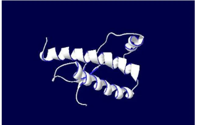
Homology Modeling of the Query PrPC and PrPSc protein
Figure 2. Modeled structure of PrPCprotein obtained from SWISS
MODEL Work Space
PrPSc is an isoform of prion protein where there is no
sequence-wise difference between PrPC and PrPSc. The main difference between these two forms is merely structural and nothing concerned with the sequence. This makes the modelling of PrPSc structure pretty difficult using in-silico technologies so this structure has to be obtained only from experimental techniques like x-ray
crystallography, so this structure is obtained from PDB directly. These structures are experimentally verified and are thoroughly validated so these won’t be any need to revalidate them. The experimentally verified structure of PrPSc obtained from Protein Data Bank is shown in figure 3.
Figure 3: Crystal structure of the prion protein namely PrPSc
obtained from PDB.
Ramachandran plot evaluation
Result obtained from What-If server using the PrPC
structure as input which reveals the Ramachandran Z- score was found to be -0.844 which was very normal so the structure is perfectly fit for further studies.
Mapping of Epitope sequences
Table 2: The most efficient epitopes mapped using BCEPRED along with their corresponding scores and starting position
Epitope Score Starting position
[NQVYYRPVDQYNNQNN] 0.93 159
[TGGSRYPGQGSPGGNR] 0.89 33
Output of IEDB conservancy analysis
Figure 3 Output for IEDB conservancy analysis.
Ligand molecule
The b-cell epitopes mapped from the antigen is extracted from PrPsc structure and the structure of this epitope is identified using SPDBV viewer. The epitope sequences were selected from the antigen protein and other residues were removed.
J. Meenupriya et al. Int. Res. J. Pharm. 2013, 4 (4)
Page 227
Docking result
Figure 5: Final output that shows the Docked prion protein
in “HARMONIC SURFACES” and in “SOLID SURFACES” view
of HEX docking having a E value of -366.29.
CONCLUSION
Immunological Study of Spongifrom Encephalopathy (ISSE) is a simple approach to study an interaction between a complex Prion protein and a misfolded prion protein using docking studies. Here B-cell epitopes are used since the mode of immune reaction followed by Prions disease is Humoral mediated. Mapping of epitopes is done with the help of BCEPRED since it used physiochemical properties to map the epitopes. It uses 5 different algorithms to map the epitopes based on their physiochemical properties. In order to refine the epitopes identified through epitope mapping, epitope conservancy analysis was done to check the conservancy of the epitope using IEDB epitope conservancy analysis tool. Docking is done with the most conserved epitope as ligand and the normal prion protein as receptor. This is a relatively small idea to simulate the interaction between the normal and misfolded protein sequence. Only epitope molecule is used for docking since it’s the b-cell epitope and only this epitope interacts or comes in contact with normal prion protein and not the
entire misfolded protein. This methodology identifies the conformational changes that could happen with prion protein and hence studies the events that could have probably occurred when the normal prion and abnormal prion protein interacts with each other.
REFERENCES
1. Devi KV, Pai RS. Antiretrovirals: Need for an Effective Drug Delivery. Indian J PharmSci 2006; 68:1-6. http://dx.doi.org/10.4103/0250-474X.22955
2. Shen HM, Zhang QF. Risk assessment of nickel carcinogenicity and occupational lung cancer. Environ Health Perspect 1994; 102 Suppl 1:275-82. http://dx.doi.org/10.1289/ehp.94102s1275
3. Payne DK, Sullivan MD, Massie MJ. Women's psychological reactions to breast cancer. Semin Oncol 1996; 23(1, Suppl 2):89-97.
4. Bastian FO, Sanders DE, Forbes WA, Hagius SD, Walker JV, Henk WG, Enright FM, Elzer PH. Spiroplasma spp. from transmissible spongiform encephalopathy brains or ticks induce spongiform encephalopathy in ruminants.J Med Microbiol. 2007;56 Suppl 9: 1235–
1242. http://dx.doi.org/10.1099/jmm.0.47159-0
5. Brown P, Preece M, Brandel JP, Sato T, McShane L, Zerr I, Fletcher A, Will RG, Pocchiari M, Cashman NR, Aignaux JH, Cervenakova L, Fradkin J, Schonberger LB, Collins SJ. Iatrogenic Creutzfeldt-Jakob disease at the millennium. Neurology 2000; 55 Suppl 8: 1075–81. http://dx.doi.org/10.1212/WNL.55.8.1075
6. Stanley B. Prusiner. Prions. PNAS.1998;95 Suppl 23 :13363-13383. 7. Hegde RS, Mastrianni JA, Scott MR, Defea KA, Tremblay P, Torchia
M, DeArmond SJ, Prusiner SB, Lingappa VR.A transmembrane form of the prion protein in neurodegenerative disease. Science 1998; 276: 827–
834. http://dx.doi.org/10.1126/science.279.5352.827
8. Brown DR. The cellular prion protein binds copper in vivo.Nature.1997; 390:684–687. http://dx.doi.org/10.1038/37733
9. Weissmann C. The State of the Prion. Nature Reviews Microbiology.2004; 2: 861–871. http://dx.doi.org/10.1038/nrmicro1025 10. Malaga E. Regulation of Embryonic Cell Adhesion by the Prion Protein.
PLoS Biology. 2009; 7: 0576-0590.
11. Chen J, Liu H, Yang J, Chou K. Prediction of linear B-cell epitopes using amino acid pair antigenicity scale. Amino Acids.2007; 33: 423-428. http://dx.doi.org/10.1007/s00726-006-0485-9
12. Qing Z, Peng W, Yohan K, Pernille H, John B,Philip B, Huynh B, Soren B,Sune F,Jason G,Ole L, Claus L, Morten N, Julia P, Alessandro S, Zhanyang Z, Bjoern P. Immune epitope database analysis resource (IEDB-AR).Nucleic Acids Research. 2008;36:513–518. http:// dx.doi.org/ 10.1093/ nar/gkn254
13. Zhang Y. Progress and challenges in protein structure prediction. Curr Opin Struct Biol.2008;18 (3): 342–348. http://dx.doi.org/10.1016 /j.sbi.2008.02.004
14. Macintosh PC, Guex N, Peitsch MC.A Fast and Easy-to-use PDB Viewer. Bioinformatics.1996; 7:77-81.
15. Lengauer T, Rarey M. Computational methods for biomolecular docking.Curr. Opin. Struct. Biol.;1996;6 suppl 3: 402–6. http://dx.doi.org/10.1016 /S0959-440X(96)80061-3
16. Kitchen DB, Decornez H, Furr JR, Bajorath J. Docking and scoring in virtual screening for drug discovery: methods and applications. Nature reviews. Drug discovery .2004;3 Suppl 11: 935–49. http://dx.doi.org/ 10.1038/nrd1549
Cite this article as:
J. Meenupriya. Immunological study of Spongiform encephalopathies. Int. Res. J. Pharm. 2013; 4(4):224-227