Oral Microbiota Shift after 12-Week
Supplementation with
Lactobacillus reuteri
DSM 17938 and PTA 5289; A Randomized
Control Trial
Nelly Romani Vestman1, Tsute Chen2, Pernilla Lif Holgerson3, Carina Öhman1,
Ingegerd Johansson1
*
1Department of Odontology/section of Cariology, UmeåUniversity, Umeå, Sweden,2Department of
Microbiology, The Forsyth Institute, Cambridge, United States of America,3Department of Odontology/ section of Pedodontics, UmeåUniversity, Umeå, Sweden
Abstract
Background
Lactobacillusspp. potentially contribute to health by modulating bacterial biofilm formation, but their effects on the overall oral microbiota remain unclear.
Methods and Findings
Oral microbiota was characterized via 454-pyrosequencing of the 16S rDNA hypervariable region V3-V4 after 12 weeks of dailyLactobacillus reuteriDSM 17938 and PTA 5289 con-sumption. Forty-four adults were assigned to a test group (n = 22) that received lactobacilli lozenges (108CFU of each strain/lozenge) or a control group that received placebo (n =
22). Presence ofL.reuteriwas confirmed by cultivation and species specific PCR. Tooth biofilm samples from 16 adults before, during, and after exposure were analyzed by pyrose-quencing. A total of 1,310,292 sequences were quality filtered. After removing single reads, 257 species or phylotypes were identified at 98.5% identity in the Human Oral Microbiome Database.Firmicutes,Bacteroidetes,Fusobacteria,Proteobacteria, and
Actinobacteria-were the most abundant phyla.Streptococcuswas the most common genus and theS. ora-lis/S.mitis/S.mitis bv2/S.infantisgroup comprised the dominant species. The number of observed species was unaffected byL.reuteriexposure. However, subjects who had con-sumedL.reuteriwere clustered in a principal coordinates analysis relative to scattering at baseline, and multivariate modeling of pyrosequencing microbiota, and culture and PCR de-tectedL.reuteriseparated baseline from 12-week samples in test subjects.L.reuteriintake correlated with increasedS.oralis/S.mitis/S.mitis bv2/S.infantisgroup andCampylobacter concisus,Granulicatella adiacens,Bergeyella sp. HOT322,Neisseria subflava,and SR1 [G-1] sp. HOT874 detection and reducedS.mutans,S.anginosus,N.mucosa,Fusobacterium
OPEN ACCESS
Citation:Romani Vestman N, Chen T, Lif Holgerson P, Öhman C, Johansson I (2015) Oral Microbiota Shift after 12-Week Supplementation with Lactobacillus reuteriDSM 17938 and PTA 5289; A Randomized Control Trial. PLoS ONE 10(5): e0125812. doi:10.1371/journal.pone.0125812
Academic Editor:Jan S Suchodolski, GI Lab, UNITED STATES
Received:December 11, 2014
Accepted:March 13, 2015
Published:May 6, 2015
Copyright:© 2015 Romani Vestman et al. This is an open access article distributed under the terms of the Creative Commons Attribution License, which permits unrestricted use, distribution, and reproduction in any medium, provided the original author and source are credited.
Data Availability Statement:The original read sequences and the OUT table is available athttp://dx. doi.org/10.6084/m9.figshare.1352069.
periodicum,F.nucleatum ss vincentii, andPrevotella maculosadetection. This effect had disappeared 1 month after exposure was terminated.
Conclusions
L.reutericonsumption did not affect species richness but induced a shift in the oral micro-biota composition. The biological relevance of this remains to be elucidated.
Trial Registration
ClinicalTrials.govNCT02311218
Introduction
The oral cavity provides distinct niches for bacterial communities, including saliva, gingival fluid, and food-flushed keratinized/non-keratinized epithelial or mineralized tooth surfaces
[1]. These communities harbor both disease- and health-associated species. In the oral cavity,
approximately 700 prevalent taxa have been identified in the mouth [www.homd.org; 2];
how-ever, the core microbiome is dominated byFirmicutes, which includes both streptococci and
lactobacilli [2]. Lactobacilli, which are commonly used in health promoting products
(probiot-ics) [3] and industrial and artisanal food and dairy fermentation [4], have been reported to
in-fluence immune responses, nutrition, and overall wellbeing [5]. Moreover, Lactobacilli are
characterized by lactic acid production and a tolerance for low pH; although these features may be advantageous for modulating the microbial ecology in some body sites, they confer
cario-genic traits on oral lactobacilli [6]. However, recent studies have suggested the following
possi-ble beneficial effects of oral lactobacilli: (i)in vitrogrowth inhibition of laboratory strains and
clinical isolates of cariogenicStreptococcus mutansandS.sobrinus[7], (ii) association between
some lactobacilli species and healthy teeth [8], and (iii)in vitrogrowth and attachment
inhibi-tion of various opportunistic oral bacteria byLactobacillus gasseriisolated from the mouths of
3–4-month-old breast-fed infants [9,10].
The bacteriumL. reuteri is indigenous in humans [11] and can be isolated from the
gastro-intestinal tract, vagina, and breast milk [12,13].L.reuteri55730 (isolated from breast milk) as
well as its daughter strain DMS 17938 (differs from 55730 by the removal of tetracycline and
lincosamide resistance gene-bearing plasmids) [14] are well-documented probiotic strains.
Thus, these strains have been reported to improve gut function in pre-term infants [15], ease
symptoms of infantile colic [16], and reduce constipation or pain in children with abdominal
dysfunction [17]. Further, genome-wide analysis and transcriptome comparisons among
lacto-bacilli support probiotic features forL.reuteri55730 [18]. PTA 5289 (human oral cavity
iso-late) is another well-documented probioticL.reuteristrain [19] that is particularly
characterized by its immunosuppressive properties [20], such as the suppression of tumor
ne-crosis factor (TNF) production by lipopolysaccharide (LPS)-activated monocytoid cells [20].
Furthermore,L.reuteriPTA 5289 can adhere to human intestinal cells [21] and bind to
saliva-coated hydroxyapatitein vitro(a model for tooth enamel) [22]. McNulty et al. [23] have shown
that probiotic bacterial species from fermented milk alter the bacteria metatranscriptome but not species profile in twins and gnotobiotic mice.
Several studies have reported reduced mutans streptococci in saliva after short-termL.
reu-tericonsumption [24,25], but this effect has not been uniformly observed [26,27]. However, it
remains unclear ifL.reutericonsumption affects the overall oral microbiota composition.
manuscript preparation. The authors declare no conflicts of interest.
Considering the complexity of oral microbiota and the unavailability of support for reducing
oral disease outcomes, recommendations regardingL.reutericonsumption for oral health
pur-poses remain unsubstantiated.
High-throughput sequencing of bacterial 16S rRNA genes, such as the 454-amplicon
pyro-sequencing technique [28] combined with taxonomic assignment in the curated 16S rRNA
Human Oral Microbiome Database (HOMD,www.homd.org) [29], facilitate bacterial
map-ping at the species level in complex oral ecosystems [30]. This level of taxonomic resolution is
desired for oral microbiota as some species within a genus, such as those inStreptococcus, are
known to be associated with health, whereas others have been linked to disease development
[31]. The current study aimed to evaluate the effects of a 12-weekL.reuteri(DSM 17938 and
PTA 5289) supplementation regimen on the oral microbiota composition using 454
pyrose-quencing and the HOMD combined withL.reuterispecific culturing and PCR detection
ac-cording to the hypothesis that probioticL.reuteriinduces a shift in oral microbiota.
Materials and Methods
The protocol for this trial and supporting CONSORT checklist are available as supporting
in-formation; seeS1 CONSORT ChecklistandS1 Protocol.
Ethics statements
The study was approved on Jan 11, 2012 by the Regional Ethical Review Board in Umeå, Swe-den (Dnr 2011-380-31M) and was conducted according to the principles expressed in the Dec-laration of Helsinki. Written informed consent was obtained from all participants.
Subjects and study design
Healthy adult volunteers, aged 20–66 years, among students and employees at the Faculty of Medicine, Umeå University, Sweden were recruited to a double-blind, randomized controlled trial (RCT) through advertisements. Subjects were recruited in January 2013, the intervention period started in March 2013, and follow-ups were done in June and August 2013. The study
has been registered athttps://clinicaltrials.gov/(NCT02311218) after the study was completed.
The reason for this is that it was requested by the journal, but it is not compulsory for approval by the Swedish Ethical Review Boards. The authors confirm that all ongoing and related trials for this intervention are registered. Inclusion criteria were a self-reported healthy status and no use of antibiotics or probiotic products during 3 months prior to the study. Based on previous
studies regarding the persistence of probiotic strains [32,33], the recruitment goal was at least
15 people per study group.
Forty-four volunteers, none of whom used tobacco products or were co-habitants, were
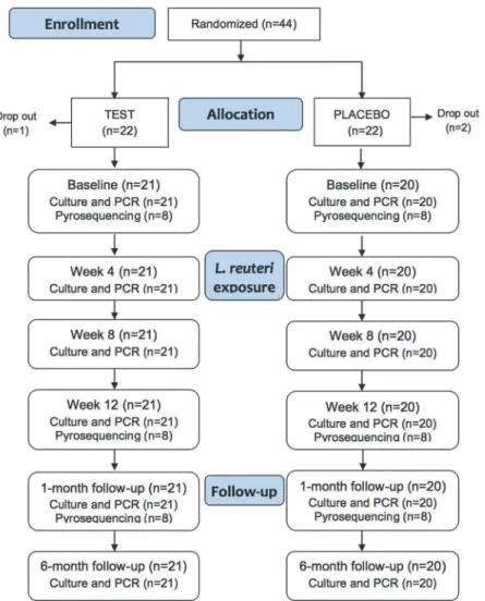
re-cruited and randomly allocated to either a test (n = 22) or placebo group (n = 22;Fig 1). SPSS
regular oral hygiene regimen except for the 48 hour preceding the sampling occasions. The test
lozenges containedL.reuteri(DSM 17938 and PTA 5289; 108CFU per strain; BioGaia AB,
Stockholm, Sweden), isomalt, hydrogenated palm oil, peppermint and menthol flavoring,
pep-permint oil, and sucralose (http://www.biogaia.com/product/biogaia-prodentis-oral-lozenges).
The placebo lozenges were identical to the test lozenges in appearance, taste, and composition except the lactobacilli. The content of the lozenges was blinded to the participants, to the per-sonal who exchanged the medical trays and sampled, and the perper-sonal who handled and ana-lyzed the samples. Compliance was monitored as the percentage of lozenges consumed of the total assigned number. The remaining lozenges were counted when the containers were
re-turned for monthly refills to assess this factor. Compliance was considered acceptable if15%
of the lozenges remained. Participants were asked to abstain from oral hygiene for 48 h and to not consume any food for at least 4 h before sampling. Participants were also instructed to not eat probiotic products throughout the study period.
Fig 1. Flow chart diagram.Number of study participants and samples analyzed in the test and placebo groups at each sampling occasion.
Saliva and biofilm sampling
Saliva and tooth biofilm samples were obtained immediately before (baseline) and after 4, 8,
and 12 weeks ofL.reuterisupplementation (Fig 1). Follow-up samples were collected 1 and 6
months after supplementation was terminated. Furthermore, whole stimulated saliva (~5 mL) was generated by chewing 1 g of paraffin and collected into ice-chilled sterile test tubes. One milliliter of saliva was used for cultural analysis, and the remaining saliva was centrifuged at
3,500 ×gfor 10 min at 4°C. The pellets were stored at−80°C until DNA extraction for
strain-specific PCR reactions. For the pyrosequencing analysis, pooled supragingival plaque was col-lected with sterilized toothpicks and transferred to Eppendorf tubes (Sarstedt, Nümbrecht,
Germany) containing 200μL of TE-buffer (10 mM Tris, 1 mM EDTA, pH 7.6). The samples
were stored at−80°C until DNA extraction.
Identification of lactobacilli by culture and PCR
Aliquots of saliva were plated onto Rogosa agar (Merck, Darmstadt, Germany) to obtain
Lacto-bacilluscounts and on selective agar for tentative identification of theL.reuteri(DSM 17938
and PTA 5289) strains [34]. All plates were anaerobically incubated at 37°C for 48–72 h, except
L.reuteriPTA 5289, which was anaerobically incubated at 40°C for 72 h.
DNA was extracted from saliva pellets as described [35].L.reuteriDSM 17938 and PTA
5289-specific PCR were identified using KAPA2G Robust HotStart PCR Ready Mix (2×)
(Kapa Biosystems, Boston, MA, USA) and strain-specific primers [34]. Briefly, 2μL of DNA
extract was added to a total reaction volume of 25μL (containing 12.5μL of master mix and
each primer pair at a concentration of 0.5μM). PCR conditions were 95°C for 3 min; 40 cycles
of 95°C for 15 s, 60°C for 15 s, and 72°C for 30 s; and 72°C for 5 min. PCR products were then verified by electrophoresis on 2% agarose gels allowed to run for 80 min at 120 V in 0.5× TBE
(Tris/Borate/EDTA) buffer, pH 8.3, followed by ethidium bromide (0.2μg/μL) staining.
Pyrosequencing analysis
For the 454 pyrosequencing analysis, 16 subjects were randomly selected among the test
(n = 8) and control (n = 8) subjects (Fig 1).
DNA was extracted from the baseline, 12-week exposure, and 1-month follow-up tooth
bio-film samples as previously described [35]. The V3-V4 hypervariable region of the 16S rRNA
gene was amplified via PCR using the forward primer 347F and reverse primer 803R [36]. For
sample identification, fusion primers were created from these primers and unique barcode
se-quences according to the Roche guidelines for experimental amplicon design (www.454.com).
DNA was amplified under the following running conditions: initial denaturation at 94°C for 3 min; 30 cycles of 94°C for 15 s, 58°C for 15 s, and 72°C for 30 s; and a final extension at 72°C for 8 min.
Amplicon processing and 454 sequencing were conducted at the Lund University Sequenc-ing Facility (Faculty of Science, Lund, Sweden). After amplicon cleanSequenc-ing to remove short frag-ments (Agencourt AMPure XP; Beckman Coulter, Brea, CA, USA) and inspection (DNA 1000 kit on a 2100 Bioanalyzer; Agilent Technologies, Palo Alto, CA, USA), amplicons were quanti-fied (Quant-iT ds DNA assay kit; Invitrogen, Carlsbad, CA, USA and Quantifluor fluorometer;
Promega, Madison, WI, USA) and diluted to obtain a total of 107copies/μL. Titration and
pyrosequencing on a 454 Life Sciences Genome Sequencer FLX+ machine (Roche Applied Sci-ences; Penzberg, Germany).
Data processing
Subject characteristic, compliance, and culture data were processed using SPSS. Descriptive sta-tistics, such as means [95% confidence intervals (CI)] and proportion distributions were calcu-lated. Differences between groups were tested with parametric or non-parametric tests
depending on the data measurement and distribution levels. P<0.05 was considered
statistically significant.
Sequences with a minimum length of 300 base pairs after primer sequence removal, correct barcode sequences, a maximum of 1 incorrect base pair in the primer sequences, and compli-ance with the default quality criteria for homopolymers and quality scores in the Quantitative
Insights into Microbial Ecology (QIIME) [37] software package (version 1.8.0) were retained
for analysis. Any sequence beyond the reversed primer were removed. Sequences beginning with the reverse primer were reverse complemented. Sequences were then clustered into opera-tional taxonomic units (OTUs) at a sequence similarity of 97% in the 16S rRNA
chimera-checked Greengene database (dated May 2013) [38] using USEARCH [39]. OTUs with a single
sequence were removed. One representative sequence per remaining OTU was taxonomically
assigned as a named or unnamed cultivable species or uncultivable phylotype at98.5%
identi-ty using the HOMD 16S rRNA RefSeq, version 12.0 (http://www.homd.org) [29].
Rarefaction curves were calculated to compare microbial richness [40]. Principal coordinate
analysis (PCoA) was applied to evaluate the phylogenetic beta diversity [41] of the bacterial
profiles at different time points. Multivariate partial least-square analysis (PLS) modeling (SIMCA P+, version 12.0; Umetrics AB, Umeå, Sweden) was performed to search if microbiota
structures were related toL.reutericonsumption (y-variables) [42,43]. Tested models included
those with pyrosequencing data only and those to which lactobacilli and streptococci culture and PCR data had been added. Variables were autoscaled to unit variance and cross-validated Y predictions were calculated. Subject clustering was displayed in score loading plots, and the importance of each x-variable was displayed in loading plots. Variables, where the 95% CI of
the PLS correlation coefficient did not inlude zero were considered statistically significant [43].
The Q2value yielded the capacities of the x-variables to predict the outcome (test or placebo
group allocation). Univariate analyses of single taxa were not applied because of the combina-tion of small groups and a high number of repeated tests.
Results
Study group and lactobacilli retrieval by culture
Saliva and plaque samples were collected on 6 different occasions from 41 subjects in either a
test group in which subjects ingested lozenges containingL.reuterior a control group in which
subjects ingested placebo lozenges (Fig 1). Three participants (1 from the test and 2 from the
placebo group) dropped out before baseline sampling for personal reasons. None reported any unintended effect from taking the lozenges. No significant differences were found in age, sex,
or compliance with the study protocol between the 2 groups (Table 1).
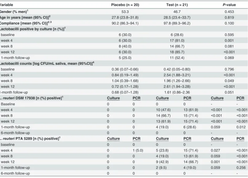
At baseline, lactobacilli were cultivated from saliva in 28.6% and 30.0% (p = 0.595) of
partic-ipants in the test and placebo groups, respectively (Table 1). During exposure, the test group
had a larger proportion of participants with cultivable lactobacilli than did the placebo group
(p<0.001). Moreover, mean numbers of log CFU/mL in saliva significantly differed between
the test group for both the proportion with cultivable lactobacilli and the mean numbers of log
CFU/mL in saliva approached the baseline values (Table 1).
L.reuteriDSM 17938 was not detected in the placebo group at any time point, whereasL.
reuteriPTA 5289 (or a highly similar strain) was detected in 1 participant at week 4 (Table 1).
Similarly, no participants in the test group carried any of theL.reuteritest strains at baseline or
at the 6-month follow-up. During exposure, mean PCR-detected prevalence rates of theL.
reu-teristrains DSM 17938 and PTA 5289 were 68.2% and 66.7%, respectively. At the 1-month
fol-low-up, DSM 17938 and PTA 5289 remained detectable in 28.6% and 19.0% of the test
Table 1. Characteristics of participants byL.reutericonsumption.
Variable Placebo (n = 20) Test (n = 21) P-value
Gender (% men)1
53.3 46.7 0.453
Age in years [mean (95% CI)]2 27.8 (23.8
–31.8) 28.5 (23.4–33.7) 0.819
Compliance [mean (95% CI)]2,3 90.2 (86.3
–94.1) 97.8 (89.3–96.2) 0.100
Lactobacilli positive by culture [n (%)]1
baseline 6 (30.0) 6 (28.6) 0.595
week 4 6 (30.0) 17 (81.0) 0.001
week 8 8 (40.0) 14 (66.7) 0.081
week 12 6 (30.0) 18 (85.7) <0.001
1-month follow-up 5 (25.0) 11 (52.4) 0.069
Lactobacilli counts [log CFU/mL saliva, mean (95%CI)]4
baseline 0.36 (0.07–0.66) 0.42 (0.05–0.80) 0.796
week 4 0.84 (0.19–1.49) 2.54 (1.88–3.21) <0.001
week 8 1.04 (0.39–1.68) 1.96 (1.26–2.66) 0.049
week 12 0.72 (0.17–1.28) 2.61 (1.94–3.28) <0.001
1-month follow-up 0.68 (0.07–1.28) 1.61 (0.86–2.36 0.051
L.reuteriDSM 17938 [n (%) positive]1 Culture PCR Culture PCR Culture PCR
Baseline 0 0 0 0 -
-week 4 0 0 10 (47.6) 13 (61.9) <0.001 <0.001
week 8 0 0 14 (66.7) 15 (71.4) <0.001 <0.001
week 12 0 0 13 (61.9) 15 (71.4) <0.001 <0.001
1-month follow-up 0 0 4 (19.0) 6 (28.6) 0.059 0.012
6-month follow-up 0 0 0 0 -
-L.reuteriPTA 5289 [n (%) positive]1 Culture PCR Culture PCR Culture PCR
baseline 0 0 0 0 -
-week 4 0 1 (5.0) 5 (23.8) 15 (71.4) 0.027 <0.001
week 8 0 0 4 (19.0) 13 (61.9) 0.059 <0.001
week 12 0 0 9 (42.9) 14 (66.7) 0.001 <0.001
1-month follow-up 0 0 2 (9.5) 4 (19.0) 0.059 0.256
6-month follow-up 0 0 0 0 -
-1Differences between group numbers tested with Chi2test 2Differences between group means tested with Student
’st-test
3% compliance = (lozenges consumed/ lozenges expected to be consumed) *100
4Difference between the test and placebo group were analyzed by non-parametric statistics (The Mann
–Whitney U-test). Numbers are expressed as log10values
CFU, colony-forming unit; CI, confidence interval
participants, respectively (Table 1).L.reuteriwas consistently more frequently detected by
PCR than by culture (Table 1).
Sequencing output
A total of 1,310,292 reads were obtained for the 48 tooth biofilm samples,i.e. 3 samples for 8
subjects in two groups. The original read sequences and the OUT table is available atftp://
www.homd.org/publication_data/20150122. Sequencing failed in 1 test group sample and that subject was excluded from all analyses. For the remaining 15 participants, quality control and denoising reduced the numbers to 1,148,923 sequences, out of which, 367,596, 422,639, and 358,688 corresponded to the baseline, 12-week exposure, and 1-month follow-up samples, re-spectively. The number of reads per sample ranged from 10,190 to 54,500 (mean, 26,690; medi-an, 22,699), and the average read length was 415 base pairs. A total of 257 named or unnamed species or uncultivable phylotypes with at least 2 sequences per cluster were taxonomically
as-signed at 98.5% identity in the HOMD. These represented 9 phyla and 66 genera (S1 Table).
Dental biofilm composition at baseline
UCLUST clustered 367,596 sequences from the baseline samples into 1,221 OTUs with2
reads per cluster against the Greengene database at97% sequence similarity. These matched
with 1,011 sequences in the HOMD at98.5% identity, representing 223 unique named or
un-named species or uncultivable phylotypes in the HOMD. Among these, 119 were un-named spe-cies, 55 were unnamed spespe-cies, and 49 were uncultivable phylotypes. Under these restrictions, an average of 101 species or phylotypes were identified per baseline sample.
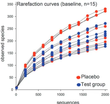
Rarefaction curves showed a 2-fold difference in sequence richness among the 15 baseline
samples (Fig 2). Although this variation was reflected in individually differing proportions at
the phylum and genus levels, the phyla and genera prevalence rankings in all subjects basically followed the pattern described below.
The 223 HOMD identified species/phylotypes in the baseline samples represented 8 phyla
and 55 genera (Fig 3andS2 Table).Firmicutes(65.1%) was the most prevalent phylum,
fol-lowed byBacteroidetes(13.0%),Fusobacteria(9.7%),Proteobacteria(8.1%), andActinobacteria
(3.9%).SR1,TM7, andSynergisteteseach represented<1% of the sample sequences. At the
genus level,Streptococcuswas most common (44.9%), followed byFusobacterium,Veillonella,
Haemophilus,Selemonas,Capnocytophaga,Abiotrophia,Leptotrichia,Prevotella, Porphyromo-nas,Actinomyces,Gemella,and Granulicatella(prevalence range: 2.1%–5.6%). The remaining
42 genera were detected at<1% abundance. At the species level, theS.oralis/S.mitis/S.mitis
bv2/S.infantisgroup was most common (26.3%), followed byS.sanguinis(7.5%),Haemophilus parainfluenzae(4.9%),S.oligofermentans(4.2%),Abiotrophia defectiva(4.2%),Veillonella
dis-par(3.6%), andFusobacterium nucleatum ss.polymorphum(3.1%). Furthermore,S.gordonii,
Porphyromonas sp. HOT279,Capnocytophaga sputigena,S.peroris,Gemella morbillorum,
Granulicatella adiacens,Veillonella parvula, andSelemonas sp. HOT137 each represented
be-tween 1% and<3% of the sequences; the remaining 208 species each represented<1% of
all sequences.
Stability in placebo-treated subjects
Microbiota compositions of the dental biofilm samples from the placebo group were stable over the 12-week treatment period, as determined by the lack of difference in the number of
observed species between the baseline and 12-week treatment samples (Fig 4), the lack of
clus-tering in PCoA modeling of beta-diversity in the baseline and 12-week samples (Fig 5), and the
(baseline versus 12-week treatment samples) was the dependent variable and phyla, genera, and species assignments were the independent variables (data not shown).
Effect of 12-week
L
.
reuteri
treatment
Similar to the placebo group, the number of observed species did not differ between baseline
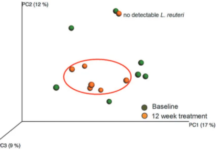
and 12-week treatment samples in the test group (Fig 4). However, PCoA modeling of the
phy-logenetic divergence among the UCLUST-identified OTUs clustered all 12-week treated
sam-ples together except for 1 sample (Fig 6). Notably, that sample was from the only participant in
the test group (used for pyrosequencing) without detectableL.reuteriDMS 17938 or PTA
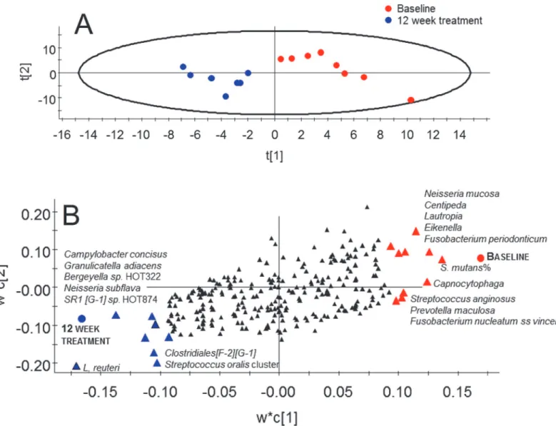
5289 at any sampling occasion. In contrast, the baseline samples did not cluster. PLS modeling using species/phylotypes and culture/PCR data yielded a model with 1 significant component
Fig 2. Rarefaction curves of baseline operational taxonomic unit prevalence rates according to the number of reads.Comparisons include all 15 subjects at baseline. Red dots indicate subjects in the placebo group; blue dots indicate subjects in the test group.
doi:10.1371/journal.pone.0125812.g002
Fig 3. Relative abundances of 9 identified bacterial phyla in 45 plaque samples.Sequences were matched to Human Oral Microbiome Database using Quantitative Insights into Microbial Ecology (QIIME) for phylum-level taxonomic identification. Data are presented as stacked bars and phyla are ordered by decreasing abundance and stratified by study group and sample occasion (i.e., baseline, 12-week exposure, and 1-month follow-up).
and a predictive power (Q2) of 43% for having ingestedL.reuteribacteria for 12 weeks relative to the baseline. This model fully separated the 12-week treatment samples of the test subjects
from their baseline samples (Fig 7A). Baseline samples were associated with a higher
propor-tion ofS.mutansamong the total streptococci count along with higher detection frequencies of
the speciesN.mucosa,F.periodicum,F.nucleatum ss vincentii,S.anginosus, andPrevotella
maculosa(Fig 7B). Presence of the test bacteria (L.reuteri) and greater detection frequency of
the speciesCampylobacter concisus,G.adiacens,Bergeyella sp. HOT322,N.subflava,SR1 [G-1]
sp. HOT874, and theS.oralis/S.mitis/S.mitis bv2/S.infantisgroup were associated with
con-sumption of theL.reuteri-containing lozenges for 12 weeks (Fig 7B). Restricting the model to
include only pyrosequencing obtained species/phylotypes did not alter the overall results.
Fig 4. Rarefaction curves of operational taxonomic unit prevalence rates according to the number of reads in the test and placebo groups.Data are presented as means with standard errors. Comparisons include 8 subjects in the placebo-treated group by sampling occasion (red symbols) and 7 subjects in the test group by sampling occasion (blue symbols). Differences within or between the groups were not
statistically significant.
doi:10.1371/journal.pone.0125812.g004
Fig 5. PCoA clustering analysis of baseline and 12-week samples in the placebo group.Red dots indicate baseline samples and blue dots 12 week samples.
Prevalence rates at the phylum and genus level as well as the species/phylotype level in the
test and placebo groups are presented inS1–S2Tables, respectively.
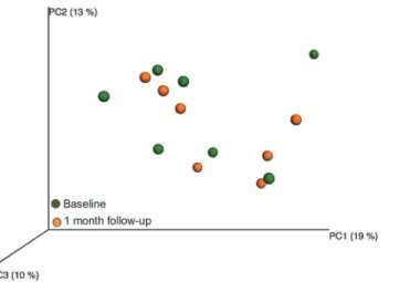
No persistent treatment effect at 1-month follow-up
Rarefaction curves showed that in the test group, species richness in the 1-month follow-up samples did not differ from that in the baseline or 12-week treatment samples (data not shown). The PCoA projection of beta diversity in the baseline samples versus the 1-month fol-low-up samples showed no sample clustering, such as that observed in the 12-week treatment
samples (Fig 8). Further, no significant components were found by PLS using
species/phylo-types with or without lactobacilli and mutans streptococci from culture and PCR as the inde-pendent block and sampling occasion as deinde-pendent variables.
Discussion
The present study evaluated if consuming the probioticL.reuteristrains DMS 17938 and PTA
5289 for 12 weeks would modify the microbiota composition and species richness in tooth
bio-films. The main findings were as follows: (i) the composition shifted but species richness
re-mained unaffected, (ii) the shift normalized within 1 month after terminating exposure, and
(iii) the test strains could not be detected in approximately 30% of the participants despite
re-ceiving daily booster doses.
The ecology of the gastrointestinal microbiota, including that of the mouth, is virtually
sta-ble once the period of variation in early childhood has passed [44]. Previous multiplex
charac-terizations of the microbiota following exposure to probiotic bacteria or dietary regimens have
essentially addressed the impact on the gut microbiome [45,46]. Studies that have addressed
effects on oral bacterial communities have mainly targeted single bacterial species, such as the
caries-associated mutans streptococci (S.mutansandS.sobrinus)[24–27].L.reuteriDSM
17938 and PTA 5289 ingestion have been previously associated with reduced oral levels of
mutans streptococci [24,25]. However, several studies found no evidence of reduced disease
de-velopment despite reductions in mutans streptococci levels [47,48], the mechanisms of which
include growth inhibition and competition for host ligands, such as gp340 [49–51]. In the
pres-ent study, the proportion ofS.mutansamong total streptococci was lower in the test group
after a 12-week period during whichL.reuteristrains DSM 17938 and PTA 5289 were ingested.
This finding partly reflected a reduction in mutans streptococci in some subjects (culture data;
Fig 6. PCoA clustering analysis of baseline and 12-week samples in the test group.Green dots indicate baseline samples and yellow dots 12 week samples.
Johanssonet al, manuscript in progress) and partly reflected the increased numbers of other
streptococci (e.g., theS.oralis/S.mitis/S.mitis bv2/S.infantisgroup). However, this result
should be interpreted with caution as mutans streptococcal colonization was not an inclusion
criterion in the present study, in contrast to the referred studies [24,25] and few subjects were
found to be colonized withS.mutans(none were colonized withS.sobrinus). A potentially
beneficial effect ofL.reuteriexposure was the reduced prevalence ofF.nucleatum ss vincentii
andF.periodicum, which may be linked to reduced biofilm formation and gingivitis
develop-ment during the ingestion of probiotic bacteria [52]. Fusobacteria are considered a key link
be-tween early and late colonizers in oral biofilm formation due to the ability to coaggregate with
a large number of bacterial species, including probiotic bacteria [1,61].
Fig 7. Partial least-squares analysis (PLS) of microbiota associated with 12 weeks consumption ofL.reuteri.(A) Scatter-loading plot illustrating clustering of baseline versus 12 week treatment samples from the test group; The scores t1 and t2 are the new PCA created variables summarizing the x-variables. The red dots indicate placebo samples and blue dots test samples. The oval circle illustrates the tolerance ellipse based on Hotelling´s of T2, any observation located outside of the elipse would be an outlier. (B) Loading plot illustrating taxa associated with baseline versus 12 week treatment in the test group. The PLS model employed test and placebo group allocation as y and pyrosequencing taxa,L.reuteriandS.mutansby cultivation and PCR as the x-block. Red triangles indicate taxa associated with the baseline microbiota in the test group and blue triangles those associated with the microbiota after 12 week treatment in the same group. Black symbols indicate variables that were non-influential in the projection.
Two previous studies evaluated oral microbiota after the short-term use (3 or 4 weeks) of
probiotic bacteria [52,53]. One study incorporated a whole-genome DNA–DNA hybridization
technique [53], whereas the other used a specially designed 16S rRNA-based microarray
(HOMIM) [52]. However, neither study found an ecological shift. The present study, which
ex-posed subjects toL.reuterifor a longer period (12 weeks) and mapped the microbiota by
se-quencing an approximately 400-base pair section of the 16S rRNA gene, revealed a transient reduction in diversity in the tooth biofilm samples, although this shift normalized within 1 month after intake was discontinued. This finding is in accordance with studies showing tran-sient fluctuations in gut bacterial profiles after introducing probiotic products or diet changes
along with a rapid reversion of the microbial community to its previous stable state [46].
The final number of species/phylotypes detected in the present study was somewhat lower than that reported in other studies; however, the phylum, genus, and species representation fol-low the patterns reported in other studies that analyzed comparable samples and populations
[54,55]. The somewhat lower taxa numbers should be considered in light of the restrictive
fil-tering criteria applied herein. Thus, only sequence clusters with at least 2 sequences and only clusters with a sequence identity of at least 98.5% with the HOMD sequences were
taxonomi-cally assigned. OTUs with identities of 97% to<98.5% could only be taxonomically assigned to
the genus level and were not retained. The numbers of OTUs reported for saliva or tooth bio-film samples in different studies have greatly varied, which partly reflects biological or ethnic variations as well as variations in filtering criteria and conditions for taxonomical assignment
[31]. In the present study, bacterial taxa were identified through alignment with the curated,
chimera-free HOMD [2]. This database contains information on approximately 700
prokary-ote species found in the human oral cavity; among these, approximately 49% taxa are named, 17% are cultivated but unnamed, and 34% are known only as uncultivated phylotypes. Regard-less, mapping from a section of the 16S rRNA gene cannot separate closely related species, such
as several members of theStreptococcusgenus. For example, the relative prevalence rates of the
4 species in the most prevalent group (S.oralis,S.mitis,S.mitis bv2,and S.infantis)cannot be
distinguished. Therefore, the species-level sequence abundance is partly due to the restrictive identification criteria and partly to the overall limitations of using 16S rRNA gene variation for
species identification [56,57].
To the best of our knowledge, this is the first study to evaluate the effects of a probiotic
bac-terium,L.reuteri, on the composition of tooth biofilm microbiota using 16S rRNA
Fig 8. PCoA clustering analysis of 12-week and 1 month follow-up samples in the test group.Yellow dots indicate baseline samples and green dots 1 month follow-up samples.
pyrosequencing. Multiplex sequencing is cost-effective for the following reasons: (i) thousands
of sequences can be simultaneously obtained from a single sample [54], (ii) open-ended view
of the microbiome is provided, and (iii) answers regarding microbial richness and ecological
stability and shift are facilitated [58]. Drawbacks of this method include (i) limitations on
taxo-nomic resolution due to the short-length sequence limit [58](ii), loss of an unknown amount
of taxa because of the rather high detection limit [59], and(iii) distortion of the true microbiota
profile at various steps, i.e. contaminations in DNA extraction and purification, and PCR and
sequencing errors and data analyses and interpretation [60,61,62]. Furthermore, Lagieret al.
[59] specified the respective detection limits in large-scale molecular studies, such as
pyrose-quencing, conventional PCR, and cultivation as 106, 104, and 102CFU/mL, respectively. Those
authors also reported that only 35% of bacterial phylotypes were detected in fecal samples by
pyrosequencing. The fact that noL.reuterior other lactobacilli (except two single sequences
that were discarded according to the filtering criteria) was detected in 12-week samples from the test group, despite culture and PCR results demonstrating that the species and specifically the test strains were present in 6 of 7 test subjects, illustrates the underestimated number of de-tected species in the present study. Accordingly, metagenomic studies may need to consider this limitation as well as additional approaches for identifying less abundant, condition-associ-ated genera/species. In contrast, the strengths of the study include the RCT design and high compliance and a double-blinded, randomized design in addition to the application of a new metagenomic approach combined with strain-specific PCR and cultivation.
Efforts to modify tooth biofilm and prevent oral diseases by consuming“beneficial bacteria”
(probiotics) have been made for decades [63]. The fact that nearly one third of the test subjects
in the present study did not present evidence ofL.reuteriDSM 17938 and PTA 5289 strains,
which is similar to findings from other studies with these strains orL.rhamnosusGG
[33,34,64,65], indicates that the expected effect is individual. Installation of probiotic
lactoba-cilli likely reflects genetic variations in host receptor epitopes for bacterial binding, such as
sali-vary gp340, MUC5, MUC7, and ABO antigen epitopes [10,66–68].
The conclusion from the present study is thatL.reutericonsumption induces a shift in oral
microbiota composition but does not affect the total species richness. The biological relevance of this finding as well as the individuality of this effect remains to be elucidated.
Supporting Information
S1 CONSORT Checklist. CONSORT 2010 checklist of information. (PDF)
S1 Protocol. Study protocol. (DOCX)
S1 Table. Phylum and genus identities and abundances of the 257 identified taxa among the 45 included pyrosequencing samples.Mean abundances (% of all sequences) for test and
control subjects at baseline and after 12 weeks exposure to anL.reuterior placebo lozenge
are listed. (DOCX)
S2 Table. Species and phylotypes identified by pyrosequencing of the 257 identified taxa among the 45 included samples.Mean abundances (% of all sequences) for test and control
subjects at baseline and after 12 weeks exposure to anL.reuterior placebo lozenge are listed.
Acknowledgments
We would like to thank Tomas Johansson for his valuable support in pyrosequencing analysis; Caroline Sörensen, Elin Jacobi, and Hanna Gedda for their help with sample collection; and Agnetha Rönnlund for her skillful technical support. Test and placebo lozenges were kindly provided by BioGaia AB, Stockholm, Sweden.
Author Contributions
Conceived and designed the experiments: NRV IJ PLH. Performed the experiments: NRV CÖ. Analyzed the data: NRV TC IJ. Contributed reagents/materials/analysis tools: IJ PLH. Wrote the paper: NRV IJ TC CÖ PLH.
References
1. Kolenbrander PE. Oral microbial communities: biofilms, interactions, and genetic systems. Annu Rev Microbiol. 2000; 54: 413–437. PMID:11018133
2. Dewhirst FE, Chen T, Izard J, Paster BJ, Tanner AC, Yu MH et al. The human oral microbiome. J Bac-teriol. 2010; 192: 5002–5017. doi:10.1128/JB.00542-10PMID:20656903
3. Stamatova I, Meurman JH. Probiotics: health benefits in the mouth. Am J Dent. 2009; 22: 329–338. PMID:20178208
4. Heller KJ. Probiotic bacteria in fermented foods: product characteristics and starter organisms. Am J Clin Nutr. 2001; 73: 374S–379S. PMID:11157344
5. Preidis GA, Versalovic J. Targeting the human microbiome with antibiotics, probiotics, and prebiotics: gastroenterology enters the metagenomics era. Gastroenterology. 2009; 136: 2015–2031. PMID: 19462507
6. Van Houte J. Bacterial specificity in the etiology of dental caries. Int. Dent J. 1980; 30:305–326. PMID: 7005104
7. Simark-Mattsson C, Emilson CG, Hakansson EG, Jacobsson C, Roos K, Holm S.Lactobacillus -medi-ated interference of mutans streptococci in caries-free vs. caries-active subjects. Eur J Oral Sci. 2007; 115: 308–314. PMID:17697171
8. Kanasi E, Johansson I, Lu SC, Kressin NR, Nunn ME, Kent R Jr, et al. Microbial risk markers for child-hood caries in pediatricians' offices. J Dent Res. 2010; 89: 378–383. doi:10.1177/0022034509360010 PMID:20164496
9. Holgerson PL, Vestman NR, Claesson R, Ohman C, Domellof M, Tanner AC, et al. Oral microbial pro-file discriminates breast-fed from formula-fed infants. J Pediatr Gastroenterol Nutr. 2013; 56: 127–136. doi:10.1097/MPG.0b013e31826f2bc6PMID:22955450
10. Vestman NR, Timby N, Holgerson PL, Kressirer CA, Claesson R, Domellöf M, et al. Characterization and in vitro properties of oral lactobacilli in breastfed infants. BMC Microbiol. 2013; 13: 193. doi:10. 1186/1471-2180-13-193PMID:23945215
11. Mitsuoka ET. The human gastrointestinal tract. The Lactic Acid Bacteria Volume 1: Springer. 1992. pp. 69–114.
12. Martin R, Langa S, Reviriego C, Jiminez E, Marin ML, Xaus J, et al. Human milk is a source of lactic acid bacteria for the infant gut. J Pediatr. 2003; 143: 754–758. PMID:14657823
13. Reuter G. TheLactobacillusandBifidobacteriummicroflora of the human intestine: composition and succession. Curr Issues Intest Microbiol. 2001; 2: 43–53. PMID:11721280
14. Rosander A, Connolly E, Roos S. Removal of antibiotic resistance gene-carrying plasmids from Lacto-bacillus reuteriATCC 55730 and characterization of the resulting daughter strain,L.reuteriDSM 17938. Appl Environ Microbiol. 2008; 74: 6032–6040. doi:10.1128/AEM.00991-08PMID:18689509
15. Indrio F, Riezzo G, Raimondi F, Bisceglia M, Cavallo L, Francavilla R. The effects of probiotics on feed-ing tolerance, bowel habits, and gastrointestinal motility in preterm newborns. J Pediatr. 2008; 152: 801–806. doi:10.1016/j.jpeds.2007.11.005PMID:18492520
16. Savino F, Pelle E, Palumeri E, Oggero R, Miniero R.Lactobacillus reuteri(American Type Culture Col-lection Strain 55730) versus simethicone in the treatment of infantile colic: a prospective randomized study. Pediatrics. 2007; 119: e124–130. PMID:17200238
18. Saulnier DM, Santos F, Roos S, Mistretta TA, Spinler JK, Molenaar D, et al. Exploring metabolic path-way reconstruction and genome-wide expression profiling inLactobacillus reuterito define functional probiotic features. PLoS One. 2011; 6: e18783. doi:10.1371/journal.pone.0018783PMID:21559529
19. Oh PL, Benson AK, Peterson DA, Patil PB, Moriyama EN, Roos S, et al. Diversification of the gut sym-biontLactobacillus reuterias a result of host-driven evolution. ISME J. 2010; 4: 377–387. doi:10.1038/ ismej.2009.123PMID:19924154
20. Jones SE, Versalovic J. ProbioticLactobacillus reuteribiofilms produce antimicrobial and anti-inflam-matory factors. BMC Microbiol 2009; 9: 35. doi:10.1186/1471-2180-9-35PMID:19210794
21. Jensen H, Grimmer S, Naterstad K, Axelsson L. In vitro testing of commercial and potential probiotic lactic acid bacteria. Int Journal of Food Microbiol. 2012; 153: 216–222. doi:10.1016/j.ijfoodmicro.2011. 11.020PMID:22177712
22. Jalasvuori H, Haukioja A, Tenovuo J. ProbioticLactobacillus reuteristrains ATCC PTA 5289 and ATCC 55730 differ in their cariogenic properties in vitro. Arch Oral Biol. 2012; 57:1633–1638. doi:10. 1016/j.archoralbio.2012.07.014PMID:23010217
23. McNulty NP, Yatsunenko T, Hsiao A, Faith JJ, Muegge BD, Goodman AL, et al. The impact of a consor-tium of fermented milk strains on the gut microbiome of gnotobiotic mice and monozygotic twins. Sci Transl Med. 2011; 26:106ra106. doi:10.1126/scitranslmed.3002701PMID:22030749
24. Caglar E, Kavaloglu SC, Kuscu OO, Sandalli N, Holgerson PL, Twetman S. Effect of chewing gums containing xylitol or probiotic bacteria on salivary mutans streptococci and lactobacilli. Clin Oral Inves-tig. 2007; 11:425–429. PMID:17574481
25. Nikawa H, Makihira S, Fukushima H, Nishimura H, Ozaki Y, Ishida K, et al.Lactobacillus reuteriin bo-vine milk fermented decreases the oral carriage of mutans streptococci. Int J Food Microbiol. 2004; 95: 219–223. PMID:15282133
26. Cildir SK, Sandalli N, Nazli S, Alp F, Caglar E. A novel delivery system of probiotic drop and its effect on dental caries risk factors in cleft lip/palate children. Cleft Palate Craniofac J. 2012; 49: 369–372. doi:10. 1597/10-035PMID:21309653
27. Keller MK, Hasslof P, Dahlen G, Stecksen-Blicks C, Twetman S. Probiotic supplements (Lactobacillus reuteriDSM 17938 and ATCC PTA 5289) do not affect regrowth of mutans streptococci after full-mouth disinfection with chlorhexidine: a randomized controlled multicenter trial. Caries Res. 2012; 46: 140–
146. doi:10.1159/000337098PMID:22472585
28. Ronaghi M, Uhlen M, Nyren P. A sequencing method based on real-time pyrophosphate. Science. 1998; 281: 363–365. PMID:9705713
29. Chen T, Yu WH, Izard J, Baranova OV, Lakshmanan A, Dewhirst FE. The Human Oral Microbiome Da-tabase: a web accessible resource for investigating oral microbe taxonomic and genomic information. Database (Oxford) 2010: baq013.
30. Zaura E, Keijser BJ, Huse SM, Crielaard W. Defining the healthy "core microbiome" of oral microbial communities. BMC Microbiol. 2009; 9: 259. doi:10.1186/1471-2180-9-259PMID:20003481
31. Wade WG. The oral microbiome in health and disease. Pharmacol Res. 2013; 69:137–143. doi:10. 1016/j.phrs.2012.11.006PMID:23201354
32. Saxelin M, Lassig A, Karjalainen H, Tynkkynen S, Surakka A, Vapaatalo H, et al. Persistence of probiot-ic strains in the gastrointestinal tract when administered as capsules, yoghurt, or cheese. Int J Food Microbiol. 2010; 144: 293–300. doi:10.1016/j.ijfoodmicro.2010.10.009PMID:21074284
33. Yli-Knuuttila H, Snall J, Kari K, Meurman JH. Colonization ofLactobacillus rhamnosusGG in the oral cavity. Oral Microbiol Immunol. 2006; 21: 129–131. PMID:16476023
34. Romani Vestman N, Hasslof P, Keller MK, Granstrom E, Roos S, Twetman S, et al.Lactobacillus reu-teriinfluences regrowth of mutans streptococci after full-Mouth disinfection: a double-blind, randomised controlled trial. Caries Res. 2013; 47: 338–345. doi:10.1159/000347233PMID:23486236
35. Lif Holgerson P, Harnevik L, Hernell O, Tanner AC, Johansson I. Mode of birth delivery affects oral microbiota in infants. J Dent Res. 2011; 90:1183–1188. doi:10.1177/0022034511418973PMID: 21828355
36. Nossa CW, Oberdorf WE, Yang L, Aas JA, Paster BJ, Desantis TZ, et al. Design of 16S rRNA gene primers for 454 pyrosequencing of the human foregut microbiome. World J Gastroenterol. 2010; 16: 4135–4144. PMID:20806429
37. Caporaso JG, Kuczynski J, Stombaugh J, Bittinger K, Bushman FD, Costello EK, et al. QIIME allows analysis of high-throughput community sequencing data. Nat Methods. 2010; 7: 335–336. doi:10. 1038/nmeth.f.303PMID:20383131
39. Edgar RC. Search and clustering orders of magnitude faster than BLAST. Bioinformatics. 2010; 26: 2460–2461. doi:10.1093/bioinformatics/btq461PMID:20709691
40. Chao A. Nonparametric estimation of the number of classes in a population. Scand J stat. 1984;265–
270.
41. Lozupone CA, Hamady M, Kelley ST, Knight R. Quantitative and qualitative beta diversity measures lead to different insights into factors that structure microbial communities. Appl Environ Microbiol. 2007; 73:1576–1585. PMID:17220268
42. Bylesjö M, Rantalainen M, Cloarec O, Nicholson JK, Holmes E. OPLS discriminant analysis: combining the strengths of PLS‐DA and SIMCA classification. J Chemom. 2006; 20: 341–351.
43. Sjöström M, Wold S, Söderström B. PLS discriminant plots; 1986. Elsevier: Amsterdam. pp. 461–470.
44. Koenig JE, Spor A, Scalfone N, Fricker AD, Stombaugh J, Knight R, et al. Succession of microbial con-sortia in the developing infant gut microbiome. Proc. Natl. Acad. Sci. U. S. A. 2011; 108:4578–4585. doi:10.1073/pnas.1000081107PMID:20668239
45. Moal Liévin-Le, Servin A. Anti-Infective Activities of Lactobacillus Strains in the Human Intestinal Micro-biota: from Probiotics to Gastrointestinal Anti-Infectious Biotherapeutic Agents. Clin Microbiol Rev. 2014; 27:167–199. doi:10.1128/CMR.00080-13PMID:24696432
46. Voreades N, Kozil A, Weir TL. Diet and the development of the human intestinal microbiome. Front Microbiol. 2014; 22:494.
47. Taipale T, Pienihäkkinen K, Alanen P, Jokela J, Söderling E. Administration of Bifidobacterium animalis subsp. lactis BB-12 in early childhood: a post-trial effect on caries occurrence at four years of age. Car-ies Res. 2013; 47:364–372. doi:10.1159/000348424PMID:23571819
48. Keller MK, Nøhr Larsen I, Karlsson I, Twetman S. Effect of tablets containing probiotic bacteria (Lacto-bacillus reuteri) on early caries lesions in adolescents: a pilot study. Benef Microbes. 2014; 5:403–407. doi:10.3920/BM2013.0089PMID:24889893
49. Haukioja A, Loimaranta V, Tenovuo J. Probiotic bacteria affect the composition of salivary pellicle and streptococcal adhesion in vitro. Oral Microbiol Immunol. 2008; 23:336–343. doi:10.1111/j.1399-302X. 2008.00435.xPMID:18582334
50. Marttinen AM, Haukioja AL, Keskin M, Söderling EM. Effects of Lactobacillus reuteri PTA 5289 and L. paracasei DSMZ16671 on the adhesion and biofilm formation of Streptococcus mutans. Curr Microbiol. 2013; 67:193–199. doi:10.1007/s00284-013-0352-3PMID:23503788
51. Vestman NR, Timby N, Holgerson PL, Kressirer CA, Claesson R, Johansson I. Characterization and in vitro properties of oral lactobacilli in breastfed infants. BMC Microbiol. 2013; 13:193. doi: 10.1186/1471-2180-13-193PMID:23945215
52. Toiviainen A, Jalasvuori H, Lahti E, Gursoy U, Salminen S, Fontana M, et al. Impact of orally adminis-tered lozenges with Lactobacillus rhamnosus GG and Bifidobacterium animalis subsp. lactis BB-12 on the number of salivary mutans streptococci, amount of plaque, gingival inflammation and the oral micro-biome in healthy adults. Clin Oral Investig. 2015; 19:77–83. doi:10.1007/s00784-014-1221-6PMID: 24638207
53. Hallström H, Lindgren S, Yucel-Lindberg T, Dahlén G, Renvert S, Twetman S. Effect of probiotic loz-enges on inflammatory reactions and oral biofilm during experimental gingivitis. Acta Odontol Scand. 2013; 71:828–833. doi:10.3109/00016357.2012.734406PMID:23294143
54. Keijser BJ, Zaura E, Huse SM, van der Vossen JM, Schuren FH, Montijn SC, et al. Pyrosequencing analysis of the oral microflora of healthy adults. J Dent Res. 2008; 87: 1016–1020. PMID:18946007
55. Griffen AL, Beall CJ, Campbell JH, Firestone ND, Kumar PS, Yang ZK, et al. Distinct and complex bac-terial profiles in human periodontitis and health revealed by 16S pyrosequencing. ISME J. 2012; 6: 1176–1185. doi:10.1038/ismej.2011.191PMID:22170420
56. Naser SM, Dawyndt P, Hoste B, Gevers D, Vandemeulebroecke K, Cleenwerck I, et al. Identification of lactobacilli by pheS and rpoA gene sequence analyses. Int J Syst Evol Microbiol. 2007; 57: 2777–
2789. PMID:18048724
57. Park HK, Yoon JW, Shin JW, Kim JY, Kim W. rpoA is a useful gene for identification and classification ofStreptococcus pneumoniaefrom the closely related viridans group streptococci. FEMS Microbiol Lett. 2010; 305: 58–64. doi:10.1111/j.1574-6968.2010.01913.xPMID:20158524
58. Zaura E. Next-generation sequencing approaches to understanding the oral microbiome. Adv Dent Res. 2012; 24: 81–85. doi:10.1177/0022034512449466PMID:22899686
59. Lagier JC, Armougom F, Million M, Hugon P, Pagnier I, Robert C, et al. Microbial culturomics: paradigm shift in the human gut microbiome study. Clin Microbiol Infect. 2012; 18: 1185–1193. doi:10.1111/ 1469-0691.12023PMID:23033984
61. Lazarevic V, Gaïa N, Girard M, François P, Schrenzel J. Comparison of DNA extraction methods in
analysis of salivary bacterial communities. PLoS One. 2013; 8:e67699. doi:10.1371/journal.pone. 0067699PMID:23844068
62. Salter SJ, Cox MJ, Turek EM, Calus ST, Cookson WO, Moffatt MF et al. Reagent and laboratory con-tamination can critically impact sequence-based microbiome analyses. BMC Biol. 2014; 12:87. doi:10. 1186/s12915-014-0087-zPMID:25387460
63. Haukioja A, Yli-Knuuttila H, Loimaranta V, Kari K, Ouwehand AC, Meurman JH, et al. Oral adhesion and survival of probiotic and other lactobacilli and bifidobacteria in vitro. Oral Microbiol Immunol. 2006; 21: 326–332. PMID:16922933
64. Meurman JH, Stamatova I. Probiotics: contributions to oral health. Oral Dis. 2007; 13: 443–451. PMID: 17714346
65. Iniesta M, Herrera D, Montero E, Zurbriggen M, Matos AR, Marín MJ, et al. Probiotic effects of orally ad-ministeredLactobacillus reuteri-containing tablets on the subgingival and salivary microbiota in patients with gingivitis. A randomized clinical trial. J Clin Periodontol. 2012; 39: 736–744. doi: 10.1111/j.1600-051X.2012.01914.xPMID:22694350
66. Loimaranta V, Jakubovics NS, Hytonen J, Finne J, Jenkinson HF, Strömberg N. Fluid- or surface-phase human salivary scavenger protein gp340 exposes different bacterial recognition properties. In-fect Immun. 2005; 73: 2245–2252. PMID:15784568
67. Uchida H, Kinoshita H, Kawai Y, Kitazawa H, Miura K, Shiiba K, et al. Lactobacilli binding human A-anti-gen expressed in intestinal mucosa. Res Microbiol. 2006; 157: 659–665. PMID:16631357