A Modified Glycosaminoglycan, GM-0111,
Inhibits Molecular Signaling Involved in
Periodontitis
Justin R. Savage1, Abigail Pulsipher1, Narayanam V. Rao1, Thomas P. Kennedy1,2, Glenn D. Prestwich1,3, Maria E. Ryan4, Won Yong Lee1*
1GlycoMira Therapeutics, Inc. Salt Lake City, UT, 84108, United States of America,2Pulmonary Diseases Critical Care and Environmental Medicine, School of Medicine, Tulane University, New Orleans, LA, 70112, United States of America,3Department of Medicinal Chemistry and Center for Therapeutic Biomaterials, University of Utah, Salt Lake City, UT, 84108, United States of America,4Department of Oral Biology and Pathology, School of Dental Medicine, Stony Brook University, Stony Brook, NY, 11794, United States of America
*lee.wonyong@glycomira.com
Abstract
Background
Periodontitis is characterized by microbial infection, inflammation, tissue breakdown, and accelerated loss of alveolar bone matrix. Treatment targeting these multiple stages of the disease provides ways to treat or prevent periodontitis. Certain glycosaminoglycans (GAGs) block multiple inflammatory mediators as well as suppress bacterial growth, sug-gesting that these GAGs may be exploited as a therapeutic for periodontitis.
Methods
We investigated the effects of a synthetic GAG, GM-0111, on various molecular events associated with periodontitis: growth ofPorphyromonas gingivalis(P.gingivalis) and Aggre-gatibacter actinomycetemcomitans(A.actinomycetemcomitans) pathogenic bacteria associated with periodontitis; activation of pro-inflammatory signaling through TLR2 and TLR4 in mouse macrophage RAW 264.7 cells and heterologously expressed HEK 293 cells; osteoclast formation and bone matrix resorption in cultured mouse pre-osteoclasts.
Results
(1) GM-0111 suppressed the growth ofP.gingivalisandA.actinomycetemcomitanseven at 1% (w/v) solution. The antibacterial effects of GM-0111 were stronger than hyaluronic acid (HA) or xylitol inP.gingivalisat all concentrations and comparable to xylitol inA. actino-mycetemcomitansat2% (w/v) solution. We also observed that GM-0111 suppressed bio-film formation ofP.gingivalisand these effects were much stronger than HA. (2) GM-0111 inhibited TLR-mediated pro-inflammatory cellular signaling both in macrophage and HEK 293 cells with higher selectivity for TLR2 than TLR4 (IC50of 1–10 ng/mLvs.>100μg/mL,
respectively). (3) GM-0111 blocked RANKL-induced osteoclast formation (as low as 300
a11111
OPEN ACCESS
Citation:Savage JR, Pulsipher A, Rao NV, Kennedy TP, Prestwich GD, Ryan ME, et al. (2016) A Modified Glycosaminoglycan, GM-0111, Inhibits Molecular Signaling Involved in Periodontitis. PLoS ONE 11(6): e0157310. doi:10.1371/journal.pone.0157310
Editor:Luc Malaval, Université de Lyon - Université Jean Monnet, FRANCE
Received:May 15, 2015
Accepted:May 30, 2016
Published:June 16, 2016
Copyright:© 2016 Savage et al. This is an open access article distributed under the terms of the Creative Commons Attribution License, which permits unrestricted use, distribution, and reproduction in any medium, provided the original author and source are credited.
Data Availability Statement:All relevant data are within the paper and its Supporting Information files.
Funding:This work was supported by grant numbers 5R44DE022216 (WYL, GDP, MER) and
ng/mL) and bone matrix resorption. While GM-0111 showed high affinity binding to RANKL, it did not interfere with RANKL/RANK/NF-κB signaling, suggesting that GM-0111 inhibits
osteoclast formation by a RANKL-RANK-independent mechanism.
Conclusions
We report that GM-0111 inhibits multiple molecular events involved in periodontitis, span-ning from the early pro-inflammatory TLR signaling, to pathways activated at the later stage component of bone loss.
Introduction
Periodontitis is a chronic inflammatory disease characterized by recurrent infection and inflammation that often progresses into alveolar bone loss. Moderate to severe forms of peri-odontitis occur in approximately 10%-15% of middle-aged adults worldwide and approxi-mately 20% of US adults [1–3]. Currently available treatment options are limited. There is a need for more effective treatments to control this common disease.
In healthy individuals, the periodontium maintains local tissue homeostasis by balancing its immune response to the local microbial ecosystem. Diseases or drastic alterations of the local microenvironment in the oral cavity can break this homeostasis, leading to dysbiotic microbial ecosystems in the periodontium. This process in turn increases the host immune responses, causes tissue destruction, enhances the proliferation of pathogenic microorganisms, and fur-ther exacerbates the host immune responses [4,5]. The development of this vicious cycle is key to the pathogenesis of periodontitis. Therapeutics for periodontitis should aim to break this continuum and restore tissue homeostasis in the periodontium.
Considerable research has focused on identifying the most pathogenic gingival microorgan-isms and their molecular targets in host tissues. Currently, over 700 different types of microor-ganisms living in the oral cavity have been identified [6]. Although protecting tissues from these massively diverse microorganisms seems daunting, animals have evolved to cope with this challenge by using innate and acquired immune systems to counter the potentially harmful invaders. Periodontal tissues are comprised of epithelial and non-epithelial cells that constantly face microorganisms and their byproducts. These cells use specialized innate pattern recogni-tion receptors (PRRs) that recognize ligands called pathogen-associated molecular patterns (PAMPs) [7,8]. Toll-like receptors (TLRs) belong to PRRs that recognize various PAMP mole-cules produced by dangerous invaders. PAMP-activated TLRSs then signal cells to produce pro-inflammatory cytokines and cellular responses.
Extensive research on various periodontal pathogenic microorganisms such as Porphyromo-nas gingivalis(P.gingivalis) andAggregatibacter actinomycetemcomitans(A. actinomycetemco-mitans) have brought significant progress to our understanding of their contributions to the pathogenesis of periodontitis [9–19]. Various bacterial molecular components have been iden-tified such as lipopolysaccharide (LPS) and fimbriae that interact with host periodontal tissues and immune systems [20–23]. Bacterial LPS and lipoproteins bind to cell surface PRRs such as TLR2 and TLR4 to activate canonical nuclear factorκ-light-chain-enhancer of activated B cells (NF-κB), cellular signaling that leads to the synthesis and release of various pro-inflammatory cytokines such as IL-1, -6 and TNFα[24–26]. Moreover, recent studies by Nussbaum [27] and Maekawa[13] have recently proposed thatP.gingivalisuses a non-canonical TLR2 signaling pathway to evade host bactericidal activities, providing important insight into the pathogenesis the manuscript. The specific roles of these authors
are articulated in the‘author contributions’section.
of chronic infection. Sustained inflammation associated with these pathogenic bacteria enhances the activity of osteoclasts, bone-demineralizing cells [28–30]. The development of osteoclasts in the periodontium is due to the increased secretion of the receptor activator of nuclear factor-kappaB ligand, also known as sRANKL, from periodontal ligament cells [31]. Upon binding sRANKL, the receptor (RANK) on pre-osteoclasts in the periodontium causes them to differentiate and demineralize alveolar bones [32–34].
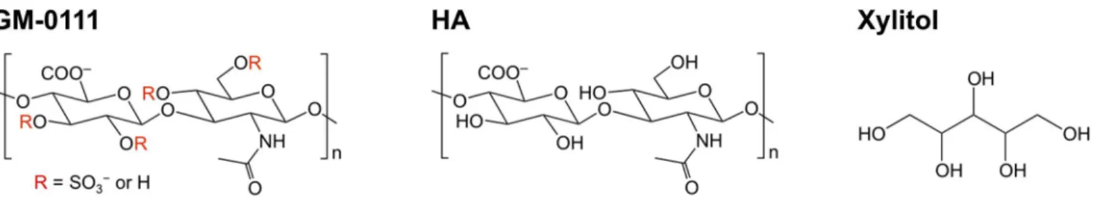
Glycosaminoglycans (GAGs), such as hyaluronic acid, heparin, and more recently synthetic GAGs, have been shown to interfere with bacterial growth, TLR-mediated signaling, and RANKL-induced osteoclast formation [35–41]. GM-0111, a highly sulfated GAG derived from hyaluronic acid (Fig 1), has been shown to reduce inflammation in animal models of bladder pain syndrome (previously known as interstitial cystitis) and rosacea [42,43]. In the present study, we investigated whether GM-0111 disrupts events associated with periodontitis such as the growth and biofilm formation ofP.gingivalis, TLR2/TLR4-mediated cellular activation and osteoclast formationin vitro. We report that GM-0111 inhibits multiple events spanning the early pro-inflammatory TLR signaling to molecular pathways activated at the later stage com-ponent of bone loss.
Materials and Methods
Bacterial culture and scanning electron microscopy
P.gingivalisstrain 2561 (ATCC 33277) was cultured at 37°C in an anaerobic chamber. We used a sterile tryptic soy broth medium containing: 3.0% (w/v) tryptic soy broth, 0.5% (w/v) yeast extract, 0.05% (w/v)L-cysteine hydrochloride, 5μg/mL hemin, and 1μg/mL vitamin K1. A.actinomycetemcomitans(ATCC 29523) was also cultured at 37°C in an anaerobic chamber with a few modifications: we used a sterile modified Todd Hewitt broth containing 450 mL of 0.037% Bacto Brain Heart Infusion broth mixed with 10 mL of 12.5% sodium bicarbonate solu-tion, pH adjusted to 7.8. The CO2level was maintained by dissolving Alka-Seltzer (Bayer
HealthCare, NJ) in a separate bottle within the culture chamber. Aliquots of an overnight cul-ture ofP.gingivalisand a 3 day culture ofA.actinomycetemcomitanswere sub-cultured in fresh medium containing varying concentrations of GM-0111 (detailed method for synthesis appears in reference [44]), HA, or xylitol. Bacterial growth was determined by measuring the absorbance at 600 nm as an approximate measure. We found the absorbance values were unre-liable due to the slightly yellow to brown color hues of compounds in culture medium and instead used flow cytometry analysis to count bacteria. These samples were diluted in sterile PBS and sonicated for 5 min in an ultrasonic water bath. Bacteria were then directly counted using preset forward- and side-scatter parameters set to detectP.gingivalisandA. actinomyce-temcomitansin the culture medium using the Guava HT-8 flow cytometer (EMD Millipore, MA) [45].
To determine the presence of biofilm formation, 100μL ofP.gingivalisovernight culture was added to 1.5 mL of tryptic broth in sterile 22 mm-glass bottom dishes (Ted Pella #14023– 20, Redding, CA). The dishes were gently rocked at 100 rpm for 36 hrs at 37°C in an anaerobic chamber. GM-0111 or HA (final concentration of 0, 0.1, 1, 5, and 10% w/v) was added to the dishes containing bacteria and gently rocked at 100 rpm for an additional 36 hrs at 37°C. The glass bottoms were removed, washed with a 0.15 M sodium cacodylate solution, and incubated in a 0.15 M sodium cacodylate solution containing 2% (w/v) glutaraldehyde, 2% (w/v) parafor-maldehyde, and 0.15% (w/v) alcian blue 8GX (dissolved in 3% acetic acid, pH 2.5) in water for 2 hrs at room temperature. These samples were washed 3 times with 0.15 M sodium cacodylate, incubated in a solution containing 0.15 M sodium cacodylate and 1% (v/v) osmium tetroxide for 1 hr at room temperature, and dehydrated in graded ethanol (70%, 20 min; 95%, 20 min; 100%, 30 min). Next, the samples were treated with hexamethyldisilizane for 5 min, desiccated, and sputter coated with gold using a Desk V HP sputterer (Denton Vacuum, Moorestown, NJ) under Ar gas at 30 mA for 30 sec. Samples were examined in blinded fashion using a Quanta 600 FEG scanning electron microscope (Field Emission, Inc., Hillsboro, OR).
GM-0111 effects on TLR2- and TLR4-mediated cellular responses
The effects of GM-0111 on TLR2- and TLR4-mediated cellular responses were determined using two different cell systems: (1) RAW 264.7 mouse macrophage cells endogenously expressing both receptors along with their signaling complexes and (2) HEK 293 cells heterolo-gously expressing TLR2 or TLR4 along with their respective co-receptors and reporter
proteins.
1. RAW 264.7 (ATCC # TIB-71) is a mouse macrophage cell line that expresses all the neces-sary molecules to activate both TLR2 and TLR4 [46–48]. RAW 264.7 cells were maintained in Dulbeco's modified Eagle medium (DMEM, #SH3028401, GE Healthcare HyClone, UT) supplemented with 50 U/mL penicillin/50μg/mL streptomycin (#15140–122, Thermo-Fisher Scientific, NY), and 10% heat inactivated fetal bovine serum (FBSi, #SH30071, GE Healthcare HyClone) in 75-cm2flasks. When the plates reached 50–70% confluency, cells were collected in PBS, washed 3 times in PBS, resuspended in DMEM supplemented with 2% FBSi. Cells were plated in 96-well plates at 3x10³cells/well and cultured overnight. The viability of cells was>85% per experiment. The following day, the cell culture medium was removed, and 180μL of fresh medium was added to each well. Cells were stimulated with serially diluted Pam3CSK4 (TLR2 agonist, tlrl-pms, InvivoGen) or LPS derived from the
Escherichia coli(E.coli) K12 strain (TLR4 agonist, tlrl-peklps). A separate set of plates was prepared with 160μL of fresh medium, 20μL of serially diluted GM-0111, and 20μL of a fixed concentration of Pam3CSK4 or LPS. After culturing for 24 hrs in the presence of ligand, the culture medium samples were collected from each plate and assayed for interleu-kin-6 (IL-6). The concentrations of IL-6 released from RAW 264.7 cells were measured using Mouse IL-6 ELISA MAX™Deluxe kits (BioLegend, CA).
at the time of harvest. To determine the effects of GM-0111 on TLR2 and TLR4 activation in cells, 20μL of varying concentrations of GM-0111 were added into 96-well plates. Then, 160μL of cells resuspended in DMEM supplemented with 10% FBSi were aliquoted into each well. HEK-Blue hTLR2 cells were stimulated with 20μL of Pam3CSK4, and HEK-Blue hTLR4 cells were stimulated with 20μL of LPS. The plates were incubated overnight at 37°C. The following day, 5–20μL of culture medium from each well was tested for TLR acti-vation by measuring SEAP activity using the Quanti-Blue assay reagent.
Osteoclast culture
The effects of GM-0111 on osteoclast formation were investigated by culturing primary precur-sor osteoclasts in the presence of varying concentrations of GM-0111. The precurprecur-sor cells derived from mouse bone marrow were obtained from a commercial source (PMC-OSC13--COS, B-Bridge International, CA) and cultured in a 96-well Osteo Assay plate coated with bone-mimicking, inorganic, crystalline calcium phosphate (Osteo Assay Surface #3989, Corn-ing, MA). Frozen precursor cells were thawed, rinsed with washing medium, and grown in the culture medium supplied by the vendor containing 50 ng/mL of macrophage-colony stimulat-ing factor (M-CSF) and 25 ng/mL of RANKL. Cell culture medium was replaced with fresh medium on day 3 or 4 after starting the culture. GM-0111 was dissolved in PBS at 100 mg/mL and further diluted in the osteoclast culture medium before adding to the cells.
Measurements of osteoclasts and activities
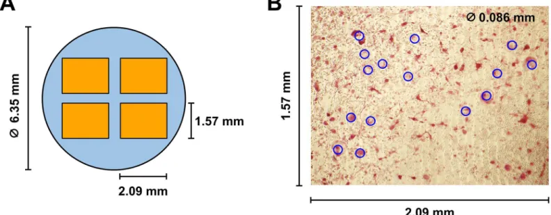
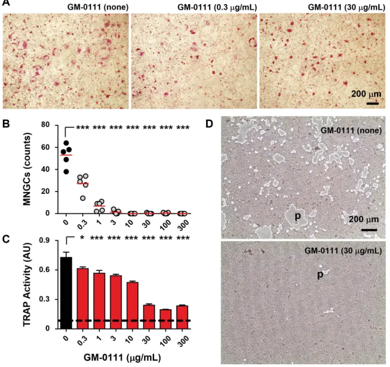
Osteoclasts secrete a large amount of tartrate-resistant acid phosphatase (TRAP) and the mea-surement of TRAP shows osteoclast differentiation and activity [49,50]. To determine the extent of osteoclast formation from precursor osteoclasts, cells in each well were fixed in for-malin and stained for TRAP using a commercially available kit (PMC-AK04F-COS, B-Bridge International). Each well was then photographed at four different areas with a frame size of 2.09 mm by 1.57 mm (Fig 2A). Osteoclasts were quantified by counting multinucleated giant cells (or TRAP-positive osteoclasts) with a diameter of86μm (blue circles,Fig 2B) in each
frame. Separate culture wells were treated with a 10% bleach solution to remove cells from the osteoplate. Culture wells were dried, and the resulting pits formed by bone resorbing osteo-clasts were photographed for qualitative comparisons. To quantify the effects of GM-0111 on the osteoclast differentiation and activity, we determined the enzymatic activity of TRAP pres-ent in cell culture medium. First, 30μL of cell culture medium from each well was mixed with 170μL of chromogenic substrate (PMC-AK04F-COS, b-bridge) and incubated for 3 hrs at 37°C. The amount of the resulting enzymatic product was then determined by measuring the absorbance at 540 nm using a microplate reader (Tecan Infinite1F200Pro, Austria).
GM-0111 binding to RANKL
To test whether 0111 inhibits RANKL binding to its receptor, RANK, we first measured GM-0111 binding to RANKL. Heparin binding plates (96-well, cat# 354676, BD Biosciences, MA) were coated with 200μL of GM-0111 (10μg/mL) dissolved in phosphate buffered saline (PBS) by incubating overnight at room temperature. The following day, the plate was washed with PBS-T (PBS containing 0.05% Tween-20), blocked with 1% bovine serum albumin (BSA) for 1 hr at 37°C, and then washed. Varying concentrations of recombinant human RANKL (cat# 390-TN, R&D systems, MN) in blocking solution were added to each well and incubated for 2 hrs at 37°C. The plate was then washed with PBS-T, and 50μL of anti-RANKL antibody solution (0.5μg/mL; AF626, R&D systems) was added to each well to detect RANKL bound to GM-0111. After 1 hr of incubation at room temperature, the plate was washed with PBS-T and 100μL of horseradish per-oxidase (HRP)-conjugated anti-goat IgG (ThermoScientific, cat# 31402) was added. After 1 hr of incubation at room temperature, the plate was washed with PBS-T, and the bound HRP was determined by incubating with 100μL of 3,30,5,50-tetramethylbenzidine solution (50-77-18, KPL
Inc., MD) as a substrate. The enzymatic reaction was stopped with 1 N HCl, and the absorbance at 450 nm was measured using a microplate reader (SpectraMax, Molecular Devices, CA). Binding affinity (KD) was calculated from the plot of the absorbancevs. the concentration of RANKL.
GM-0111 effects on RANKL binding to RANK
The effects of GM-0111 on RANKL binding to its receptor RANK were determined by measur-ing the interaction between RANKL and RANK in the presence of varymeasur-ing concentrations of GM-0111. Polyvinyl 96-well plates were coated with 500 ng/well of recombinant human RANK (cat# 683-RK, R&D systems). Separately, 100μL of RANKL (500 ng/mL in PBS-T sup-plemented with 0.1% BSA) was incubated with 100μL of serially diluted GM-0111 for approxi-mately 16 hrs at 4°C. The RANKL-GM-0111 mixtures were then transferred to the RANK-coated wells and incubated for 1 hr at 37°C. The plates were then washed with PBS-T, and the RANK-bound RANKL was measured following the same steps described for the GM-0111/ RANKL binding studies to detect RANKL. The resulting absorbance of RANKL-RANK bind-ing was plotted against the GM-0111 concentration.
GM-0111 effects on RANKL-mediated cellular response
and washed with PBS 4 times. The viability of the cells was>90% per experiment. To stimulate cells, 20μL of serially diluted RANKL solutions were added into 96-well plates, and 180μL of cell suspension was then added into each well. In separate sets of experiments, 20μL of serially diluted 10x RANKL solutions and 20μL of a GM-0111 solution were added into 96-well plates, and 160μL of cell suspension (5x10⁵cells/well) was added into each well. Cell suspensions and compound dilutions were made with DMEM supplemented with 5% FBSi. Cells were stimu-lated with variable concentrations of RANKL for 48 hrs. Aliquots (5–20μL) of culture medium from each well were tested for SEAP activity using the QUANTI-Blue™(rep-qb, InvivoGen) alkaline phosphatase detection reagent.
Data Analysis
Biochemical measurements and the number of multinucleated giant cell counts were expressed as the mean and standard of error of the mean (SEM). All measurement data were tested for homogeneity with either Bartlett's test or Fligner Killeen test for equal variances as well as visual inspection of histograms to check the distribution of data [51]. All data sets showed homoge-neous distribution supporting the use of parametric tests. The mean differences among treated groups were tested for statistical significance byone-wayanalysis of variance test followed by Dunnet'st-test or Tukey's multiple comparison test aspost hoc. Ligand binding data—RANKL, RANK, and GM-0111—were plotted and fitted by the equation Y = a × [Ligand] + b + (Bmax×
[Ligand])/(KD+ [Ligand]) to estimate KD(equilibrium binding constant). Statistical analyses
and curve-fits were performed with GraphPad Prism 5.0.4 (GraphPad Software, Inc.) or R (ver-sion 3.0.2, The R Foundation for Statistical Computing).
Results
Antibacterial effects of GM-0111 on
P
.
gingivalis
and
A
.
actinomycetemcomitans
To test whether GM-0111 suppresses bacterial growth, we measured the effects of GM-0111 on
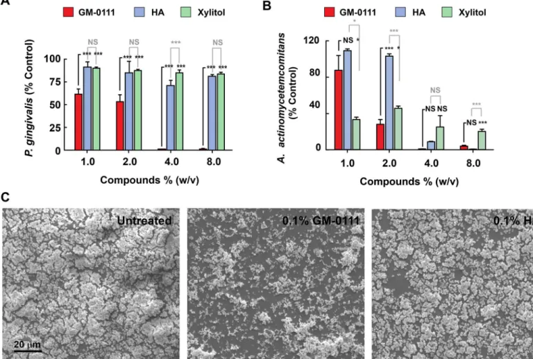
P.gingivalisandA.actinomycetemcomitansgrowth. At 1–2% (w/v), GM-0111 suppressedP.
gingivalisby 50–60% andA.actinomycetemcomitansgrowth by 20–80%, and completely inhib-ited the growth of both organisms at 4% (w/v) in the medium (Fig 3A and 3B). The antibacte-rial effects of 0111 were compared with HA, the starting mateantibacte-rial for the synthesis of GM-0111, and xylitol, commonly used in the oral cavity to prevent dental caries [52]. HA or xylitol had negligible effects onP.gingivalisgrowth, but did inhibitA.actinomycetemcomitanslike GM-0111 (Fig 3A and 3B). These data suggest that GM-0111 is more effective than HA and xylitol in suppressing the growths ofP.gingivalisand is as effective as HA and xylitol in sup-pressingA.actinomycetemcomitansgrowth.
GM-0111 suppresses the biofilm formation of
P
.
gingivalis
To determine whether GM-0111 reduces biofilm formation,P.gingivaliswas cultured as a bio-film on a glass surface in the presence of 0–10% GM-0111 and HA. Biobio-film formation was markedly reduced by 0.1% (w/v) GM-0111 (Fig 3C, middle panel), in contrast to 0% GM-0111 and 0.1% HA (Fig 3C, left and right panels). GM-0111 therefore suppressesP.gingivalisgrowth both in liquid culture and as a biofilm.
GM-0111 blocks TLR2- and TLR4-mediated cellular responses
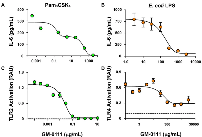
Upon stimulation with the TLR2- or TLR4-specific agonists (Pam3CSK4 or LPS from theE.
coliK strain, respectively), RAW 264.7 cells produced and secreted IL-6 (Fig 4A and 4B). How-ever, the amount of IL-6 released from RAW 264.7 cells was dose-dependently decreased when these cells were treated with GM-0111. These data suggest that GM-0111 inhibits TLR-medi-ated cytokine release.
To delineate the specificity of GM-0111 on TLR2 and TLR4, we measured TLR2- and TLR4-mediated cellular activation of SEAP reporter protein by NF-κB and AP-1 in HEK-Blue hTLR2 and HEK-Blue hTLR4 cells. Treatment with GM-0111 demonstrated dose-dependent inhibition of TLR2- and TLR4-mediated cellular signaling (Fig 4C and 4D) in HEK-Blue cells. These data are consistent with our RAW 264.7 cell results showing that GM-0111 blocks both TLR2 and TLR4 signaling. In addition, our data indicate that GM-0111 blocks TLR2-mediated signaling at sub-μg/mL concentrations compared to the hundreds ofμg/mL inhibitory concen-tration for TLR4-mediated signaling, illustrating that GM-0111 is a TLR inhibitor with high selectivity to TLR2. Overall, our data demonstrate that GM-0111 inhibits TLR-mediated pro-inflammatory cellular signaling, which is part of the pathogenesis in periodontitis.
Fig 3. GM-0111 suppresses the growth ofP.gingivalisandA.actinomycetemcomitansand inhibits the biofilm formation ofP.gingivalis.Flow cytometry-assistedP.gingivalis(A) andA.actinomycetemcomitans(B) counts, grown in the presence of GM-0111, HA, or xylitol show increased growth suppression by GM-0111.C, Scanning electron microscopy images ofP.gingivaliscultures grown with GM-0111 (0.1% w/v, middle panel) show reduced biofilm formation compared to control and HA (left and right panels). InAandB, bars and error bars represent mean±SEM (n = 4).*p<0.05,
GM-0111 inhibits RANKL-induced osteoclast formation
Pre-osteoclasts derived from mouse bone marrow differentiate into TRAP-positive, multi-nucleated giant cells (MNGCs or osteoclasts) within four days when cultured in the presence of M-CSF and recombinant RANKL (rRANKL). When treated with GM-0111, the differentiation of pre-osteoclasts into MNGCs was markedly suppressed (Fig 5A). The anti-osteoclastic effect of GM-0111 was apparent even at 300 ng/mL (Fig 5B). These observations are consistent with our biochemical analysis of TRAP activity in culture medium (Fig 5C), which showed signifi-cantly reduced osteoclastic activity in GM-0111-treated cells. In addition, the extent of the bone resorption pits formed by osteoclasts was reduced considerably with GM-0111 treatment (Fig 5D). These results suggest that GM-0111 blocks RANKL-induced osteoclast formation and also reduces consequential bone resorption.
Fig 4. GM-0111 blocks TLR2- and TLR4-mediated NF-κB signaling.Mouse macrophage RAW 264.7 cells secrete IL-6 when stimulated with the TLR2
agonist Pam3CSK4 (1 ng/mL) (A) or the TLR4 agonist LPS (1 ng/mL) derived fromE.coli(B) (n = 4). GM-0111 blocks these cellular responses by inhibiting TLR2/TLR4-induced NF-κB activation. HEK-Blue hTLR2 (C) and HEK-Blue hTLR4 (D) cells release reporter protein SEAP (activity measured in Relative Absorbance Unit, RAU) in response to agonist-induced NF-κB activation (n = 5). Symbols and error bars are mean±SEM. Dotted horizontal lines represent the baseline values of SEAP activities in the culture medium.
Fig 5. GM-0111 prevents multinucleated giant osteoclast formation.Mouse bone marrow derived pre-osteoclast cells cultured with M-CSF (50 ng/ mL) and RANKL (25 ng/mL) transformed into TRAP-positive MNGCs in 4 days (A). The number of MNGCs was significantly decreased when pre-osteoclast cells were cultured in the presence of GM-0111 (AandB). GM-0111 also reduced TRAP secretion from MNGCs (C) and the resorption of bone mimetic matrix with the resulting pit formation (empty gray areas, p inD). Red lines inBrepresent mean values. InC, bars and error bars are mean±SEM (n = 5) and the dotted horizontal line represent the baseline value of TRAP activity (measured in Absorbance Unit, AU) in culture medium.
GM-0111 does not block RANKL-RANK-mediated signaling
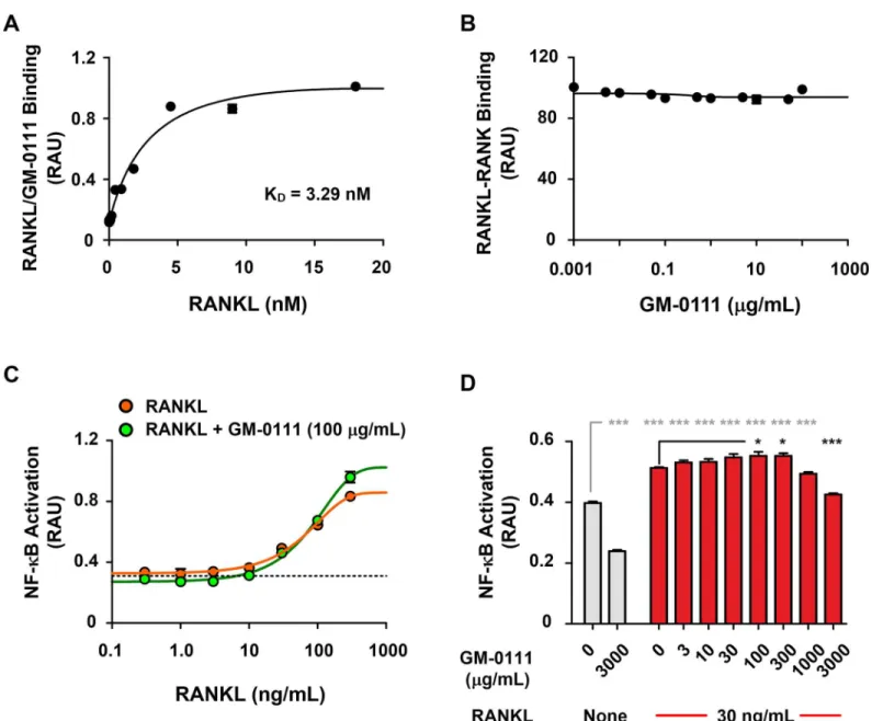
To test the possibility that GM-0111 reduces, RANK activation, we measured the concentration of RANKL bound to a given amount of GM-0111 coated onto 96-well microplates. We found that the interaction between GM-0111 and rRANKL was strong, with a dissociation constant (KD) of 3.29 nM (Fig 6A). This binding affinity is similar to monomeric OPG-RANKL binding
(~ 4.24 nM), but is approximately 500-fold stronger than RANKL to RANK binding [53]. The high affinity binding of GM-0111 to RANKL may be a major contributing mechanism, interfering with the interaction between RANKL and RANK. To further identify whether GM-0111/RANKL binding can reduce the RANKL-RANK interaction, we first mixed varying
Fig 6. GM-0111 inhibits osteoclast formation independent of RANKL-RANK interaction.GM-0111 binds to RANKL with high affinity (KD= 3.29 nM) (A), but does not inhibit the interaction between RANKL and its receptor RANK (B) (n = 3). RANKL induces NF-κB-mediated cell signaling in a dose-dependent manner (C), but GM-0111 does not reduce RANKL-induced NF-κB signaling (CandD) (n = 4). Symbols and bars are mean values. Error bars represent SEM.*p<0.05 and***p<0.001 (compared to the controls).
concentrations of GM-0111 with rRANKL and added these mixtures to a 96-well microplate coated with rRANK. We then measured GM-0111/rRANKL-bound rRANK. As illustrated in
Fig 6B, GM-0111 premixed with rRANKL did not alter the binding properties of rRANKL to rRANK. These results indicate that GM-0111 does not interfere with RANKL-RANK interac-tion. The absence of blocking effects of GM-0111 on the RANKL-RANK interaction could be a consequence of the recombinant RANK protein lacking transmembrane and intracellular sig-naling domains present in the native protein. We therefore cannot rule out the possibility that GM-0111 inhibits the RANKL-RANK interaction. We therefore used RAW-Blue cells to moni-tor the level of downstream SEAP expression to monimoni-tor whether GM-0111 inhibits the RANKL-RANK-NF-κB signaling cascade [33]. When stimulated with RANKL, RAW-Blue cells produced and secreted SEAP in a dose-dependent manner as demonstrated inFig 6C. However, the effects of GM-0111 on RANKL-induced RANK activation were weak, with reduced activa-tion only observable at very high concentraactiva-tions (3000μg/mL,Fig 6C and 6D). These data dem-onstrate that although GM-0111 does bind to RANKL with high affinity, it does not interfere with RANKL-RANK binding or RANKL-induced RANK signaling. Thus, at present, the spe-cific mechanism by which GM-0111 suppresses osteoclast formation remains unclear.
Discussion
Disrupting the balance between a host and its oral microbiota is a critical element contributing to periodontitis. Therefore, therapeutic strategies to restore host-commensal homeostasis is a logical approach. Current therapy for periodontitis focuses on removing inflammatory sources in the peritoneum by scaling and root planing (SRP), which is often followed by supplemental topical antibiotic treatments to reduce recurring pathogenic bacterial growth in the periodon-tium. Despite problems of antibiotic resistant microorganisms [54–56], the development of antibiotics such as tetracyclines, quinolones and nitronidazoles continues as an adjunct treat-ment to SRP [57–60]. Prolonged use of antibiotics can alter the oral microbiome, leading to the establishment of an abnormal microbial ecosystem [61,62]. Strategies to reduce the inflamma-tory mediators will therefore likely reduce such unintended consequences [25].
Emerging evidence supports the idea that periodontitis could be treated by inhibiting pro-inflammatory molecular signaling pathways such as NF-κB or poly (ADP-ribose) polymerase (PARP) activation, as demonstrated in an animal model of periodontitis [63,64]. GAGs such as HA and synthetic GAGs are anti-inflammatory by inhibiting multiple molecular targets of inflammation. One of these molecular targets include the family of TLRs [65,66]. Bacterial products such as LPS and lipopetides activate TLRs, which induce pro-inflammatory cytokine productions through NF-κB activation [25,67–69]. Our data demonstrate that GM-0111 can inhibit pro-inflammatory cytokine release by blocking TLR2 and TLR4 (Fig 4andS1 Figand
S1 File). The enhanced expression of TLRs in the periodontium with periodontitis [7,70], as well as significant contribution of TLR2 to alveolar bone loss [71–73] suggest that the inhibi-tory effects of GM-0111 on TLRs may provide a therapeutic benefit in periodontitis.
Our study did not clarify how GM-0111 inhibits the differentiation of pre-osteoclast to bone-resorbing osteoclasts. One possibility is that GM-0111, as well as other types of GAGs, directly binds to RANKL and reduces the activation of RANK. This hypothesis is based on the mecha-nism of OPG-RANKL interactions, in which free RANKL is directly sequestered by OPG [75,76]. In this scenario, GAGs may function as a scavenger to reduce the amount of free RANKL. Such a possibility has been proposed as a mechanism of heparin to reduce RANKL-induced osteoclast formation [77]. An alternative possibility is that GAG-bound RANKL may block the interaction between RANKL and RANK. Crystal structure studies of OPG bound RANKL suggest that the RANK interacting regions of RANKL become inaccessible upon binding OPG [53,78]. Our data demonstrates that GM-0111 does not inhibit RANKL to RANK binding, which contradicts this scenario. RANKL-induced oligomerization of RANK is a critical step for osteoclastogenesis [74]. It is possible that the GM-0111 may prevent oligomerization of RANKL or inhibit RANKL-induced oligomerization of RANK [74]. It remains difficult to explain the lack of inhibitory effects of GM-0111 on RANKL-induced NF-κB activation in RAW cells.
Recent advances in our understanding of various diseases affecting bone homeostasis pro-vided insights into solving these discrepancies. Although RANKL-RANK signaling is critical for osteoclast formation, cytokines also contribute to osteoclast formation. For example, IL-1β, IL-6, and TNF-αstimulate RANKL production in osteoblasts [79]. GAGs such as heparin reduce the effects of these cytokines [80–82]. GM-0111 shares various biochemical properties of heparin due to its structural similarities and may interact with cytokines affecting osteoclast formation. Furthermore, recent studies indicate that TLRs play important roles in pathogen-mediated osteoclast formation. Activation of both TLR2 and TLR4 receptors are linked to increased expression of RANKL in fibroblast-like synoviocytes in rheumatoid arthritis [83]. Additionally, TLR2 is a necessary component forP.gingivalis-mediated inflammation and bone loss in mice [84]. Thus, GM-0111 inhibition of TLR2 signaling may provide a clue to the inhibitory effect of this modified GAG on osteoclastogenesis.
Our data suggest that GM-0111 can reduce RANKL-induced osteoclast formation. But what is the source of RANKL and which cells express RANK? More specifically, which cells express high levels of RANKL-RANK that are highly activated by RANKL-RANK interaction? The Crotti and Giannopoulou groups found that the inflammatory cells within the periodontium from periodontitis patients express abnormally higher amounts of RANKL and RANK [85,86]. More specifically, both activated B cells and Th1 lymphocytes were suggested as the major source of RANKL to increase osteoclastogenesis [87]. These cells play important roles in chronic inflammatory lesions. Therefore, prolonged periodontal inflammation consequent to bacterial infection likely contributes to the lymphocytic infiltration and the resulting increased RANKL-RANK promotes osteoclastogenesis.
Biofilm formation by pathogenic bacteria enhances their survival and infection, which con-tributes to chronic periodontitis. The contributions of different bacterial species in oral micro-bial ecosystem have been extensively discussed by Socransky and Haffajee [61]. These authors discuss how successive additions of different types of microbial species contribute to periodon-titis. For example, an initial colonization of bacteria such as yellow/green/purple microbial complexes create a microenvironment favorable for orange and red microbial complexes that are more pathogenic [61,88]. This sequential colonization model has been regarded as impor-tant for designing therapeutic strategies that should focus on creating a microenvironment favorable for less harmful symbiotic microorganisms such as removing dental plaques supple-mented with broad spectrum antibiotics.
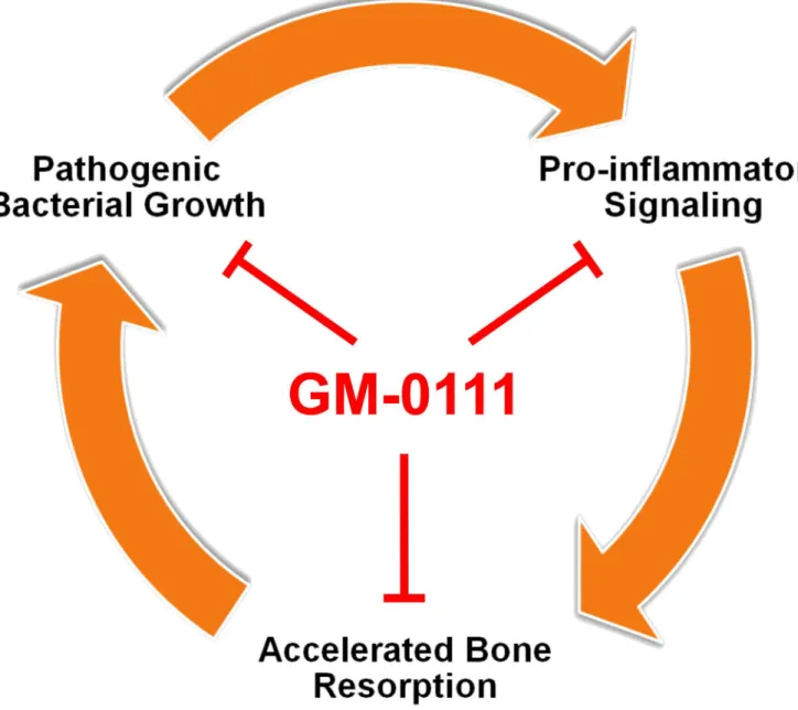
degree of colonization ofP.gingivaliswithout the successive colonization of other bacterial spe-cies in rodents [90]. The keystone pathogen hypothesis provides a new insight into designing therapeutic strategies. In a ligature-induced periodontitis model, vaccination of non-human primates against a specific cysteine protease fromP.gingivaliswas effective in reducing peri-odontitis [91]. These findings suggest that dysbiosis caused byP.gingivaliscould be a mecha-nism of periodontitis and targeting a set of specific pathogens may provide therapeutic benefit without the risks of using antibiotics. Our data demonstrate that GM-0111 can suppress the growth ofP.gingivaliscommonly associated with periodontitis. We also showed that GM-0111 suppresses the growth ofA.actinomycetemcomitansassociated mostly with localized aggressive periodontitis [19,92]. When used as a prophylactic, GM-0111 may suppress pathogenic bacte-rial growth and consequently reduce periodontal inflammation (Fig 7).
Fig 7. Proposed mechanism of GM-0111 to treat or to prevent periodontitis.GM-0111 (1) reduces the source of inflammatory mediators by suppressingP.gingivalisgrowth and biofilm formation; (2) directly blocks pro-inflammatory signaling mediated by TLRs; and (3) also inhibits alveolar bone destruction by preventing osteoclast formation.
Conclusion
In the present study, we investigated the effects of GM-0111 on molecular events associated with periodontitis, ranging from the anti-inflammatory effects mediated by TLR2 and TLR4, RANKL-induced osteoclast formation, and pathogenic bacterial growth. We propose that the combined effects of GM-0111 on these multiple mediators of periodontitis will be beneficial as a prophylactic or therapeutic for periodontitis. Further studies in animal models of periodonti-tis will delineate the efficacy as well as the significance of these molecular targets.
Supporting Information
S1 Fig. Flow cytometry analysis showing that GM-0111 does not directly interact with Pam3CSK4 to inhibit TLR2-mediated cell signaling.(A) GM-0111 functionalizes into pLL-coated microbeads. GM-0111CF633 functionalized microbeads were highly fluorescent com-pared to pLL-coated beads (solid line vs. red histogram). (B) GM-0111-functionalized microbeads were incubated with Pam3CSK4Rhodamine. Histograms show slight increase in fluorescence of GM-0111-functionalized microbeads mixed with 1000 ng/mL of Pam3CSK4R-hodamine. (solid line vs. cyan vs. red histogram). (C) Pam3CSK4RPam3CSK4R-hodamine. nonspecifically binds to pLL-coated beads. pLL-coated microbeads were mixed with 0 or 1000 ng/mL of Pam3CSK4Rhodamine (solid line vs. red histogram). (D) GM-0111 does not quench Pam3CSK4Rhodamine fluorescence. GM-0111 (without the beads) was mixed with
Pam3CSK4Rhodamine and the resulting fluorescence measured. Pam3CSK4Rhodamine fluo-rescence intensity did not change with GM-0111 (red vs. black bars). Bars are mean and error bars are S.D. values (n = 4).
(TIF)
S1 File. Does GM-0111 bind to Pam3CSK4? (DOCX)
Author Contributions
Conceived and designed the experiments: JRS AP TPK MER WYL. Performed the experi-ments: JRS AP NVR WYL. Analyzed the data: JRS AP NVR WYL. Wrote the paper: WYL JRS AP MER TPK GDP.
References
1. Acharya A, VanWormer JJ, Waring SC, Miller AW, Fuehrer JT, Nycz GR. Regional Epidemiologic Assessment of Prevalent Periodontitis Using an Electronic Health Record System. Am J Epidemiol. 2013; 177: 700–707. doi:10.1093/aje/kws293PMID:23462966
2. Eke PI, Dye BA, Wei L, Thornton-Evans GO, Genco RJ. Prevalence of Periodontitis in Adults in the United States: 2009 and 2010. J DENT RES. 2012; 91: 914–920. doi:10.1177/0022034512457373 PMID:22935673
3. Petersen PE, Ogawa H. Strengthening the Prevention of Periodontal Disease: The WHO Approach. Journal of Periodontology. 2005; 76: 2187–2193. doi:10.1902/jop.2005.76.12.2187PMID:16332229
4. Hajishengallis G. Immuno-microbial pathogenesis of periodontitis: Keystones, pathobionts, and the host response. Trends Immunol. 2014; 35: 3–11. doi:10.1016/j.it.2013.09.001PMID:24269668
5. Paquette DW, Williams RC. Modulation of host inflammatory mediators as a treatment strategy for peri-odontal diseases. Periodontology 2000. 2000; 24: 239–252. doi:10.1034/j.1600-0757.2000.2240112.x PMID:11276870
7. Beklen A, Hukkanen M, Richardson R, Konttinen YT. Immunohistochemical localization of Toll-like receptors 1–10 in periodontitis. Oral Microbiol Immunol. 2008; 23: 425–431. doi:10.1111/j.1399-302X. 2008.00448.xPMID:18793367
8. Gribar SC, Richardson WM, Sodhi CP, Hackam DJ. No Longer an Innocent Bystander: Epithelial Toll-Like Receptor Signaling in the Development of Mucosal Inflammation. Mol Med. 2008; 14: 645–659. doi:10.2119/2008-00035.GribarPMID:18584047
9. Maezono H, Noiri Y, Asahi Y, Yamaguchi M, Yamamoto R, Izutani N, et al. Antibiofilm effects of azithro-mycin and erythroazithro-mycin on Porphyromonas gingivalis. Antimicrob Agents Chemother. 2011; 55: 5887–
5892. doi:10.1128/AAC.05169-11PMID:21911560
10. Mah T-F, Pitts B, Pellock B, Walker GC, Stewart PS, O’Toole GA. A genetic basis for Pseudomonas aeruginosa biofilm antibiotic resistance. Nature. 2003; 426: 306–310. doi:10.1038/nature02122PMID: 14628055
11. Noguchi N, Noiri Y, Narimatsu M, Ebisu S. Identification and Localization of Extraradicular Biofilm-Forming Bacteria Associated with Refractory Endodontic Pathogens. Appl Environ Microbiol. 2005; 71: 8738–8743. doi:10.1128/AEM.71.12.8738-8743.2005PMID:16332869
12. Mysak J, Podzimek S, Sommerova P, Lyuya-Mi Y, Bartova J, Janatova T, et al. Porphyromonas gingi-valis: Major Periodontopathic Pathogen Overview. J Immunol Res. 2014; 2014. doi:10.1155/2014/ 476068
13. Maekawa T, Krauss JL, Abe T, Jotwani R, Triantafilou M, Triantafilou K, et al. Porphyromonas gingivalis Manipulates Complement and TLR Signaling to Uncouple Bacterial Clearance from Inflammation and Promote Dysbiosis. Cell Host & Microbe. 2014; 15: 768–778. doi:10.1016/j.chom.2014.05.012
14. Scapoli L, Girardi A, Palmieri A, Testori T, Zuffetti F, Monguzzi R, et al. Microflora and periodontal dis-ease. Dent Res J (Isfahan). 2012; 9: S202–206. doi:10.4103/1735-3327.109755
15. Siddiqui H, Yoder-Himes DR, Mizgalska D, Nguyen K-A, Potempa J, Olsen I. Genome Sequence of Porphyromonas gingivalis Strain HG66 (DSM 28984). Genome Announc. 2014; 2. doi:10.1128/ genomeA.00947-14
16. Nelson KE, Fleischmann RD, DeBoy RT, Paulsen IT, Fouts DE, Eisen JA, et al. Complete genome sequence of the oral pathogenic Bacterium porphyromonas gingivalis strain W83. J Bacteriol. 2003; 185: 5591–5601. PMID:12949112
17. Andrian E, Grenier D, Rouabhia M. Porphyromonas Gingivalis-Epithelial Cell Interactions in Periodonti-tis. J DENT RES. 2006; 85: 392–403. doi:10.1177/154405910608500502PMID:16632751
18. Kelk P, Abd H, Claesson R, Sandström G, Sjöstedt A, Johansson A. Cellular and molecular response of human macrophages exposed to Aggregatibacter actinomycetemcomitans leukotoxin. Cell Death Dis. 2011; 2: e126. doi:10.1038/cddis.2011.6PMID:21390060
19. Raja M, Ummer F, Dhivakar C. Aggregatibacter Actinomycetemcomitans—A Tooth Killer? J Clin Diagn Res. 2014; 8: ZE13–ZE16. doi:10.7860/JCDR/2014/9845.4766
20. Coats SR, Jones JW, Do CT, Braham PH, Bainbridge BW, To TT, et al. Human Toll-like receptor 4 responses to P. gingivalis are regulated by lipid A 1- and 4’- phosphatase activities. Cell Microbiol. 2009; 11: 1587–1599. doi:10.1111/j.1462-5822.2009.01349.xPMID:19552698
21. Yoshimura A, Kaneko T, Kato Y, Golenbock DT, Hara Y. Lipopolysaccharides from Periodontopathic Bacteria Porphyromonas gingivalis and Capnocytophaga ochracea Are Antagonists for Human Toll-Like Receptor 4. Infect Immun. 2002; 70: 218–225. doi:10.1128/IAI.70.1.218-225.2002PMID: 11748186
22. Zhou Q, Desta T, Fenton M, Graves DT, Amar S. Cytokine Profiling of Macrophages Exposed to Por-phyromonas gingivalis, Its Lipopolysaccharide, or Its FimA Protein. Infect Immun. 2005; 73: 935–943. doi:10.1128/IAI.73.2.935-943.2005PMID:15664935
23. Enersen M, Nakano K, Amano A. Porphyromonas gingivalis fimbriae. J Oral Microbiol. 2013; 5. doi:10. 3402/jom.v5i0.20265
24. Garlet GP. Destructive and protective roles of cytokines in periodontitis: a re-appraisal from host defense and tissue destruction viewpoints. J Dent Res. 2010; 89: 1349–1363. doi:10.1177/ 0022034510376402PMID:20739705
25. Deo V, Bhongade ML. Pathogenesis of periodontitis: role of cytokines in host response. Dent Today. 2010; 29: 60–62, 64–66; quiz 68–69. PMID:20973418
26. Endo Y, Tomofuji T, Ekuni D, Irie K, Azuma T, Tamaki N, et al. Experimental periodontitis induces gene expression of proinflammatory cytokines in liver and white adipose tissues in obesity. J Periodontol. 2010; 81: 520–526. doi:10.1902/jop.2009.090574PMID:20367095
28. Zhang W, Ju J, Rigney T, Tribble G. Porphyromonas gingivalis infection increases osteoclastic bone resorption and osteoblastic bone formation in a periodontitis mouse model. BMC Oral Health. 2014; 14: 89. doi:10.1186/1472-6831-14-89PMID:25027664
29. Scheres N, de Vries TJ, Brunner J, Crielaard W, Laine ML, Everts V. Diverse effects of Porphyromonas gingivalis on human osteoclast formation. Microb Pathog. 2011; 51: 149–155. doi:10.1016/j.micpath. 2011.04.006PMID:21539907
30. Bodet C, Chandad F, Grenier D. [Pathogenic potential of Porphyromonas gingivalis, Treponema denti-cola and Tannerella forsythia, the red bacterial complex associated with periodontitis]. Pathol Biol. 2007; 55: 154–162. doi:10.1016/j.patbio.2006.07.045PMID:17049750
31. Krajewski AC, Biessei J, Kunze M, Maersch S, Perabo L, Noack MJ. Influence of lipopolysaccharide and interleukin-6 on RANKL and OPG expression and release in human periodontal ligament cells. APMIS. 2009; 117: 746–754. doi:10.1111/j.1600-0463.2009.02532.xPMID:19775343
32. Wada T, Nakashima T, Hiroshi N, Penninger JM. RANKL–RANK signaling in osteoclastogenesis and bone disease. Trends in Molecular Medicine. 2006; 12: 17–25. doi:10.1016/j.molmed.2005.11.007 PMID:16356770
33. Ariyoshi W, Takahashi T, Kanno T, Ichimiya H, Takano H, Koseki T, et al. Mechanisms Involved in Enhancement of Osteoclast Formation and Function by Low Molecular Weight Hyaluronic Acid. J Biol Chem. 2005; 280: 18967–18972. doi:10.1074/jbc.M412740200PMID:15757905
34. Jeganathan S, Fiorino C, Naik U, Sun H song, Harrison RE. Modulation of Osteoclastogenesis with Macrophage M1- and M2-Inducing Stimuli. PLoS ONE. 2014; 9: e104498. doi:10.1371/journal.pone. 0104498PMID:25101660
35. Ebid R, Lichtnekert J, Anders H-J. Hyaluronan Is Not a Ligand but a Regulator of Toll-Like Receptor Signaling in Mesangial Cells: Role of Extracellular Matrix in Innate Immunity. ISRN Nephrol. 2014; 2014. doi:10.1155/2014/714081
36. Hirabara S, Kojima T, Takahashi N, Hanabayashi M, Ishiguro N. Hyaluronan inhibits TLR-4 dependent cathepsin K and matrix metalloproteinase 1 expression in human fibroblasts. Biochem Biophys Res Commun. 2013; 430: 519–522. doi:10.1016/j.bbrc.2012.12.003PMID:23232115
37. Salbach J, Kliemt S, Rauner M, Rachner TD, Goettsch C, Kalkhof S, et al. The effect of the degree of sulfation of glycosaminoglycans on osteoclast function and signaling pathways. Biomaterials. 2012; 33: 8418–8429. doi:10.1016/j.biomaterials.2012.08.028PMID:22954516
38. Pollari S, Käkönen RS, Mohammad KS, Rissanen JP, Halleen JM, Wärri A, et al. Heparin-like Polysac-charides Reduce Osteolytic Bone Destruction and Tumor Growth in a Mouse Model of Breast Cancer Bone Metastasis. Mol Cancer Res. 2012; 10: 597–604. doi:10.1158/1541-7786.MCR-11-0482PMID: 22522458
39. Ling L, Murali S, Stein GS, van Wijnen AJ, Cool SM. Glycosaminoglycans modulate RANKL induced osteoclastogenesis. J Cell Biochem. 2010; 109: 1222–1231. doi:10.1002/jcb.22506PMID:20135643
40. Pirnazar P, Wolinsky L, Nachnani S, Haake S, Pilloni A, Bernard GW. Bacteriostatic Effects of Hyal-uronic Acid. Journal of Periodontology. 1999; 70: 370–374. doi:10.1902/jop.1999.70.4.370PMID: 10328647
41. Rosett W, Hodges GR. Antimicrobial activity of heparin. J Clin Microbiol. 1980; 11: 30–34. PMID: 6766462
42. Lee WY, Savage JR, Zhang J, Jia W, Oottamasathien S, Prestwich GD. Prevention of Anti-microbial Peptide LL-37-Induced Apoptosis and ATP Release in the Urinary Bladder by a Modified Glycosamino-glycan. PLoS ONE. 2013; 8: e77854. doi:10.1371/journal.pone.0077854PMID:24204996
43. Zhang J, Xu X, Rao NV, Argyle B, McCoard L, Rusho WJ, et al. Novel Sulfated Polysaccharides Disrupt Cathelicidins, Inhibit RAGE and Reduce Cutaneous Inflammation in a Mouse Model of Rosacea. PLoS One. 2011; 6. doi:10.1371/journal.pone.0016658
44. Prestwich GD, Zhang J, Kennedy TP, Rao NV. Alkylated semi synthetic glycosaminoglycosan ethers, and methods for making and using thereof. US8329673 B2, 2012.
45. Gunasekera TS, Attfield PV, Veal DA. A Flow Cytometry Method for Rapid Detection and Enumeration of Total Bacteria in Milk. Appl Environ Microbiol. 2000; 66: 1228–1232. PMID:10698799
46. Park BS, Lee J-O. Recognition of lipopolysaccharide pattern by TLR4 complexes. Exp Mol Med. 2013; 45: e66. doi:10.1038/emm.2013.97PMID:24310172
47. Applequist SE, Wallin RPA, Ljunggren H-G. Variable expression of Toll-like receptor in murine innate and adaptive immune cell lines. Int Immunol. 2002; 14: 1065–1074. doi:10.1093/intimm/dxf069PMID: 12202403
49. Kremer M, Judd J, Rifkin B, Auszmann J, Oursler MJ. Estrogen modulation of osteoclast lysosomal enzyme secretion. J Cell Biochem. 1995; 57: 271–279. doi:10.1002/jcb.240570211PMID:7759564
50. Alatalo SL, Halleen JM, Hentunen TA, Mönkkönen J, Väänänen HK. Rapid Screening Method for Oste-oclast Differentiation in Vitro That Measures Tartrate-resistant Acid Phosphatase 5b Activity Secreted into the Culture Medium. Clinical Chemistry. 2000; 46: 1751–1754. PMID:11067809
51. de Smith MJ. STATSREF: Statistical Analysis Handbook—a web-based statistics resource. [Internet]. 2015 [cited 28 Oct 2015]. Available:http://www.statsref.com/HTML/inde
52. Ardizzoni A, Neglia RG, Baschieri MC, Cermelli C, Caratozzolo M, Righi E, et al. Influence of hyaluronic acid on bacterial and fungal species, including clinically relevant opportunistic pathogens. J Mater Sci Mater Med. 2011; 22: 2329–2338. doi:10.1007/s10856-011-4408-2PMID:21892787
53. Nelson CA, Warren JT, Wang MWH, Teitelbaum SL, Fremont DH. RANKL employs distinct binding modes to engage RANK and the OPG decoy receptor. Structure. 2012; 20: 1971–1982. doi:10.1016/j. str.2012.08.030PMID:23039992
54. Ardila CM, Granada MI, Guzmán IC. Antibiotic resistance of subgingival species in chronic periodontitis patients. J Periodont Res. 2010; 45: 557–563. doi:10.1111/j.1600-0765.2010.01274.xPMID: 20546113
55. Jungermann GB, Burns K, Nandakumar R, Tolba M, Venezia RA, Fouad AF. Antibiotic resistance in pri-mary and persistent endodontic infections. J Endod. 2011; 37: 1337–1344. doi:10.1016/j.joen.2011. 06.028PMID:21924178
56. Serrano C, Torres N, Valdivieso C, Castaño C, Barrera M, Cabrales A. Antibiotic resistance of peri-odontal pathogens obtained from frequent antibiotic users. Acta Odontol Latinoam. 2009; 22: 99–104. PMID:19839485
57. Chhina S, Rathore AS, Juneja S. Alpha-2-Macroglobulin Levels in Gingival Crevicular Fluid Pre- and Post-scaling and Root Planing with Adjunctive Tetracycline Fibers in Chronic Periodontitis: A Random-ized Controlled Trial. J Contemp Dent Pract. 2015; 16: 474–478. PMID:26323451
58. Khan G, Yadav SK, Patel RR, Nath G, Bansal M, Mishra B. Development and Evaluation of Biodegrad-able Chitosan Films of Metronidazole and Levofloxacin for the Management of Periodontitis. AAPS PharmSciTech. 2015; doi:10.1208/s12249-015-0466-y
59. Priyanka N, Kalra N, Saquib S, Saquib S, Malgaonkar N, Nikhil M, et al. Efficacy of Subgingivally Deliv-ered Satranidazole in the Treatment of Type 2 Diabetes Subjects with Chronic Periodontitis: A Ran-domized Controlled Clinical Trial. J Int Acad Periodontol. 2015; 17: 42–48. PMID:26242010
60. Mombelli A, Cionca N, Almaghlouth A, Cherkaoui A, Schrenzel J, Giannopoulou C. Effect of Periodon-tal Therapy With Amoxicillin-Metronidazole on Pharyngeal Carriage of Penicillin- and Erythromycin-Resistant Viridans Streptococci. J Periodontol. 2015; 1–13. doi:10.1902/jop.2015.150494
61. Socransky SS, Haffajee AD. Periodontal microbial ecology. Periodontology 2000. 2005; 38: 135–187. doi:10.1111/j.1600-0757.2005.00107.xPMID:15853940
62. Kumar PS, Mason MR. Mouthguards: does the indigenous microbiome play a role in maintaining oral health? Front Cell Infect Microbiol. 2015; 5: 35. doi:10.3389/fcimb.2015.00035PMID:26000251
63. MuiàC, Mazzon E, Maiere D, Zito D, Di Paola R, Domenico S, et al. Pyrrolidine dithiocarbamate
reduced experimental periodontitis. European Journal of Pharmacology. 2006; 539: 205–210. 16/j. ejphar.2006.03.072 PMID:16696968
64. Di Paola R, Mazzon E, MuiàC, Terrana D, Greco S, Britti D, et al. 5-aminoisoquinolin-1(2H)-one, a
water-soluble poly (ADP-ribose) polymerase (PARP) inhibitor reduces the evolution of experimental periodontitis in rats. Journal of Clinical Periodontology. 2007; 34: 95–102. doi:10.1111/j.1600-051X. 2006.01016.xPMID:17309584
65. Campo GM, Avenoso A, D’Ascola A, Prestipino V, Scuruchi M, Nastasi G, et al. Hyaluronan differently modulates TLR-4 and the inflammatory response in mouse chondrocytes. BioFactors. 2012; 38: 69–
76. doi:10.1002/biof.202PMID:22287316
66. Campo GM, Avenoso A, Campo S, Traina P, D’Ascola A, Calatroni A. Glycosaminoglycans reduced inflammatory response by modulating toll-like receptor-4 in LPS-stimulated chondrocytes. Arch Bio-chem Biophys. 2009; 491: 7–15. doi:10.1016/j.abb.2009.09.017PMID:19800307
67. Kempe S, Kestler H, Lasar A, Wirth T. NF-κB controls the global pro-inflammatory response in endothe-lial cells: evidence for the regulation of a pro-atherogenic program. Nucl Acids Res. 2005; 33: 5308–
5319. doi:10.1093/nar/gki836PMID:16177180
68. Makimura Y, Asai Y, Taiji Y, Sugiyama A, Tamai R, Ogawa T. Correlation between chemical structure and biological activities of Porphyromonas gingivalis synthetic lipopeptide derivatives. Clinical & Exper-imental Immunology. 2006; 146: 159–168. doi:10.1111/j.1365-2249.2006.03182.x
70. Mori Y, Yoshimura A, Ukai T, Lien E, Espevik T, Hara Y. Immunohistochemical localization of Toll-like receptors 2 and 4 in gingival tissue from patients with periodontitis. Oral Microbiol Immunol. 2003; 18: 54–58. PMID:12588460
71. Myneni SR, Settem RP, Sharma A. Bacteria take control of tolls and T cells to destruct jaw bone. Immu-nol Invest. 2013; 42: 519–531. doi:10.3109/08820139.2013.822761PMID:24004056
72. Zhang P, Liu J, Xu Q, Harber G, Feng X, Michalek SM, et al. TLR2-dependent Modulation of Osteoclas-togenesis by Porphyromonas gingivalis through Differential Induction of NFATc1 and NF-κB. J Biol Chem. 2011; 286: 24159–24169. doi:10.1074/jbc.M110.198085PMID:21566133
73. Ukai T, Yumoto H, Gibson FC, Genco CA. Macrophage-Elicited Osteoclastogenesis in Response to Bacterial Stimulation Requires Toll-Like Receptor 2-Dependent Tumor Necrosis Factor-Alpha Produc-tion. Infect Immun. 2008; 76: 812–819. doi:10.1128/IAI.01241-07PMID:17998311
74. Iwamoto K, Miyamoto T, Sawatani Y, Hosogane N, Hamaguchi I, Takami M, et al. Dimer formation of receptor activator of nuclear factor kappaB induces incomplete osteoclast formation. Biochem Biophys Res Commun. 2004; 325: 229–234. doi:10.1016/j.bbrc.2004.10.024PMID:15522223
75. Boyle WJ, Simonet WS, Lacey DL. Osteoclast differentiation and activation. Nature. 2003; 423: 337–
342. doi:10.1038/nature01658PMID:12748652
76. Glantschnig H, Fisher JE, Wesolowski G, Rodan GA, Reszka AA. M-CSF, TNFαand RANK ligand pro-mote osteoclast survival by signaling through mTOR/S6 kinase. Cell Death Differ. 2003; 10: 1165–
1177. doi:10.1038/sj.cdd.4401285PMID:14502240
77. Ariyoshi W, Takahashi T, Kanno T, Ichimiya H, Shinmyouzu K, Takano H, et al. Heparin inhibits osteo-clastic differentiation and function. J Cell Biochem. 2008; 103: 1707–1717. doi:10.1002/jcb.21559 PMID:18231993
78. Luan X, Lu Q, Jiang Y, Zhang S, Wang Q, Yuan H, et al. Crystal structure of human RANKL complexed with its decoy receptor osteoprotegerin. J Immunol. 2012; 189: 245–252. doi:10.4049/jimmunol. 1103387PMID:22664871
79. Jung SM, Kim KW, Yang C-W, Park S-H, Ju JH, Jung SM, et al. Cytokine-Mediated Bone Destruction in Rheumatoid Arthritis, Cytokine-Mediated Bone Destruction in Rheumatoid Arthritis. Journal of Immu-nology Research, Journal of ImmuImmu-nology Research. 2014; 2014, 2014: e263625. doi:10.1155/2014/ 263625,
80. Hasan M, Najjam S, Gordon MY, Gibbs RV, Rider CC. IL-12 is a heparin-binding cytokine. J Immunol. 1999; 162: 1064–1070. PMID:9916734
81. Mummery RS, Rider CC. Characterization of the Heparin-Binding Properties of IL-6. J Immunol. 2000; 165: 5671–5679. doi:10.4049/jimmunol.165.10.5671PMID:11067924
82. Muñoz EM, Linhardt RJ. Heparin-Binding Domains in Vascular Biology. Arterioscler Thromb Vasc Biol.
2004; 24: 1549–1557. doi:10.1161/01.ATV.0000137189.22999.3fPMID:15231514
83. Kim K-W, Cho M-L, Lee S-H, Oh H-J, Kang C-M, Ju JH, et al. Human rheumatoid synovial fibroblasts promote osteoclastogenic activity by activating RANKL via TLR-2 and TLR-4 activation. Immunol Lett. 2007; 110: 54–64. doi:10.1016/j.imlet.2007.03.004PMID:17467812
84. Burns E, Bachrach G, Shapira L, Nussbaum G. Cutting Edge: TLR2 is required for the innate response to Porphyromonas gingivalis: activation leads to bacterial persistence and TLR2 deficiency attenuates induced alveolar bone resorption. J Immunol. 2006; 177: 8296–8300. PMID:17142724
85. Crotti T, Smith MD, Hirsch R, Soukoulis S, Weedon H, Capone M, et al. Receptor activator NF kappaB ligand (RANKL) and osteoprotegerin (OPG) protein expression in periodontitis. J Periodont Res. 2003; 38: 380–387. PMID:12828654
86. Giannopoulou C, Martinelli-Klay CP, Lombardi T. Immunohistochemical expression of RANKL, RANK and OPG in gingival tissue of patients with periodontitis. Acta Odontol Scand. 2012; 70: 629–634. doi: 10.3109/00016357.2011.645064PMID:22214279
87. Chen B, Wu W, Sun W, Zhang Q, Yan F, Xiao Y. RANKL Expression in Periodontal Disease: Where Does RANKL Come from? BioMed Research International. 2014; 2014: e731039. doi:10.1155/2014/ 731039
88. Socransky SS, Haffajee AD, Cugini MA, Smith C, Kent RL. Microbial complexes in subgingival plaque. Journal of Clinical Periodontology. 1998; 25: 134–144. doi:10.1111/j.1600-051X.1998.tb02419.x PMID:9495612
89. Hajishengallis G, Darveau RP, Curtis MA. The keystone-pathogen hypothesis. Nat Rev Micro. 2012; 10: 717–725. doi:10.1038/nrmicro2873
91. Page RC, Lantz MS, Darveau R, Jeffcoat M, Mancl L, Houston L, et al. Immunization of Macaca fasci-cularis against experimental periodontitis using a vaccine containing cysteine proteases purified from Porphyromonas gingivalis. Oral Microbiology and Immunology. 2007; 22: 162–168. doi:10.1111/j. 1399-302X.2007.00337.xPMID:17488441