52 53
52 53
ABSTRACT: The presence of DNA of Actinobacillus actinomycetemcomitans, Porphyromonas gingivalis, and Prevo-tella intermedia in the peri-implant sulcus samples of 19 partially edentulous patients was analyzed by polymerase chain reaction (PCR) and related to the depth of the peri-implant sulcus, bleeding on probing, and probable risk of disease. Ten of those patients presented a history of periodontal disease and nine of those did not. The DNA am-plification of these pathogens was observed in seven samples, of which four were from patients without history of periodontal disease. The results suggest that even when significant inflammatory signs are absent the qualitative detection may indicate risk of peri-implantitis, requiring more strict postoperative control.
DESCRIPTORS:Actinobacillus actinomycetemcomitans; Porphyromonas gingivalis; Prevotella intermedia; PCR. RESUMO: A presença dos ADN de Actinobacillus actinomycetemcomitans, Porphyromonas gingivalis e Prevotella intermedia em amostras coletadas de sulco periimplantar de 19 pacientes parcialmente desdentados foi analisada pela reação em cadeia da polimerase (PCR). Dentre esses 19 pacientes, dez apresentavam histórico de doença pe-riodontal e nove não apresentavam antecedentes. Os resultados obtidos nesta análise foram relacionados com a profundidade do sulco periimplantar, o sangramento à sondagem e o provável risco de doença. Constatou-se que houve a amplificação do ADN das bactérias-alvo em sete amostras, sendo quatro de pacientes sem histórico de periodontopatia. Este resultado sugere que mesmo na ausência de sinais inflamatórios significantes, essa detecção qualitativa pode indicar risco de periimplantite, requerendo manutenção pós-operatória mais rigorosa.
DESCRITORES:Actinobacillus actinomycetemcomitans; Porphyromonas gingivalis; Prevotella intermedia; PCR.
INTRODUCTION
Peri-implantitis occurs in a few dental im-plants caused by infection and/or by the action of excessive load, jeopardizing osseointegration. Microbiologic exams can greatly help in the treat-ment of infectious lesions, because they allow the recognition of the pathogens and thus of the ap-propriate antimicrobial medication that can be chosen to fight the disease.
Only 20 years after the pioneer research of Rams, Link25 (1983), there are several scientific pa-pers in the literature that demonstrate that: a) the microbiota of healthy peri-implant sites is similar to that of healthy periodontal sites (large number of Gram-positive facultative saccharolytic cocci and
rods); b) the microbiota of peri-implantitis sites is similar to that of periodontitis sites, present-ing expressive prevalence of Gram-negative an-aerobic proteolytic rods and spirochetes (bacterial shift); c) the periodontal pocket can be a reservoir of pathogens capable of infecting the peri-implant area, putting partially edentulous patients at greater risk than totally edentulous patients.
However, despite these researches, according to Quirynen et al.24 (2002), the effect of periodontal disease in implant users is still unknown. This opinion justifies the propositions of our study, which are:
* Chief Professor, Specialization Course in Periodontology; **Professor, Discipline of Microbiology and Oral Immunology, Master’s Program in Implantology; ****Coordinator, Master’s Program in Implantology – University of Santo Amaro.
*** Associate Professor, Department of Microbiology, Institute of Biomedical Sciences, University of São Paulo.
Analysis of the presence of pathogens which predict the risk of
disease at peri-implant sites through polymerase chain
reaction (PCR)
Análise por reação em cadeia da polimerase (PCR) da presença
de patógenos preditores de risco em sítios periimplantares
Joely Ângela de Oliveira Leitão* José Luiz De Lorenzo**
52 53
52 53
1. To identify, by using the polymerase chain reaction (PCR), the presence of Actinobacil-lus actinomycetemcomitans, Porphyromonas gingivalis, and Prevotella intermedia, signs of the risk of developing the disease, in clinically healthy peri-implant sulci of patients with and without a history of periodontal disease, in order to determine if this condition has a de-cisive influence on the colonization of these bacteria;
2. To confirm the importance of postoperative control in relation to the risk of developing the disease, especially in patients with a history of periodontitis.
MATERIAL AND METHODS
Patients and peri-implant clinical exam
After the approval of the Research Ethics Com-mittee of Experimentation on Humans, University of Santo Amaro (São Paulo, Brazil), 19 partially edentulous patients were selected; these patients had been dental implant users for at least one year and had not received treatment with antibiot-ics or immunosuppressants during the last three months or periodontal treatment during the last two months previous to the study. Ten patients had a history of periodontitis. Seven out of the 19 patients were males and the ages of the 19 patients ranged from 30 to 64. All selected patients signed a specific consent form of agreement and after the formal procedures they were examined with a millimeter teflon probe (Hu-Friedy, Chicago, USA) to determine the depth of the peri-implant sulcus and the presence of bleeding on probing.
Sample collection
After the partial isolation of the area with cot-ton rolls and the removal of the supra-gingival bio-film with teflon curettes (Hu-Friedy, Chicago, USA) we proceeded to collect the material. Two cones of
sterilized absorbent paper #40 (Dentsply®, Petrópo-lis, RJ, Brazil) were introduced in the peri-implant sulcus as deep as possible for 60 seconds7 and transferred to an Eppendorf tube (Fisher Scientific, Pittsburgh, PA, USA) containing 400 µl of Milli-Q ultra-pure sterilized water (Millipore Ltda., São Paulo, Brazil), maintained at 4°C or transported immediately to the Laboratory of Anaerobes, De-partment of Microbiology, Institute of Biomedical Sciences, University of São Paulo.
Polymerase chain reaction (PCR)
PCR was used to detect the DNA of the target bacteria with species-specific initiators (Invitrogen do Brasil Ltda., São Paulo, SP, Brazil) derived from the sequence of the gene 16S rDNA. The DNA of the bacteria present in the samples was extracted by boiling for 15 minutes5. Then the samples were centrifuged at 14,000 g for 10 minutes and the su-pernatant containing DNA was used immediately or stocked at –20°C. The amplification of the DNA was accomplished in final volumes of 25 µl con-taining 10 µl of the DNA extracted from each ma-terial, and the following substances: 2.5 µl of PCR buffer (10 X) (Boehringer Mannheim, Indianapolis, IN, USA), 1.25 µl of MgCl2 (50 mM) (Invitrogen do Brasil Ltda., São Paulo, SP, Brazil), 1.0 µl of the dNTP mixture (0.2 mM) (Invitrogen do Brasil Ltda., São Paulo, SP, Brazil), 1.0 µl of each specific ini-tiator (0.4 µM), 0.25 µl of Taq DNA polymerase (0.5 IU) (Invitrogen do Brasil Ltda., São Paulo, SP, Brazil), and 8.0 µl of Milli-Q sterilized H2O. The pairs of initiators were synthesized according to Ashimoto et al.3 (1996) and Avila-Campos et al.4 (1999) (Table 1).
The reaction was accomplished in a thermo-cycler (Perkins Elmer, Gene Ampl PCR System 9700, São Paulo, Brazil) programmed for a cycle of 94°C for 5 minutes; 30 cycles of 94°C for 30
TABLE 1 - Specific Initiators, hybridization temperature and amplified products used in the PCR test for the target bacteria selected.
Initiators’ sequence
5’ → 3’ Hybridization temperature Amplified products Actinobacillus
actinomycetemcomitans GCA GGA TCC ATA TTA AAT CTC CTT GTGCG GTC GAC AAC CTG ATA ACA GTA TT 55°C 0.5 kb
Porphyromonas gingivalis GGC TTG AGT TCA GCG GCG GCA GCCC CGA AGG AAG ACG GTT TTC ACC ATC AG 60°C 0.6 kb
54 55
54 55
seconds; 50°C, 55°C or 60°C (according to each pair of specific initiators) for 30 seconds; and a cycle of 72°C for 5 minutes. The products of the amplification were submitted to electrophoresis (Biorad®, São Paulo, Brazil) in agarose gel (Invitro-gen do Brasil Ltda., São Paulo, SP, Brazil) at 1%, in current source, at 70 V for 150 minutes. After this procedure, the gel was colored with bromide ethidium (0.5 µg/ml) (Invitrogen do Brasil Ltda., São Paulo, SP, Brazil), submitted to a trans-illu-minator with ultraviolet light (Invitrogen do Brasil Ltda., São Paulo, SP, Brazil) and photographed (Digital Kodak Science System-DC 120, Rochester, NY, USA). For the negative control of the amplifica-tion, the DNA was substituted by water and, for the positive control, the DNA of recognized samples of each researched species was used. As a marker of molecular weight, 1 kb DNA ladder (Gibco-BRL®, São Paulo, Brazil) was used.
RESULTS
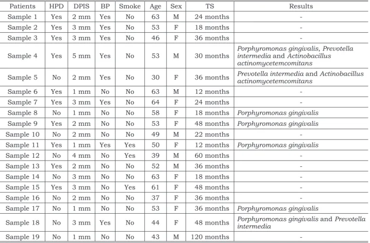
The results of the analysis of the clinical pa-rameters and of the microbiologic procedure are described in Table 2.
Seven peri-implant samples were positive for the target microorganisms.
Associations of these pathogens were observed in only three of the 19 samples; the DNAs of Por-phyromonas gingivalis, Prevotella intermedia and
Actinobacillus actinomycetemcomitans were detect-ed only in sample 4 (history of periodontal disease, sulcus depth of 5 mm and bleeding on probing). In sample 5 (without history of periodontal dis-ease, depth of 2 mm and bleeding on probing), the DNAs ofboth Prevotella intermedia and Actinoba-cillus actinomycetemcomitans were found. Sample 18 (without history of periodontal disease, depth of 3 mm and bleeding on probing) contained the DNAs of Porphyromonas gingivalis and Prevotella intermedia.
TABLE 2 - Anamnesis, clinical and microbiologic data (PCR) of the 19 patients analyzed.
Patients HPD DPIS BP Smoke Age Sex TS Results
Sample 1 Yes 2 mm Yes No 63 M 24 months
-Sample 2 Yes 3 mm Yes No 53 F 18 months
-Sample 3 Yes 3 mm Yes No 46 F 36 months
-Sample 4 Yes 5 mm Yes No 53 M 30 months Porphyromonas gingivalis, Prevotella intermedia and Actinobacillus actinomycetemcomitans
Sample 5 No 2 mm Yes No 30 F 36 months Prevotella intermedia and Actinobacillus actinomycetemcomitans
Sample 6 Yes 1 mm No No 63 M 12 months
-Sample 7 Yes 3 mm Yes No 64 F 24 months
-Sample 8 No 1 mm No No 58 F 18 months Porphyromonas gingivalis Sample 9 Yes 2 mm No No 53 F 48 months Porphyromonas gingivalis
Sample 10 No 2 mm No No 49 M 22 months
-Sample 11 Yes 1 mm Yes Yes 50 F 12 months Porphyromonas gingivalis
Sample 12 No 4 mm No Yes 39 M 60 months
-Sample 13 Yes 2 mm No No 52 M 36 months
-Sample 14 No 3 mm No No 63 F 18 months
-Sample 15 Yes 3 mm No Yes 61 F 48 months
-Sample 16 No 2 mm No No 37 F 36 months
-Sample 17 No 1 mm No No 53 F 36 months Porphyromonas gingivalis
Sample 18 No 3 mm Yes No 44 F 48 months Porphyromonas gingivalis and Prevotella intermedia
Sample 19 No 1 mm No No 43 M 120 months
54 55
54 55
Porphyromonas gingivalis was the only target pathogen detected in sample 8 (without history of periodontal disease, depth of 1 mm and no bleeding on probing), sample 9 (history of periodontal dis-ease, depth of 2 mm and no bleeding on probing), sample 11 (history of periodontal disease, depth of 1 mm and bleeding on probing), and sample 17 (without history of periodontal disease, depth of 1 mm and no bleeding on probing).
In summary, the DNA of at least one of the target pathogens was detected in only three of the 10 peri-implant samples of patients with history of periodontitis. In the other seven samples, those species were not detected, but they were found in four of the nine patients without history of peri-odontal disease.
DISCUSSION
Biofilms associated with periodontitis and peri-implantitis frequently harbor high levels of
Porphyromonas gingivalis, Prevotella intermedia
and Actinobacillus actinomycetemcomitans7,10,11,24,29. These pathogens are also related to the risk of periodontitis11,26,29. These observations led us to research the presence of these mentioned bacteria in seemingly healthy peri-implant sites and to try to establish a relationship between them and the risk of disease.
With this purpose, we chose the PCR, a highly sensitive molecular method that detects minimum amounts of the target DNA. PCR is also repro-ducible and relatively easy to execute as well as particularly valuable in the detection of non-cul-tivable organisms or those that are difficult to be distinguished in the cultivation4,28.
The implant sites of patients with peri-odontitis can present high concentrations of patho-gens in the periodontal pockets2,10,19,30. Therefore, periodontal lesions considered irreversible can determine the indication of extraction of un-healthy teeth to avoid infection of future peri-im-plant sites8. The presence of severe periodontitis should not hinder the insertion of implants; how-ever, this is a high risk situation that should be minimized by previous periodontal treatment as well as appropriate post-surgical control, which, ideally, should include the analysis of the micro-biota6,10,12,15. The need of that analysis was em-phasized by Alcoforado et al.1 (1991), who, besides isolating periodontopathogens from sites with peri-implantitis, showed the presence of high propor-tions of Candida albicans and exogenous bacteria (Gram-negative enteric rods, Staphylococcus spp.
and Pseudomonas aeruginosa), probably due to the inadequate administration of antibiotics that affect the resident microbiota. The microbiologic exam is often indicated when clinical and radiographic signs of disease exist23, but our results suggest that it should also be indicated to identify the risk of the disease.
We should consider that pathogens are usu-ally found at a proportion lower than 1.0% (supple-mental microbiota) in healthy sites9 where they are controlled by the host’s defense2,9. Likewise, the observation of pathogens at peri-implant sites does not necessarily mean failure of the implant18. That statement partially explains some unexpected results found in this research, such as the detec-tion of the target pathogens in clinically healthy sites and in patients without history of periodontal disease.
Another aspect worthy of emphasis is that hosts with similar levels of pathogens can present different clinical manifestations due to individual variations in their inflammatory response. People with polymorphism in genes that codify the pro-duction of interleukins and the tumor necrosis factor-alpha (response mediators) are more sus-ceptible to periodontitis17. This fact can probably be extrapolated to include peri-implantitis, which is equally dependent on the interaction between the levels of microbial aggression and the response of the host. Thus, even if pathogens are present in higher proportions, clinical signs are not always present or do not arise with the same intensity. However, inadequate biofilm control allows the gradual installation of larger numbers of patho-gens that produce metabolites, which, in addition to destroying the tissues, incite defense cells to secrete mediators that contribute to an increase in tissue destruction.
56 57
56 57
established a modern protocol seeking to preserve osseointegration.
The absence of relationship amongst the pres-ence of pathogens, clinical signs of severe inflam-mation and history of periodontitis, observed in this research, contradicts reports of several au-thors2,8,13,14,16,20,22,27 and the following considerations try to explain that discrepancy:
1. undetection does not mean that microorgan-isms are not present in the examined material, due to the possibility of technical failure in col-lection, transport and laboratorial processing of the material, sometimes making it neces-sary to repeat the exam;
2. PCR detects low amounts of DNA of target microorganisms, including even the non-vi-able ones; therefore, the result also shows the past presence of this microorganism in the examined material;
3. the possibility of random collection of some clones with defective or absent genes that cod-ify the production of virulence factors should be considered. The pathogenic species show variations in the degree of production of those
factors and supplementary genetic exams are necessary to distinguish the most virulent clones.
CONCLUSIONS
1. PCR proved efficient to identify the DNA of
Actinobacillus actinomycetemcomitans, Por-phyromonas gingivalis and Prevotella interme-dia even in peri-implant sulci with reduced depths.
2. The detection of DNA of the bacterial species studied in apparently healthy peri-implant sites can indicate peri-implantitis risk, making the establishment of a more strict preventive control in those patients compulsory, in order to guarantee the success of the treatment.
ACKNOWLEDGMENT
The authors thank Prof. Dr. Roberto Fraga M. Lotufo for the suggestions that enriched this research.
REFERENCES
1. Alcoforado GAP, Rams TE, Feik D, Slots J. Microbial as-pects of failing osseointegrated dental implants in humans. J Parodontol 1991;10:11-8.
2. Apse P, Ellen RP, Overall CM, Zarb GA. Microbiota and crevicular fluid collagenase activity in the osseointegrated dental implant sulcus: a comparison of sites in edentu-lous and partially edentuedentu-lous patients. J Periodontal Res 1989;24:96-105.
3. Ashimoto A, Chen C, Bakker I, Slots J. Polymerase chain reaction detection of 8 putative periodontal pathogens in subgingival plaque of gingivitis and advanced periodontitis lesions. Oral Microbiol Immunol 1996;11:266-73. 4. Avila-Campos MJ, Sacchi CT, Whitney AM, Steigerwalt
AG, Mayer LW. Specific primer for AP-PCR identification of Actinobacillus actinomycetemcomitans. J Clin Periodontol 1999;26:699-704.
5. Avila-Campos MJ, Velásquez-Meléndez G. Prevalence of putative periodontopathogens from periodontal patients and healthy subjects in São Paulo, SP, Brazil. Rev Inst Med Trop S Paulo 2002;44:1-5.
6. Bauman GR, Mills M, Rapley JW, Hallmon WW. Plaque-in-duced inflammation around implants. Int J Oral Maxillofac Implants 1992;7:330-7.
7. Becker W, Becker BE, Newman MG, Nyman S. Clinical and microbiologic findings that may contribute to dental implant failure. Int J Oral Maxillofac Implants 1990;5:31-8. 8. Danser MM, van Winkelhoff AJ, van der Velden U.
Peri-odontal bacteria colonizing oral mucous membranes in edentulous patients wearing dental implants. J Periodontol 1997;68:209-16.
9. De Lorenzo JL. O Ecossistema Bucal. In: De Lorenzo JL. Microbiologia para o Estudante de Odontologia. São Paulo: Atheneu; 2004. p. 55-72.
10. De Lorenzo JL, Cavenague M. Microbiologia Perim-plantar. In: De Lorenzo JL. Microbiologia para o Estudante de Odontologia. São Paulo: Atheneu; 2004. p. 151-62. 11. De Lorenzo JL, Mayer MPA. Microbiologia das
Do-enças Periodontais. In: De Lorenzo JL. Microbiologia para o Estudante de Odontologia. São Paulo: Atheneu; 2004. p. 127-50.
12. De Lorenzo JL, Simionato MRL, De Lorenzo A. Infec-ção: principal causa de insucessos em implantes dentários. Rev ABO Nac 1997;5:321-4.
13. Ellen RP. Microbial colonization of peri-implant envi-ronment and its relevance to long-term success of osseo-integrated implants. Int J Prosthodont 1998;11:433-41. 14. Gouvoussis J, Sindhusake D, Yeung S.
Cross-infec-tion from periodontitis sites to failing implant sites in the same mouth. Int J Maxillofac Implants 1997;12:666-73. 15. Gromatzky A, Sendyk WR. Preservação da
osseointe-gração através de um programa de controle e manutenção. Rev Periodontia 2002;13:11-6.
16. Koka S, Razzoog ME, Bloem TJ, Syed S. Microbial colonization of dental implants in partially edentulous sub-jects. J Prosthet Dent 1993;70:141-4.
17. Kornman I, Newman MG. Papel da genética na verifi-cação do risco e na abordagem da periodontite do adulto. In: Rose LF, Genco RJ, Mealey BL, Cohen DW. Medicina Periodontal. São Paulo: Santos; 2002. p. 11-33.
56 57
56 57
using clinical, radiographic and microbiological param-eters. Clin Oral Implants Res 2002;13:127-32.
19. Mombelli A. Microbiology and antimicrobial therapy of peri-implantitis. Periodontol 2000 2002;28:177-89. 20. Mombelli A, Marxer M, Gaberthuel T, Grunder U, Lang
NP. The microbiota of osseointegrated implants in patients with the history of periodontal disease. J Clin Periodontol 1995;22:124-30.
21. Mombelli A, van Oosten MAC, Schürch E, Lang NP. The microbiota associated with successful or failing os-seointegrated titanium implants. Oral Microbiol Immunol 1987;2:145-51.
22. Papaioannou W, Quirynen M, van Steenberghe D. The influence of periodontitis on the subgingival flora around implants in partially edentulous patients. Clin Oral Im-plants Res 1996;7:405-9.
23. Pfau EA. Incidência de espécies dos gêneros Prevotella e Porphyromonas em pacientes submetidos a implantes dentais [Dissertação de Mestrado]. São Paulo: Instituto de Ciências Biomédicas da USP; 2002.
24. Quirynen M, de Soete M, van Steenberghe D. In-fectious risks for oral implants. Clin Oral Implants Res 2002;13:1-19.
25. Rams TE, Link CC Jr. Microbiology of failing dental implants in humans: electron microscopic observations. J Oral Implantol 1983;11:93-100.
26. Rams TE, Robert TW, Tatum H, Keyes P. Utility of major putative periodontal pathogens and selected clinical parameters to predict periodontal breakdown in patients on maintenance care. J Clin Periodontol 1996;23:346-54. 27. Rutar A, Lang NP, Buser D, Bürgin W, Mombelli A.
Retrospective assessment of clinical and microbiological factors affecting periimplant tissue conditions. Clin Oral Implants Res 2001;12:189-95.
28. Slots J, Ashimoto A, Flynn MJ, Li G, Chen C. Detection of putative periodontal pathogens in subgingival specimens by 16S ribosomal DNA amplification with the Polymerase Chain Reaction. Clin Infect Dis 1995;20 Suppl 2:S304-7. 29. Slots J, Bragd L, Wikström M, Dahlén G. The occur-rence of Actinobacillus actinomycetemcomitans, Bacteroides gingivalis and Bacteroides intermedius in destructive dis-ease in adults. J Clin Periodontol 1986;13:570-7. 30. Sumida S, Ishihara K, Kishi M, Okuda K.
Transmis-sion of periodontal disease-associated bacteria from teeth to osseointegrated implant regions. Int J Oral Maxillofac Implants 2002;17:696-702.