Chaperone HtpG Predict Health in Periodontitis
Susceptible Patients
Charles E. Shelburne1*, P. Sandra Shelburne1, Vishnu M. Dhople1¤, Domenica G. Sweier2, William V. Giannobile3, Janet S. Kinney3, Wilson A. Coulter4, Brian H. Mullally4, Dennis E. Lopatin1
1Department of Biologic and Materials Sciences, The University of Michigan School of Dentistry, Ann Arbor, Michigan, United States of America,2Department of Cariology, Restorative Sciences and Endodontics, The University of Michigan School of Dentistry, Ann Arbor, Michigan, United States of America,3Department of Periodontics and Oral Medicine and The Michigan Center for Oral Health Research, The University of Michigan School of Dentistry, Ann Arbor, Michigan, United States of America,4Centre for Oral Research, Queen’s University, Belfast, Northern Ireland
Abstract
Background: Chaperones are ubiquitous conserved proteins critical in stabilization of new proteins, repair/removal of defective proteins and immunodominant antigens in innate and adaptive immunity. Periodontal disease is a chronic inflammatory infection associated with infection by Porphyromonas gingivalis that culminates in the destruction of the supporting structures of the teeth. We previously reported studies of serum antibodies reactive with the human chaperone Hsp90 in gingivitis, a reversible form of gingival disease confined to the oral soft tissues. In those studies, antibodies were at their highest levels in subjects with the best oral health. We hypothesized that antibodies to the HSP90 homologue ofP. gingivalis(HtpG) might be associated with protection/resistance against destructive periodontitis.
Methodology/Principal Findings: ELISA assays using cloned HtpG and peptide antigens confirmed gingivitis subjects colonized with P. gingivalis had higher serum levels of anti-HtpG and, concomitantly, lower levels of attachment loss. Additionally, serum antibody levels to P. gingivalis HtpG protein were higher in healthy subjects compared to patients with either chronic or aggressive periodontitis. We found a negative association between tooth attachment loss and anti-P. gingivalis HtpG (p = 0.043) but not anti-Fusobacterium nucleatum (an oral opportunistic commensal) HtpG levels. Furthermore, response to periodontal therapy was more successful in subjects having higher levels of anti-P. gingivalis HtpG before treatment (p = 0.018). There was no similar relationship to anti-F. nucleatum HtpG levels. Similar results were obtained when these experiments were repeated with a synthetic peptide of a region of P. gingivalis HtpG.
Conclusions/Significance:Our results suggest: 1) anti-P. gingivalisHtpG antibodies are protective and therefore predict health periodontitis-susceptable patients; 2) may augment the host defence to periodontitis and 3) a unique peptide ofP. gingivalis HtpG offers significant potential as an effective diagnostic target and vaccine candidate. These results are compatible with a novel immune control mechanism unrelated to direct binding of bacteria.
Citation:Shelburne CE, Shelburne PS, Dhople VM, Sweier DG, Giannobile WV, et al. (2008) Serum Antibodies toPorphyromonas gingivalisChaperone HtpG Predict Health in Periodontitis Susceptible Patients. PLoS ONE 3(4): e1984. doi:10.1371/journal.pone.0001984
Editor:Debbie Fox, The Research Institute for Children at Children’s Hospital New Orleans, United States of America
ReceivedJanuary 15, 2008;AcceptedFebruary 28, 2008;PublishedApril 23, 2008
Copyright:ß2008 Shelburne et al. This is an open-access article distributed under the terms of the Creative Commons Attribution License, which permits unrestricted use, distribution, and reproduction in any medium, provided the original author and source are credited.
Funding:Supported by NIH/NIDCR DE11117 (DEL) and patient sample collections were supported by NIH grants NIH/NIDCR U01-DE-14961 and NIH/NCRR M01-RR000042 (WVG). The sponsors of this work had no role in the design and conduct of the study, in the collection, analysis, and interpretation of the data and in the preparation, review, or approval of the manuscript.
Competing Interests:The authors have declared that no competing interests exist.
* E-mail: ceshelbu@umich.edu
¤ Current address: The Centre for Cellular and Molecular Biology, Hyderabad, India
Introduction
Porphyromonas gingivalisis a gram negative obligate anaerobe that has a major etiological role in human periodontitis. The bacterium is found with high frequency in persons with periodontitis where it participates in the initiation and establishment of chronic, infectious biofilms [1,2]. These biofilms facilitate the long term survival ofP. gingivalisand induces an inflammatory reaction that is responsible for the destruction of the hard and soft tissue supporting structures of the teeth. In additionP. gingivaliscan invade and persist in the cells of the
gingival tissue[3]. It also can escape the oral cavity and has been found in atherosclerotic plaque where it may have a role in the pathophysiology of cardiovascular disease [4].
may have consequences in the pathogenesis of periodontitis [9]. These functions are important in the establishment and perpet-uation of chronic inflammatory diseases.
Recent studies have shown that antibodies to periodontal disease associated pathogens may have protective effects although the exact mechanism is still unclear. Rams et al [10] demonstrated that serum levels of IgG antibodies againstA. actinomycetemcomitans or P. gingivalis in periodontitis-stable patients were higher than those in patients with active periodontitis suggesting that these antibodies have a protective effect against periodontal infections. Yamasaki et al have shown that antibodies toP. gingivalisHsp60 homologue increases with successful treatment [11]. We have recently described experiments that indicate that antibodies to HtpG may mitigate some of the induction of inflammatory chemokines through TLR4 and CD91 [12], a receptor expressed in human atherosclerotic lesions [13]. Taken together, these findings suggest a role for antibodies toP. gingivalischaperones in both periodontal and cardiovascular disease.
The possible protective role of antibodies to chaperones in periodontitis is controversial. It has been suggested that these antibodies simply reflected the high level of homology between human and bacterial proteins, a hallmark of these evolutionarily conserved molecules [14]. Other results suggest that the conserved nature of the chaperones might lead to autoimmune phenomenon due to ‘‘immune mimicry’’. Earlier results from this laboratory suggested that high levels of anti-Hsp90 antibodies could have protective qualities [9]. However, that study utilized a group of individuals with minimal periodontal disease. Here we describe findings from a study of periodontitis subjects with more extensive disease that are similar to those reported earlier and support the notion of a protective role for anti-HtpG antibodies in untreated subjects. In addition, these studies suggest that the levels of these antibodies also may predict the response to treatment. These results may also reflect an immune control mechanism in chronic infections unrelated to direct binding of bacteria.
Results
Colonization of plaque byP. gingivalis andF. nucleatum
Initially we reported [9] that in gingivitis subjects probing depth and the levels ofP. gingivaliswere both associated with serum anti-Hsp90 antibody levels. Subjects from the gingivitis group were tested for the presence ofP. gingivalisby slot immunoblot; 82 % of the subjects were positive forP. gingivalis[15,16]. Genomic DNA of pooled plaque samples from all teeth for each subject in the CP and AP groups were tested; 71% of the healthy subjects and 86% of individuals with periodontitis were positive forP. gingivalis16S rRNA genes by PCR. The low end sensitivity of the assay was
between 10 and 100 bacterial cells per sample. The mean percentage ofP. gingivaliswas 0.11 and 0.98 (p,0.001,t-test) in the controls and periodontitis subjects respectively. Both CP groups were 96% positive for F. nucleatum; the mean percentage of F. nucleatumwas 2.77 and 3.78 (p = 0.07; t-test) in the controls and periodontitis subjects respectively (Table 1). Plaque samples from the normal controls for the AP group were not available for F. nucleatumtesting and as reported previously 17% of the AP subjects were positive forP. gingivalis16S rRNA [17].
Antibodies toP. gingivalisandF. nucleatumtotal cell lysates
The level of antibodies to total cell lysates ofP. gingivalisandF. nucleatum was determined as a measure of the long term colonization experience that each subject had with these bacteria. These two bacteria have different roles in periodontitis,P. gingivalis as a pathogen whileF. nucleatumis an opportunistic commensal in the subgingival biofilm. Anti-P. gingivalisantibodies were detected in 38 of 45 samples obtained from gingivitis subjects. All CP and AP subjects had levels of anti-P. gingivalisand anti-F. nucleatummore than 3 standard deviations above the background of the assay. In the CP group there was a statistically significant elevated levels of antibodies toP. gingivalis(p = 0.01,t-test) andF. nucleatum(p = 0.02, t-test) in the diseased subjects compared to the control group; there was no difference between levels of antibody to totalP. gingivalisin the AP group (Table 2). Similar results were found in CP subjects that were colonized byP. gingivalis(Table 3) and CP subjects not colonized byP. gingivalis(Table 4) at the time the serum samples were obtained. These results suggest that most subjects in the study were likely to have been colonized withP. gingivalisprior to their participation in this study.
Total IgG Antibodies toP. gingivalisHtpG andF. nucleatumHtpG
Levels of IgG-class antibodies that specifically reacted with recombinant HtpG proteins of P. gingivalis and F. nucleatum were measured. Anti-HtpG antibody levels were measured in gingivitis subjects; subjects with more CAL (more tissue loss) had lower levels of anti-P. gingivalisHtpG.
In the CP subjects we hypothesized that the pattern of anti-HtpG would be different for the two bacteria. However, there was a trend, both in CP individuals colonized (Table 3) or un-colonized (Table 4) byP. gingivalis, to higher antibody levels to HtpG from both species in healthy subjects than in subjects with disease even though this trend did not reach statistical significance. There were substantial and significant differences between the healthy and diseased AP subjects in all the variables tested. The three clinical measurements were all significantly higher in the
Table 1.Research Subject Demographics
Group (n) Gingvitis (50) CP-Healthy (49) CP-Diseased (50) AP-Healthy (21) AP-Diseased (24)
% Female 58 54 58 62 68
Age Range in years (mean) 18-66 (31.6) 20-78(43.0) 30-77(51.3) 18-35 (34.4) 22-40(32.0)
Attachment Loss (mm, mean6SD) 1.5660.70 0.6860.52 2.2461.07 0.7260.52 3.2560.63
Probing Depth (mm, mean6SD) 2.6460.54 1.6060.21 2.6060.71 1.5060.22 2.8361.32
Bleeding on Probing (%, mean6SD) ND 0.2460.12 0.5760.17 0.1360.06 0.3360.16
Gingival Index (%, mean6SD ) 0.260.17 0.1860.19 0.5260.23 ND ND
ND–not determined
diseased groups (Table 5) while the anti-HtpG levels were significantly lower in the diseased subjects (p,0.002, ANOVA).
Antibodies toP. gingivalis HtpG peptide p18
Previous studies [18] suggested that a peptide antigen, p18, of the HtpG molecule was responsible for eliciting the apparent protective effect found in the serum of healthy subjects. Therefore the serum response to a synthetic peptide antigen of this region was investigated in the CP and AP subjects. We expected that the levels of antibody to the peptide would be higher in control subjects compared to periodontitis subjects of both groups and that
elimination of the majority of the epitopes in the HtpG molecules, most of which are highly conserved between bacterial species, would clarify the trend observed using the whole HtpG proteins as ELISA target antigens. As expected, subjects in both the CP (Table 3 & 4) and AP (Table 5) groups diagnosed with periodontitis had lower levels of anti-p18 than their respective control groups. Although the same trend was observed in both colonized and un-colonized individuals using the HtpG molecule only the differences in p18 antibody levels in CP subjects colonized byP. gingivalisreached statistical significance (Table 3 & 4).
Cluster Analysis by Disease Subgroups
There were trends supporting our hypothesis when the results of the assays were compared between groups as they were recruited into the studies, basically health or disease status. However, examination of those groupings showed that there was overlap between the groups based on clinical measures, especially in the CP subjects, the most complete study. Therefore the subjects were regrouped in line with their clinical measures by K-means clustering, irrespective of recruitment groups. The clusters were used as a basis for ANOVA analysis of differences in the clinical measures and antibody levels.
Gingivitis subjects were regrouped into three clusters and differences in four clinical measures (PD, CAL, GI, PL) and two antibody levels (anti-P. gingivalisHtpG, anti-P. gingivalis whole cell lysate) sought. Cluster 1 had the greatest PD and CAL; there were significant differences between the clusters for PD and CAL. Antibody levels against P. gingivalis HtpG were lowest in the subjects with the highest disease measures, similar to earlier findings [9] for human Hsp90, but did not reach statistical significance (Table 6).
CP Subjects were regrouped into 4 clusters and differences sought in four clinical measures (PD, CAL, BOP, GI) and three antibody levels (anti-P. gingivalisHtpG, anti-Fn HtpG and anti-Pg p18). There were significant differences between the groups in all clinical measures (Table 7). Cluster 2 had the highest levels of all clinical measures and the lowest levels of anti-P. gingivalis HtpG and anti-P. gingivalis HtpG p18, the latter being significant (p = 0.00056, ANOVA).
AP subjects were regrouped into 3 clusters and differences sought in three clinical measures (PD, CAL, BOP) and three antibody levels (anti-Pg whole bacteria, anti-P. gingivalisHtpG and anti-Pg P18). There were significant differences in all the clinical measures between groups and in anti-P. gingivalisHtpG and anti-P. gingivalis HtpG p18, but not anti-Pg whole bacteria (Table 8). Cluster 2 had the highest levels of clinical measures and lowest levels of anti-P. gingivalis HtpG p18. Both clusters 1 and 2 had substantially lower levels of anti-P. gingivalisHtpG compared to cluster 3.
Table 2.Antibody responses toP. gingivalisandF. nucleatumin subjects.
Group (n) Gingivitis (50) CP-Healthy (49) CP-Diseased (50) AP-Healthy (21) AP-Diseased (24)
% positive forP. gingivalis 82 71 86 ND 17
% positive forF. nucleatum ND 96 96 ND ND
Antibody to totalP. gingivalis(Log mean ELISA Units6S.D.) 4.3960.17 3.6860.23 3.9260.26 4.4760 .07 4.3960.15
Antibody to totalF. nucleatum(Log mean ELISA Units6S.D.) ND 3.9560.62 4.8660.81 ND ND
ND–not determined
doi:10.1371/journal.pone.0001984.t002
Table 3.Serum antibody (IgGc) levels to bacterial antigens in CP subjects colonized byP. gingivalis.
ELISA Units
Health (mean)
Disease (mean)
p-value (t-test)
Valid N (Health/ Disease)
Anti-P.gingivalisHtpG 10,200 9,882 NS 35/41
Anti-F.nucleatumHtpG 19,959 19,817 NS 35/38
Anti-P.gingivalisHtpG p18 7,138 6,254 0.047 35/40
Anti-P.gingivalisW83 5,654 10,076 0.002 35/40
Anti-F.nucleatum22586 27,735 13,701 0.009 35/41
NS-not significant
doi:10.1371/journal.pone.0001984.t003
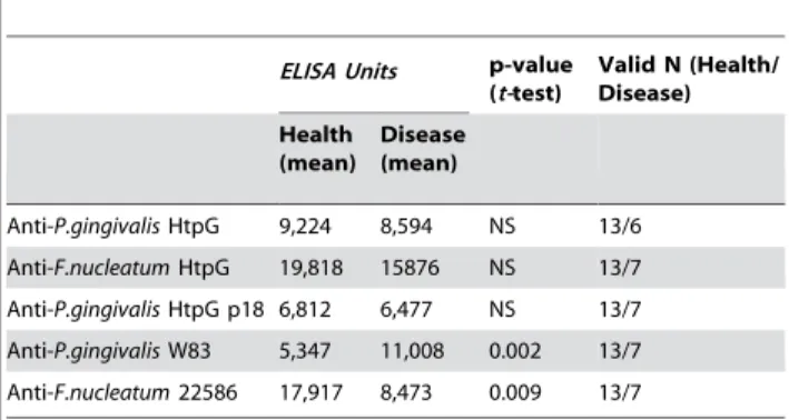
Table 4.Serum antibody (IgGc) levels to bacterial antigens in CP subjects not colonized byP. gingivalis.
ELISA Units p-value
(t-test)
Valid N (Health/ Disease)
Health (mean)
Disease (mean)
Anti-P.gingivalisHtpG 9,224 8,594 NS 13/6
Anti-F.nucleatumHtpG 19,818 15876 NS 13/7
Anti-P.gingivalisHtpG p18 6,812 6,477 NS 13/7
Anti-P.gingivalisW83 5,347 11,008 0.002 13/7
Anti-F.nucleatum22586 17,917 8,473 0.009 13/7
NS-not significant
Correlation of Anti-P. gingivalisHtpG antibodies to clinical measures in Chronic and Aggressive Periodontitis
The supposition that antibodies to P. gingivalis HtpG may be protective is based on the hypothesis that high levels of antibodies should be found in subjects exhibiting healthier clinical signs and lower levels in subjects exhibiting more periodontitis related damage. Clinical measurements from each site in all the subjects were obtained and the average value determined for each subject. Relationships between those measurements and the levels of P.
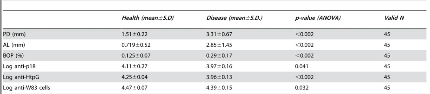
gingivalisandF. nucleatumanti-HtpG were sought using Pearson’s R to compare baseline antibody levels in both AP and CP groups with the clinical signs obtained at the baseline were compared. When anti-P. gingivalis HtpG levels from CP subjects were analyzed with four clinical measurements in each of the same patients there was a trend for pocket depth and attachment loss measurements to inversely correlate with antibody levels, but the correlations were small and not statistically significant. Similar results were found with anti-F. nucleatumHtpG values. However, in the AP subjects there were substantial negative associations three Table 5.Serum antibody (IgGc) levels to bacterial antigens in AP subjects.
Health (mean6S.D) Disease (mean6S.D.) p-value (ANOVA) Valid N
PD (mm) 1.5160.22 3.3160.67 ,0.002 45
AL (mm) 0.71960.52 2.8561.45 ,0.002 45
BOP (%) 0.12560.07 0.2960.17 ,0.002 45
Log anti-p18 4.1160.27 3.9760.16 0.041 45
Log anti-HtpG 4.2560.04 3.9660.13 ,0.002 45
Log anti-W83 cells 4.4760.07 4.3960.15 0.032 45
doi:10.1371/journal.pone.0001984.t005
Table 6.Analysis of clustered subjects in the Gingivitis group.
Cluster 1 Cluster 2 Cluster 3
p-value (ANOVA) Mild perio. n = 22 Gingivitis n = 10 Healthy n = 18
PD 3.160.2 2.860.4 2.0560.2 ,0.001
AL 2.260.6 1.060.0 1.1160.3 ,0.001
GI 27.365.5 30.064.8 5.562.7 NS
Pl 45.565.9 8.066.3 27.864.6 NS
Anti-P. gingivalisHtpG 982564720 14973610178 1267666928 NS
Anti-P. gingivalis W83 310066898 13016177 7488611008 NS
NS–not significant
Subjects were clustered by K-means clustering and differences sought in means (6S.D.) of variables by ANOVA. PD and AL measured in mm; GI is the % sites with gingival redness; PI values are % sites with accumulated plaque. Antibody values are expressed in ELISA units.
doi:10.1371/journal.pone.0001984.t006
Table 7.Cluster analysis of CP subjects.
Cluster 1 Cluster 2 Cluster 3 Cluster 4
p-value (ANOVA) Moderate CP n = 31 Severe CP n = 10 Gingivitis n = 29 Healthy n = 27
PD 2.460.4 3.660.8 1.760.2 1.660.2 ,0.001
AL 1.960.1 4.060.9 1.260.3 0.360.2 ,0.001
BOP 56.8616.2 68.4616.6 27.76.12.7 24.6614.6 ,0.001
GI 54.3621.1 60.6624.6 20.2617.9 21.4621.0 ,0.001
Anti-P. gingivalisHtpG 1053164791 787763906 931163833 1035864443 NS
Anti-F.nucleatumHtpG 200536806 1853069346 1823165752 2074866540 NS
Anti-P. gingivalisHtpG p18 648561920 479362120 717161782 695761068 0.006
NS–not significant
Subjects were clustered by K-means clustering and differences sought in means (6S.D.) of variables by ANOVA. PD and AL measured in mm; GI is the % sites with gingival redness. Antibody values are expressed in ELISA units.
clinical measurements and anti-HtpG levels, all of which were statistically significant (Table 9). Similar experiments using HtpG cloned from F. nucleatum gave no significant correlations to the clinical signs (data not shown).
Correlation of Anti-P. gingivalisHtpG p18 antibodies to clinical measures in Chronic and Aggressive Periodontitis
We then sought relationships between the same measurements and the levels ofP. gingivalisanti-HtpG p18 by comparing antibody levels in both AP and CP groups with the clinical measurement using Pearson’s R analysis. Sera from the CP group and controls showed that levels of anti-p18 were significantly (p = 0.047) inversely correlated to CAL but not correlated to the other indices. In the AP group antibodies to p18 were significantly inversely correlated to PD (p = 0.008), CAL (p = 0.018) and BOP
(p = 0.046), Table 10. Lastly, associations were sought between changes in clinical measurements 6 months after SRP treatment and the anti-p18 levels 6 months before treatment to determine if they might predict treatment success or failure. There was a significant positive correlation (p = 0.05) between the pre-treat-ment levels of anti-HtpG p18 and reduction in CAL.
Discussion
Chaperones are simultaneously highly conserved and immuno-dominant antigens which may have important roles in the pathogenesis of numerous human diseases. Although a subject of intense investigation, the role of the immune response to chaperones in human disease is currently not well understood. Periodontal diseases present a unique opportunity to examine the Table 8.Cluster analysis of AP subjects.
Cluster 1 Cluster 2 Cluster 3 p-value (ANOVA)
Localized AP n = 11 Generalized AP n = 12 Healthy n = 21
PD 3.060.6 3.660.7 1.560.2 ,0.001
AL 2.261.2 3.461.5 0.760.5 ,0.001
BOP 21.4611.2 37.9618.1 12.565.7 ,0.001
Anti-P. gingivalis HtpG p18 1018262437 837162114 1239065318 0.0283
Anti-P. gingivalisHtpG 885362493 1016563479 1774261349 ,0.001
Anti-P. gingivalis W83 2689969509 26040612191 3004164977 NS
NS–not significant
Subjects were clustered by K-means clustering and differences sought in means (6S.D.) of variables by ANOVA. PD and AL measured in mm; BOP is the % sites bleeding on probing. Antibody values are expressed in ELISA units. NS–not significant.
doi:10.1371/journal.pone.0001984.t008
Table 9.Correlation of Anti-P. gingivalisHtpG antibody levels with clinical measurements.
A. Pretreatment B. Post-treatment
Anti-P. gingivalisHtpG Anti-F. nucleatumHtpG Anti-P. gingivalisHtpG Anti-F. nucleatumHtpG Pearson’s R p-value Pearson’s R p-value Pearson’s R p-value Pearson’s R p-value Gingivitis
PD 20.111 0.440
AL 20.341 0.012
GI 20.101 0.483
PI 20.130 0.367
CP
PD 20.050 NS 20.019 NS 0.361 0.002 0.076 NS
AL 20.108 NS 20.113 NS 0.273 0.020 0.144 NS
BOP 0.144 NS 0.057 NS 0.381 0.001 0.113 NS
GI 0.093 NS 0.001 NS 0.263 0.026 0.140 NS
AP
PD 20.787 ,0.001 ND
AL 20.628 ,0.001 ND
BOP 20.388 ,0.01 ND
NS–not significant ND–not determined
immune response to chaperones in a distinctive and readily accessible human environment. A few reports of antibody levels to P. gingivalischaperones (particularly GroEL) have been published but this is the first comprehensive analysis of anti-P. gingivalisHtpG in multiple periodontal disease states. Results reported here and earlier [9,12] suggest that anti-P. gingivalisHtpG antibodies predict health in patients susceptible to periodontal disease and are protective in the untreated periodontal disease patient. These results may also reflect a previously uncharacterized immune control mechanism unrelated to direct binding of bacteria. Further, these antibodies may augment periodontitis treatment and P. gingivalis HtpG might be an attractive vaccine candidate. These results appear to be unique to P. gingivalis as they are directed at a segment of HtpG unique to P. gingivalis; parallel experiments with the HtpG homologue of F. nucleatum did not manifest these same qualities. In addition, there is reason to believe that these results may be extended to other chronic infectious diseases.
Earlier studies of a population of subjects with minimal periodontal disease suggested that antibodies to human Hsp90 related chaperones might have a protective effect. This report expands on that notion by examining the response of periodontitis subjects to the Hsp90 homologue in P. gingivalis, HtpG. In gingivitis subjects as a whole it was found that there was a distinctive, but not statistically significant, trend in support of the hypothesis. Interestingly, when anti-P. gingivalis HtpG levels and clinical measures are compared on an individual basis there is a discernable correlation with CAL. At the opposite end of the periodontitis disease spectrum, the AP subjects, there is consider-able support for the hypothesis. At the group level, there are substantial and significant differences between both anti-P. gingivalis HtpG and anti-P. gingivalis HtpG p18 between clusters based on clinical measures, but no differences in the response to the whole bacterium. On an individual basis, both anti-P. gingivalis
HtpG and anti-P. gingivalis HtpG p18 are inversely and significantly correlated to the clinical measures.
Support of the hypothesis was somewhat tenuous in the initial examination of the CP subjects. Those recruited as either healthy or with CP and colonized by P. gingivalis possessed a humoral response to Pg HtpG p18 higher in subject groups with less periodontal destruction and inversely related to the level of attachment loss (p,0.05). A similar relationship was not found in un-colonized individuals. This was the opposite of antibody levels to the whole bacterium which were significantly higher is subjects with disease than controls (p#0.01), a finding reported by many other laboratories. Similar trends were found when we examined the humoral response to HtpG fromF. nucleatumbut there was no relationship to disease status. In addition, correlations between the same antibodies and clinical measures when considered in the context of individual subjects were not found, except for a correlation between ant-P. gingivalis HtpG p18 and CAL (R =20.205, p,0.05). This dilemma was resolved when the subjects were clustered by clinical measures. In a cluster of 10 subjects with the most severe disease there was a substantial and significantly lower level of anti-P. gingivalis HtpG and anti-P. gingivalis HtpG p18 compared to the other clusters in the CP subjects. As might be expected, the clinical measures of these subjects resemble those of the AP subjects. In summary, there is a trend for both anti-P. gingivalisHtpG and anti-P. gingivalisHtpG p18 to be lower in subjects with the more serious disease in each of the 3 periodontal disease groups. Neither trend reaches statistical significance in the gingivitis group. In the CP group anti-P. gingivalis HtpG p18 is significantly lower in the most diseased cluster; anti-P. gingivalis HtpG trends in the same direction but does not reach statistical significance. In the AP group both anti-P. gingivalis HtpG and anti-P. gingivalis HtpG p18 are significantly lower in the diseased clusters of subjects. Our initial observations (16) were probably due to the response of the most diseased individuals in that group of subjects. However, we believe that the current results are not fortuitous because 1) the relationship between the antibody levels and disease is evident not only in the original group but in groups with more serious disease in an almost dose dependent manner; 2) there is a relationship between these antibody levels and response to periodontal treatment; 3) there is a link through these antibodies to cellular receptors involved in antigen recognition and inflammation; 4) similar results have been noted in other chronic bacterial infections. In addition, there is no evidence that the lower levels of anti-P. gingivalisHtpG are due to adsorption by high circulating levels of either human Hsp90. There was no significant correlation between individual serum levels of human Hsp90 protein and anti-P. gingivalisHtpG, anti-F. nucleatumHtpG or anti-P. gingivalisHtpG p18 (Table S1). In fact, there was a trend to higher levels of anti-P. gingivalis HtpG in subjects with higher levels of human Hsp90, the opposite of what be expected if such absorption was taking place. There was also no significant difference between levels of human Hsp90 in healthy subjects with high levels of anti-P. gingivalisHtpG antibodies and severe periodontitis subjects with low levels of anti-P. gingivalis HtpG (Table S2). Lastly, while it is possible that a subset of anti-P. gingivalisHtpG antibodies might bind other HSP90 homologues, including human Hsp90, the P. gingivalis HtpG p18 epitope is unique and antibodies to that peptide could not be adsorbed by other HSP90 family proteins (see discussion below).
Subjects with CP who were given periodontal treatment responded more effectively to that treatment the higher their original pretreatment levels of anti-P. gingivalis HtpG. There was no similar effect with antibodies toF. nucleatumHtpG. Notably, the serum samples, which are time-averaged values, correlate best Table 10.Correlation of Anti-P. gingivalisHtpG p18 antibody
levels with clinical measurements.
Pre-treatment Post-treatment
Anti-P. gingivalisHtpGp18 Anti-P. gingivalisHtpGp18
Pearson’s R p-value Pearson’s R p-value Gingivitis
PD 20.135 NS
AL 0.066 NS
GI 20.081 NS
PI 0.136 NS
CP
PD 20.063 NS 0.072 NS
AL 20.204 0.047 0.153 NS
BOP 20.100 NS 20.119 NS
GI 20.111 NS 20.084 NS
AP
PD 20.393 0.008
AL 20.361 0.015
BOP 20.298 0.046
NS–not significant
with clinical indices related to tissue destruction and less with indices related to inflammation. This suggests that these anti-bodies are found in subjects predisposed to a better treatment outcome or they may somehow facilitate healing after treatment. Interestingly, drugs that inhibit Hsp90 have been shown to prolong survival, attenuate inflammation, and reduce lung tissue injury in murine sepsis [19].
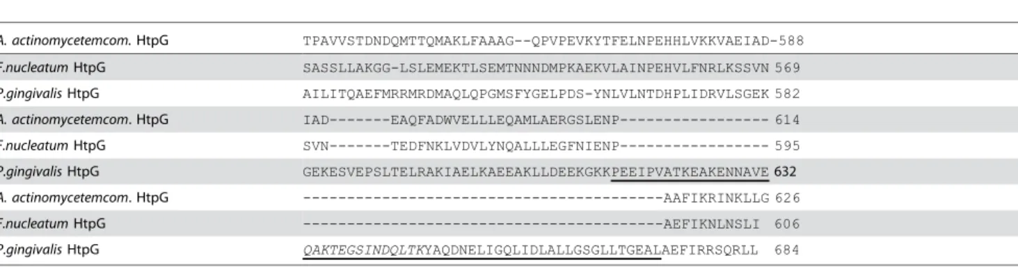
These findings are also worthy of note because despite high homology among chaperones in diverse organisms, theP. gingivalis HtpG is dissimilar from A. actinomycetemcomitans and F. nucleatum HtpG proteins: the C-terminus contains an extra inserted sequence (Table 11. bold in alignment). This insert appears to be exclusive to the Cytophaga-Flavobacterium-Bacteroides group HtpG proteins sequenced to date, and contains both a portion unique to each species (p18, underlined in alignment) and a conserved portion (p19). In contrast, the DnaK and GroEL ofP. gingivalis, A. actinomycetemcomitans,andF. nucleatumalign closely and display high homology. Examination of an alignment of Bacertoidacae HtpG molecules (Table 12) shows the insert can be roughly divided into 2 sections of about 30 amino acids each. The first-which contains p18-has relatively little sequence homology to the other species, only 6 of 36 (17%) amino acids are exact matches to the other Bacteroidacae HtpGs. The adjacent segment, p19, has 13 of 25 (52%) amino acids that are identical,
similar to that between whole HtpG molecules from all 5 species (60-63%). BLAST analysis of p18 against the entire non-redundant protein database at GeneBank results in no hits except P. gingivalisHtpG. Similar searches using p19 produce significant hits with species in each of the Bacteroidacae genera (B. fragilis group; Non-B. fragilisgroup, Prevotella, Porphyromonas, Taner-ella and ‘‘other’’). Taken together we have concluded it is reasonable to assume that p18 is or contains an epitope that is unique toP. gingivalisHtpG and are currently investigating that hypothesis.
The mechanism that may connect anti-HtpG antibodies and progression of periodontitis is not known, but we speculate that it may well be that these antibodies block an interaction between HtpG and cellular receptors on macrophage [12]. Interactions between HtpG and the TLR4 and CD91 receptors induce the chemokine CXCL8, a chemoattractant for phagocytic cells in periodontitis [20,21] and thus perpetuating the uncontrolled inflammation characteristic of periodontitis. Additionally, interac-tions between TLR4 and CD91 and HtpG induce inflammatory cytokines, including TNFa, as part of the innate immune response. Serum antibodies in 8 subjects with titers toP. gingivalisHtpG were found to reduce CXCL8 production in human monocytes in a dose dependent manner while serum antibodies from subjects without anti-HtpG activity did not [12].
Table 11.Alignment of oral bacterial species HtpG amino acid sequences.
A. actinomycetemcom.HtpG TPAVVSTDNDQMTTQMAKLFAAAG--QPVPEVKYTFELNPEHHLVKKVAEIAD-588
F.nucleatumHtpG SASSLLAKGG-LSLEMEKTLSEMTNNNDMPKAEKVLAINPEHVLFNRLKSSVN 569
P.gingivalisHtpG AILITQAEFMRRMRDMAQLQPGMSFYGELPDS-YNLVLNTDHPLIDRVLSGEK 582
A. actinomycetemcom. HtpG IAD---EAQFADWVELLLEQAMLAERGSLENP--- 614
F.nucleatumHtpG SVN---TEDFNKLVDVLYNQALLLEGFNIENP--- 595
P.gingivalisHtpG GEKESVEPSLTELRAKIAELKAEEAKLLDEEKGKKPEEIPVATKEAKENNAVE632
A. actinomycetemcom. HtpG ---AAFIKRINKLLG 626
F.nucleatumHtpG ---AEFIKNLNSLI 606
P.gingivalisHtpG QAKTEGSINDQLTKYAQDNELIGQLIDLALLGSGLLTGEALAEFIRRSQRLL 684
Alignment of oral bacterial species HtpG amino acid sequences in the region of the 65 amino acid ‘‘Bacteroidacae insert’’ (Bold, underlined characters). The insert has not been described outside the Cytophaga, Flavobacteria and Bacteroides (CFB) group. Italic, bold–p18.
doi:10.1371/journal.pone.0001984.t011
Table 12.Alignment of Bacteroidiacae HtpG C-terminal inserts
B. thetaiotaomicron LKDSQKDKKEEDIPTAEKDELNELDKKWDELKNKKEGIFAG 643
B. fragilis LKKKQEGKKDEDIPTAEKDELNDLDKKWDELKQQKDSIFAG 643
P. intermedia LRQEQGKKKAEEITQEEKDDLKNTEESLSKQRTEKNDVIAN 646
T. forsythia LREAQNKKKEDEITASEKEDLTNTNRKLTELRGQQNAILAE 647
P. gingivalis LLDEEKGKKPEEIPVATKEAKENNAVEQAKTEGSINDQLTK 646
Consensus * . : ** ::*. *: : . : . . : ::
B. thetaiotaomicron YASNNKVIRQLIDLALLQNNMLKGEALNNFVKRSIELI- 681
B. fragilis YAGKNKVVRQLIDLALLQNNMLKGEALNNFVKRSIELI- 681
P. intermedia YAKGNNAIHQLIDLALLQNGMLKGAALDKFIKRSVDLIK 685
T. forsythia YAAGNPIIGQLIDLALLGNGLLKGEALSRFIRRSVDLIR 686
P. gingivalis YAQDNELIGQLIDLALLGSGLLTGEALAEFIRRSQRLL- 684
Consensus ** * : ******** ..:*.* ** .*::** *:
Alignment of 65 amino insert found in most CFB group bacteria, but not other bacterial groups. Bacteroidiacae HtpG C-terminal inserts shows p18 is much less conserved than p19. Underline/bold–p18 sequence. Dashed underline–p19 sequence. Amino acid identities across all species-*.
The results reported here are similar to those of other chronic infections includingHeliobacter pylori. World wide as much as 75% of the population has evidence of H. pylori infection but only subsets of these individuals manifest the peptic ulcers and stomach cancers associated with those chronic infections. The H. pylori chaperone hsp60 induces CXCL8 production in monocytic cells [22] and humoral immune response to a peptide epitope (pH9) of hsp60 is unique and seems to be associated with protection against H. pyloriinfection [23]. Intriguingly, when subjects with mucosa-associated lymphoid tissue lymphoma were examined for anti-bodies to the H. pylori hsp60 chaperone it was found that pre-treatment titers in patients whose tumors regressed after pre-treatment were significantly higher than in patients whose tumors did not regress [24]. Low levels of antibodies to chaperones in disease subjects compared to controls has also been reported in inflammatory bowel disease [25], fungal infections [26] and other chronic bacterial infections [27].
The unique restriction of theP. gingivalisHtpG p18 peptide to the most important pathogen in periodontitis may have applica-tions in vaccine and diagnostic arenas. The use of chaperones as vaccine candidates has been suggested by a number of investigators. Antibody to the Hsp90 homologue of C. albicans (MycograbH, Novartis) has been shown to be effective in the treatment of disseminated fungal infections [28] in combination with amphotericin B. Other chaperones have been suggested as vaccine targets for diverse diseases in humans, cattle [29] and fish [30].P. gingivalisGroEL has been used as a vaccine in a rat model of periodontitis that resulted in prevention of attachment loss [31], similar to that described here in humans. However, the authors cautioned that extensive homology between human and microbial chaperones may require use of peptides that do not induce cross-reactive antibodies with human hsp60 molecules. We believe this is the case with p18, which appears unique toP. gingivaliseven to the exclusion of other Bacteroides species. The data presented here also suggests that serum antibodies to p18 may be useful in diagnosing periodontitis patients with extensive treatment require-ments, identification of which would have substantial economic and epidemiological impact on the practice of periodontology.
Materials and Methods
Subjects
All work with human subjects was approved by the University of Michigan Institutional Review Board or the Queen’s University, Belfast Ethics Committee, respectively. Each subject gave written individual informed consent and was advised that withdrawal from the study was available at their discretion at any time. The clinical condition of each subject was determined by examination of five clinical measurements: Probing pocket depth (PD) was determined to the nearest mm at six sites around each tooth and then averaged for all sites in each subject [32]. Bleeding on probing (BOP), was reported as a dichotomous measure and recorded as a percentage of the sites [33]. Clinical attachment loss (CAL) was determined at the same sites by measuring the distance between the cemento-enamel junction and the bottom of each pocket to the nearest mm and averaged [34]. The gingival index (GI) value represents the percentage of sites in each patient exhibiting redness associated with inflammation[35]. The plaque index value (PI) represents the percentage of sites found to have plaque biofilm accumulated at the gingival margin.
Gingivitis Subjects
This group was examined from archived samples described in a previous report and was included here to address the potential
differences between response to P. gingivalis HtpG and human Hsp90. This retrospective study focused on a population of individuals living in southwestern Michigan [15,16] with gingivitis, a form of periodontal disease with no or minimal tissue destruction that is reversible with active oral hygiene. Subjects were recruited into the original study based on their membership in the rural community, not their oral health status, and all essentially presented with gingivitis or mild periodontitis. The clinical data and archived serum samples collected from 50 subjects were used. The average age was 31.6 years (range: 18–66) and 58% were female. Other characteristics of these subjects are found in Table 1.
Chronic Periodontitis Subjects
Subjects were recruited at the Michigan Center for Oral Health Research. Subject inclusion was based on: possession of at least 20 teeth, no periodontal treatment or antibiotic-related therapy for medical or dental reasons for 3 months before study inclusion, no history of long-term treatment with medications known to affect periodontal status, and no history of metabolic bone diseases. Healthy control subjects (n = 49) were recruited who had less than 3 mm of CAL, no pocket depth (PD)s greater than 4 mm, no radiographic bone loss, and less than 20 sites with bleeding on probing. Chronic periodontitis (CP) subjects (n = 50) exhibited at least 4 sites with evidence of radiographic bone loss, mean CAL .3mm, PD.4 mm and bleeding on probing (Table 1).
Aggressive Periodontitis Subjects
Studies were done with serum samples from an ongoing study of Aggressive Periodontitis (AP) subjects (n = 24) and age matched control subjects (n = 21) living in Northern Ireland. AP subjects included in this investigation were 30.364.0 years of age at the time of clinical and radiographic examination, diagnosed with severe periodontitis, and had a minimum of 4 sites with a probing depth of at least 5 mm and CAL loss of at least 2 mm. Age matched controls without periodontitis were recruited from regular attendees at the Queen’s University School of Dentistry. Colonization of AP subjects byP. gingivaliswas determined by PCR using primers specific for theP. gingivalis16S rRNA gene [17].
Bacterial strains and culture conditions
Porphyromonas gingivalis(ATCC 33277) andFusobacterium nucleatum (ATCC 25586) were obtained from the American Type Culture Collection.Porphyromonas gingivalisstrain W83 was a gift from Dr. Donald Clewell, University of Michigan School of Dentistry. The bacteria were maintained by weekly transfer in an anaerobe chamber (Coy Manufacturing, Grass Lake, MI) at 37uC on PRAS Brucella agar plates (Anaerobe Systems, Morgan Hill, CA) in a 5% hydrogen, 10% carbon dioxide, 85% nitrogen atmosphere. Broth cultures were grown in a mixture of 50% Brain Heart Infusion Broth, 50% Trypticase Soy Broth and 5 gram/L Yeast Extract supplemented with 0.01 gm/L Sodium Bisulfite, 5 mg/L hemin and 5ug/L Vitamin K.
Antigen preparation
(Invitrogen) transformed with the vector were ampicillin selected and then screened by PCR. Inserts that produced amplicons of the correct size were sequenced to verify the full-length insert (Biomedical Research Core Facilities, University of Michigan, Ann Arbor, MI). Plasmids with in-frame inserts were used to transform TOP10F BL21(DE3)pLysS E.coli (Invitrogen) cells that were subsequently induced with 100mM isopropyl b -D-1-thiogalactopyranoside for 4 hours to produce a fusion protein with 6 consecutive histidine (6xhis) residues preceding the N-terminal of the cloned proteins. The protein was purified to .95%, as determined by SDS-PAGE electrophoresis, by Ni-agarose chromatography (Ni-NTATMAgarose, Qiagen, Valencia, CA).
Preparation of HtpG peptide (p18). A 36 amino aid peptide (KKPEEIPVATKEAKENNAVEQAKTEGSINDQLT-KYA) was synthesized on an Applied Biosystem 433A peptide synthesizer at the University of Michigan department of Chemistry using Fmoc amino acids from AnaSpec (San Jose, CA). The crude peptide was purified by HPLC and the purity (,98%) confirmed by MALDI-TOF analysis.
ELISA assays
All assays were carried out in 384-well microtiter plates (NUNCTMblack MaxiSorp, Rochester, NY) using 4-methyumbli-ferone phosphate as a substrate for alkaline phosphatase-labeled tracers. Antigens were coated onto the plates in 25ml/well volumes in sodium carbonate/bicarbonate buffer (0.05M, pH 9.5) and incubated overnight at 4uC. Plates were washed 3 times with 0.02 M phosphate buffered saline (PBS, pH 7.4) and PBS with 1% bovine serum albumin (PBS-BSA) added to block unoccupied protein binding sites (100mL/well). After an additional hour of incubation at room temperature plates were washed with PBS with 0.125% NP40 three times and human serum (or control rabbit serum) diluted 1:100 in PBS-BSA (25 uL/well) added to the plates in triplicate and incubated overnight at 4uC. Plates were then washed 3 times with PBS with 0.125% NP40 and detection reagents added as described below. Blocked wells not coated with antigen were used as negative controls for each individual serum sample. Data is expressed as net relative florescent units (RFU) calculated by subtracting the average of 3 control wells from the average of 3 antigen coated wells and were repeated at least 3 times each.
Antibodies toP. gingivalis Whole Cell Lysate
P. gingivalis cells (strain W83) were grown to mid-log phase, washed by centrifugation at 10,0006g 3 times with sterile PBS and resuspended at an OD600of 1.0 in water. The cells were sonicated for 2 minutes on ice three times and centrifuged at 10,0006g for 20 minutes. Total protein was determined and the clarified lysate diluted to 10mg/mL for coating. Serum IgG binding was determined as described above using alkaline phophatase-labeled anti-human IgG(c) antibodies and 4-methylubelliferyl phosphate (1mg/mL in 0.2 M TRIS, pH 9.5) (Sigma, St. Louis).
Antibodies toP. gingivalis HtpG andF. nucleatumHtpG Recombinant HtpG proteins were dissolved in carbonate buffer to 1mg/mL for plate coating. Total IgG(c) binding was determined using the same second antibodies as described for the whole cell lysate assay above.
Antibodies toP. gingivalisHtpG peptide 18
The peptide was dissolved in carbonate buffer to 10mg/mL for plate coating. Total serum IgG(c) binding was determined using the same second antibodies as above.
Detection ofP. gingivalisandF. nucleatumcolonization in plaque samples
The detection ofP. gingivalisin pooled plaque samples from the gingivitis subjects was done using a slot immunoblotting method as described previously [36]. Colonization of plaque samples collected simultaneously with the serum in the CP groups were evaluated by real-time PCR as described [37] using primers specific forP. gingivalis (forward:59-CATAGATATCACG-AGGAACTCCGATT-39; re-verse 59-AAACTGTTAGCAACTACCGATGTGG-39) and F. nucleatum (forward: 59 -AAATATGTTGAATTCTGGAAAGAGT-TTG-39; reverse: 59 -TGAACTCCAGCTTTTATACTTCTAC-CAA-39). Percentage of the total flora for each species was calculated by dividing the number of target organisms by the total number of bacteria as determined by realtime PCR using 16S rRNA primers that reacted with all bacterial species (forward: 59 -CCATGAA-GTCGGAATCGCTAG-39; reverse: 59 -GCTTGACGGGCGG-TGT-39). The presence of P. gingivalis in plaque from the AP subjects was determined by PCR as described earlier [17] using the same primers.
Statistical Analysis of anti-HtpG levels
Statistical computations were done using STATISTICATM v. 6.0 (StatSoft, Omaha, NE). Comparison of two means was performed usingt-tests. The relationships between specific indices of periodontal disease, antibody levels, and colonization were assessed by ANOVA. Fisher’s method for multiple comparisons was used for group comparisons. Log transformation of data was performed where appropriate. Results with p-values of # 0.05 were considered significant.
Supporting Information
Table S2 Antibody to P. gingivalis HtpG and human Hsp90 serum levels in CP subjects.
Found at: doi:10.1371/journal.pone.0001984.s001 (0.03 MB DOC)
Table S1 Correlation of human Hsp90 levels to Anti-HtpG levels in 95 CP patients and healthy controls.
Found at: doi:10.1371/journal.pone.0001984.s002 (0.03 MB DOC)
Acknowledgments
The authors wish to acknowledge the technical assistance of Florence Y-P An in cloning theF. nucleatumHtpG. The authors thank Dr. Vincent L. Pecoraro (University of Michigan Department of Chemistry) for use of the ABI 433A Peptide Synthesizer.
Author Contributions
Conceived and designed the experiments: DL CS. Performed the experiments: PS CS. Analyzed the data: WG CS. Contributed reagents/ materials/analysis tools: WG VD DS JK WC BM DL CS. Wrote the paper: CS.
References
1. Socransky SS, Haffajee AD (1997) The nature of periodontal diseases. AnnPeriodontol 2: 3–10.
3. Progulske-Fox A, Kozarov E, Dorn B, Dunn W Jr, Burks J, et al. (1999) Porphyromonas gingivalis virulence factors and invasion of cells of the cardiovascular system. J Periodontal Res 34: 393–399.
4. Kozarov E, Sweier D, Shelburne C, Progulske-Fox A, Lopatin D (2006) Detection of bacterial DNA in atheromatous plaques by quantitative PCR. Microbes Infect 8: 687–693.
5. Goulhen F, Grenier D, Mayrand D (2003) Oral microbial heat-shock proteins and their potential contributions to infections. Crit Rev Oral Biol Med 14: 399–412. 6. Prohaszka Z, Fust G (2004) Immunological aspects of heat-shock proteins-the
optimum stress of life. Mol Immunol 41: 29–44.
7. Shamaei-Tousi A, D’Aiuto F, Nibali L, Steptoe A, Coates AR, et al. (2007) Differential regulation of circulating levels of molecular chaperones in patients undergoing treatment for periodontal disease. PLoS ONE 2: e1198. 8. Young D, Lathigra R, Hendrix R, Sweetser D, Young RA (1988) Stress proteins
are immune targets in leprosy and tuberculosis. ProcNatlAcadSciUSA 85: 4267–4270.
9. Lopatin DE, Shelburne CE, Van Poperin N, Kowalski CJ, Bagramian RA (1999) Humoral immunity to stress proteins and periodontal disease. J Periodontol 70: 1185–1193.
10. Rams TE, Listgarten MA, Slots J (2006) Actinobacillus actinomycetemcomitans and Porphyromonas gingivalis subgingival presence, species-specific serum immunoglobulin G antibody levels, and periodontitis disease recurrence. J Periodontal Res 41: 228–234.
11. Yamazaki K, Ueki-Maruayama K, Honda T, Nakajima T, Seymour GJ (2004) Effect of periodontal treatment on the serum antibody levels to heat shock proteins. Clin Exp Immunol 135: 478–482.
12. Shelburne CE, Coopamah MD, Sweier DG, An FY, Lopatin DE (2007) HtpG, the Porphyromonas gingivalis HSP-90 homologue, induces the chemokine CXCL8 in human monocytic and microvascular vein endothelial cells. Cell Microbiol 9: 1611–1619.
13. Luoma J, Hiltunen T, Sarkioja T, Moestrup SK, Gliemann J, et al. (1994) Expression of alpha 2-macroglobulin receptor/low density lipoprotein receptor-related protein and scavenger receptor in human atherosclerotic lesions. J Clin Invest 93: 2014–2021.
14. Kinane DF, Mooney J, Ebersole JL (1999) Humoral immune response to Actinobacillus actinomycetemcomitans and Porphyromonas gingivalis in periodontal disease. Periodontol 2000 20: 289–340.
15. Bagramian RA, Farghaly MM, Lopatin D, Sowers M, Syed SA, et al. (1993) Periodontal disease in an Amish population. JClinPeriodontol 20: 269–272. 16. Bagramian RA, Farghaly MM, Lopatin D, Sowers M, Syed SA, et al. (1994) A
comparison of periodontal disease among rural Amish and non-Amish adults. JClinPeriodontol 21: 386–390.
17. Mullally BH, Dace B, Shelburne CE, Wolff LF, Coulter WA (2000) Prevalence of periodontal pathogens in localized and generalized forms of early-onset periodontitis. JPeriodontal Res 35: 232–241.
18. Sweier DG, Shelburne CE, Cameron J, Lopatin DE (2004) Localizing antibody-defined immunoreactivity in Porphyromonas gingivalis HtpG recognized by human serum utilizing selective protein expression. J Immunol Methods 285: 165–170.
19. Chatterjee A, Dimitropoulou C, Drakopanayiotakis F, Antonova G, Snead C, et al. (2007) Heat shock protein 90 inhibitors prolong survival, attenuate inflammation, and reduce lung injury in murine sepsis. Am J Respir Crit Care Med 176: 667–675.
20. Hosokawa Y, Hosokawa I, Ozaki K, Nakae H, Matsuo T (2006) Proin-flammatory effects of tumour necrosis factor-like weak inducer of apoptosis (TWEAK) on human gingival fibroblasts. Clin Exp Immunol 146: 540–549. 21. Okada H, Murakami S (1998) Cytokine expression in periodontal health and
disease. Crit Rev Oral Biol Med 9: 248–266.
22. Lin SN, Ayada K, Zhao Y, Yokota K, Takenaka R, et al. (2005) Helicobacter pylori heat-shock protein 60 induces production of the pro-inflammatory cytokine IL8 in monocytic cells. J Med Microbiol 54: 225–233.
23. Yamaguchi H, Osaki T, Kai M, Taguchi H, Kamiya S (2000) Immune response against a cross-reactive epitope on the heat shock protein 60 homologue of Helicobacter pylori. InfectImmun 68: 3448–3454.
24. Takenaka R, Yokota K, Mizuno M, Okada H, Toyokawa T, et al. (2004) Serum antibodies to Helicobacter pylori and its heat-shock protein 60 correlate with the response of gastric mucosa-associated lymphoid tissue lymphoma to eradication of H. pylori. Helicobacter 9: 194–200.
25. Huszti Z, Bene L, Kovacs A, Fekete B, Fust G, et al. (2004) Low levels of antibodies against E. coli and mycobacterial 65kDa heat shock proteins in patients with inflammatory bowel disease. Inflamm Res 53: 551–555. 26. Matthews RC, Burnie JP, Howat D, Rowland T, Walton F (1991) Autoantibody
to heat-shock protein 90 can mediate protection against systemic candidosis. Immunology 74: 20–24.
27. Zugel U, Kaufmann SH (1999) Role of heat shock proteins in protection from and pathogenesis of infectious diseases. ClinMicrobiolRev 12: 19–39. 28. Burnie JP, Carter TL, Hodgetts SJ, Matthews RC (2006) Fungal heat-shock
proteins in human disease. FEMS Microbiol Rev 30: 53–88.
29. Koets A, Hoek A, Langelaar M, Overdijk M, Santema W, et al. (2006) Mycobacterial 70 kD heat-shock protein is an effective subunit vaccine against bovine paratuberculosis. Vaccine 24: 2550–2559.
30. Sudheesh PS, LaFrentz BR, Call DR, Siems WF, LaPatra SE, et al. (2007) Identification of potential vaccine target antigens by immunoproteomic analysis of a virulent and a non-virulent strain of the fish pathogen Flavobacterium psychrophilum. Dis Aquat Organ 74: 37–47.
31. Lee JY, Yi NN, Kim US, Choi JS, Kim SJ, et al. (2006) Porphyromonas gingivalis heat shock protein vaccine reduces the alveolar bone loss induced by multiple periodontopathogenic bacteria. J Periodontal Res 41: 10–14. 32. Loe H, Theilade E, Jensen SB (1965) Experimental gingivitis man. JPeriodontol
36: 177–187.
33. Offenbacher S (2005) Commentary: clinical implications of periodontal disease assessments using probing depth and bleeding on probing to measure the status of the periodontal-biofilm interface. J Int Acad Periodontol 7: 157–161. 34. Wolff LF, Liljemark WF, Pihlstrom BL, Schaffer EM, Aeppli DM, et al. (1988)
Dark-pigmented Bacteroides species in subgingival plaque of adult patients on a rigorous recall program. J Periodontal Res 23: 170–174.
35. Silness J, Loe H (1966) Periodontal disease in pregnancy. 3. Response to local treatment. Acta OdontolScand 24: 747–759.
36. Van Poperin N, Lopatin DE (1991) Slot immunoblot assay for detection and quantitation of periodontal disease-associated microorganisms in dental plaque. JClinMicrobiol 29: 2554–2558.