359
Yolk sac volume obtained by multiplanar method
Radiol Bras. 2009 Nov/Dez;42(6):359–362
Original Article • Artigo Original
Correlation of yolk sac volume obtained by
three-dimensional ultrasonography with the gestational age
at 7–10 weeks utilizing the multiplanar method*
Correlação do volume da vesícula vitelínica obtida por meio da ultrassonografia tridimensional com a idade gestacional entre a 7ª e a 10ª semanas usando o método multiplanar
Liliam Cristine Rolo1, Luciano Marcondes Machado Nardozza2, Edward Araujo Júnior3, João Bortoletti Filho1, Paulo Martin Nowak1, Antonio Fernandes Moron4
OBJECTIVE: To evaluate the correlation between the yolk sac volume measured by three-dimensional ultrasonography with gestational age at 7–10 weeks. MATERIALS AND METHODS: A cross-sectional study involving 72 healthy pregnant women at 7th–10th gestational weeks. The multiplanar method with 1.0 mm intervals was utilized. Regression models were constructed to analyze the correlation between yolk sac volume and gestational age, adjusted by the determination coefficient (R2
). Mean, median, standard deviation, maximum and minimum values for yolk sac volume were calculated for each gestational age. RESULTS: A poor correlation was observed between yolk sac volume (YSV) and gestational age (GA). The quadratic regression was the model that best expressed this correlation: YSV = 0.9757 – 0.2499 × GA + 0.0172 × GA2
(R2
= 0.234). Mean yolk sac volume ranged from 0.07 cm3
(0.02–0.11) to 0.20 cm3
(0.02–0.74) between the 7th and 10th weeks (mean, 0.11 cm3
, ± 0.10 cm3
). CONCLUSION: There was a poor correlation between yolk sac volume and gestational age.
Keywords: Yolk sac; First trimester of pregnancy; Gestational age; Prenatal ultrasonography; Three-dimensional imaging.
OBJETIVO: Avaliar a correlação do volume da vesícula vitelínica aferida por meio da ultrassonografia tridi-mensional com a idade gestacional entre a 7ª e a 10ª semanas. MATERIAIS E MÉTODOS: Realizou-se um estudo do tipo corte transversal envolvendo 72 gestantes normais entre a 7ª e a 10ª semanas de gestação. Para o cálculo do volume da vesícula vitelínica, utilizou-se o método multiplanar com intervalo de 1,0 mm entre os planos. Para o volume da vesícula vitelínica foram determinadas médias, medianas, desvios-padrão e valores máximo e mínimo. Para avaliar a correlação entre o volume da vesícula vitelínica e a idade gesta-cional, foram criados modelos de regressão, sendo os ajustes realizados pelo coeficiente de determinação (R2
). RESULTADOS: O volume da vesícula vitelínica (VV) mostrou-se fracamente correlacionado com a idade gestacional (IG), melhor representado pela regressão quadrática, representada pela equação: volume VV = 0,9757 – 0,2499 × IG + 0,0172 × IG2
(R2
= 0,234). O volume médio da vesícula vitelínica variou de 0,07 cm3
(0,02–0,11) a 0,20 cm3
(0,02–0,74) entre a 7ª e a 10ª semanas de gestação, com média de 0,11 cm3
(± 0,10 cm3
). CONCLUSÃO: O volume da vesícula vitelínica correlacionou-se fracamente com a idade ges-tacional.
Unitermos: Vesícula vitelínica; Primeiro trimestre de gravidez; Idade gestacional; Ultrassonografia pré-natal; Imagem tridimensional.
Abstract
Resumo
* Study developed at Unit of Three-Dimensional Ultrasonog-raphy, Department of Obstetrics – Universidade Federal de São Paulo/Escola Paulista de Medicina (Unifesp/EPM), São Paulo, SP, Brazil.
1. Masters, MDs, Division of Fetal Medicine, Department of Obstetrics – Universidade Federal de São Paulo/Escola Paulista de Medicina (Unifesp/EPM), São Paulo, SP, Brazil.
2. Private Docent, Associate Professor and Head of Division of Fetal Medicine, Department of Obstetrics – Universidade Fe-deral de São Paulo/Escola Paulista de Medicina (Unifesp/EPM), São Paulo, SP, Brazil.
3. Fellow PhD degree, Affiliate Professor, Department of Obstetrics – Universidade Federal de São Paulo/Escola Paulista de Medicina (Unifesp/EPM), São Paulo, SP, Brazil.
toring of the first gestational trimester and the recognition of early signs suggestive of miscarriage(1).
The yolk sac (YS) is an extra-amniotic structure detectable at two-dimensional ultrasonography (2DUS) even before the embryo can be identified, at about the 5th gestational week. The YS is the origin of blood vessels and promotes nutrients trans-fer to the embryo(2). As the embryo grows, Rolo LC, Nardozza LMM, Araujo Júnior E, Bortoletti Filho J, Nowak PM, Moron AF. Correlation of yolk sac volume obtained by three-dimensional ultrasonography with the gestational age at 7–10 weeks utilizing the multiplanar method. Radiol Bras. 2009;42(6):359–362.
0100-3984 © Colégio Brasileiro de Radiologia e Diagnóstico por Imagem
4. Private Docent, Titular Professor at Department of Obstet-rics – Universidade Federal de São Paulo/Escola Paulista de Medicina (Unifesp/EPM), São Paulo, SP, Brazil.
Mailing address: Dr. Edward Araujo Júnior. Rua Carlos Weber, 950. ap. 113, Visage, Vila Leopoldina. São Paulo, SP, Brazil, 05303-000. E-mail: [email protected]
Received May 9, 2009. Accepted after revision September 14, 2009.
INTRODUCTION
moni-360
Rolo LC et al.
Radiol Bras. 2009 Nov/Dez;42(6):359–362 the YS endoderm passively forms the
em-bryonic bowel and, progressively, the pre-viously existing communication between the YS and the embryos is closed. From the 12th week, the presence of the YS is not detected anymore, since it has already been completely incorporated into the umbilical cord.
During the first gestational trimester, some parameters, such as small diameter of the gestational sac(3) and absence, or even
YS deformity(4), were observed at 2DUS
and associated with a poor gestational prognosis. Additionally 2DUS images have demonstrated that the YS diameter expo-nentially increases in the period between the 5th and the 11th weeks(5), and then
pre-sents a significant decrease, as the embryo develops. Other studies have evaluated the YS characteristics and sizes in the setting of gestational loss, particularly in cases of abnormal YS diameter(5–7).
The first study developed with three-di-mensional ultrasonography (3DUS) for evaluating the YS volume by means of the multiplanar method has observed an expo-nential increase in volume between the 5th and 8th gestational weeks, with later de-crease between the 8th and 10th weeks(8).
A study in the literature, has utilized the virtual organ computer-aided analysis (VO-CAL) in the determination of the YS vol-ume(9).
The increasing relevance of 3DUS as a complementary obstetric imaging method, particularly at the first gestational trimes-ter, has guided the present study whose objective is to evaluate the correlation of the YS volume measured by the multi-planar method with the gestational age be-tween the 7th and 10th weeks.
MATERIALS AND METHODS
In the period from November 2006 to March 2008, a cross-sectional study was developed with 72 healthy pregnant women between the 7th and 10th gesta-tional weeks. Such study was approved by the Committee for Ethics in Research of Universidade Federal de São Paulo/Escola Paulista de Medicina (Unifesp/EPM), un-der No. 1492/06.
The selected patients accepted to volun-tarily participate in the study and signed a
term of free and informed consent. The patients came from the SUS – Sistema Único de Saúde (the Brazilian health sys-tem) in the city of São Paulo, and from the Division of Low-Risk Prenatal Follow-up of Unifesp/EPM. Inclusion criteria were the following: singleton gestation with live embryo, and gestational age calculation based on the first day of the last menstrual period, confirmed by ultrasonography per-formed up to the 10th week utilizing the crown-rump length as a parameter. Exclu-sion criteria were the following: vaginal bleeding up to the 20th week, pregnancy resulting from infertility treatment, women with chronic clinical disease (chronic arte-rial hypertension, diabetes mellitus, colagenosis, among others), uterine malfor-mations, tobacco or illicit drugs users, and use of either abortive or teratogenic drugs. All the studies were performed at the Unit of Three-Dimensional Ultrasonogra-phy, Department of Obstetrics –Unifesp/ EPM, São Paulo, SP, Brazil, and all the patients were evaluated only once (cross-sectional study) in a SA-8000LIVE equip-ment (Medison; Seoul, Korea) with a mul-tifrequency volumetric endocavitary trans-ducer. Two sonographers with experience in obstetric 3DUS performed the examina-tions, but only one of them performed the off-line 3D reconstructions and the YS volume calculations with the aid of the software Sonoview Pro version 1.03 (Medison; Seoul, Korea).
Initially, a 2D, real-time US scan was performed to measure the crown-rump
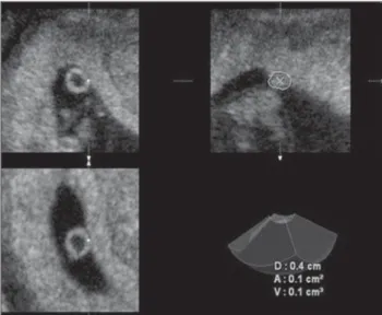
length, the mean gestational sac diameter (arithmetic mean of the three largest diam-eters) and evaluating the embryonic heart frequency. Subsequently, the 3D function was selected and the scan window (BOX) was positioned perpendicularly to the plane of the gestational sac at a 30° scanning angle (region of interest). After automatic scanning (regular mode) the all the three or-thogonal planes were simultaneously dis-played on the screen: transverse or axial plane (A) (at the upper left corner of the screen); longitudinal or sagittal plane (B) (at the upper right corner of the screen); and frontal or coronal plane (C) (lower left corner of the screen).
The YS volume was calculated by the multiplanar method after the 3D images capture, utilizing the B (sagittal) plane as a reference. On the axial (A) plane, the cursor was displaced at 1.0 mm intervals from one extremity to the other of the YS, with simultaneous manual delimitation of the outer YS surface in the sagittal plane (B) and determination of the respective areas. Subsequently, and based on these areas, the device automatically calculated the volume in cm3 (Figure 1).
The data were stored on an Excel work-sheet (Microsoft Corp.; Redmond, EUA) and statistically analyzed through the Win-dows SPSS version 13.0 (SPSS Inc.; Chi-cago, USA). For the YS volume, mean, median, standard deviations, and minimum and maximum values for each gestational age were calculated. Polynomial regression was utilized for evaluating the correlation
361
Yolk sac volume obtained by multiplanar method
Radiol Bras. 2009 Nov/Dez;42(6):359–362
sac(3) and YS absence or deformity(4)
dur-ing this gestational period were correlated with abnormal gestational results(2).
Several studies utilizing 2DUS have correlated alterations in the YS diameter and shape with adverse gestational re-sults(4,6,7). Küçük et al.(6) have evaluated
250 singleton pregnancies at the first tri-mester, with 31 presenting abnormal re-sults. Among these 31 pregnant women, 20 had YS diameter > two standard deviations. The advent of 3DUS has allowed a more accurate volumetric evaluation of irregu-larly shaped objects because of the better delimitation of their outer surface(11–13).
Studies approaching new normality pa-rameters for the first gestational trimester at 3DUS, such as embryo volume(14,15) and
YS volume(8–10), already can be found in
the literature, and some of these studies have also correlated 3DUS parameters at the first trimester with adverse gestational results.
Cho et al.(4) have reported that, in the
presence of a heart beating embryo, with ir-regular or hydropic YS, or even an early disappearance of the YS, the gestation could result in miscarriage.
Kupesic et al.(8) have evaluated pregnant
women between 6th and 10th gestational weeks and measured the YS volume by means of 3DUS with the multiplanar method, observing a correlation between YS volume and gestational age, with an exponential increase in volume up to the 10th week and later decrease.
Figueras et al.(9) have analyzed pregnant
women between the 6th and 10th gesta-tional weeks, also utilizing the VOCAL method. Their results have demonstrated a correlation between spontaneous miscar-riage and YS diameters below the 5th per-centile and above the 95th perper-centile for the respective gestational ages.
The multiplanar method with volumet-ric technique was chosen because its avail-ability from the first generation of 3DUS systems (Combison 330) and proved in vitro accuracy(12). Additionally, this
tech-nique has already been utilized for measur-ing the volume of first trimester structures such as gestational sac(10) and placenta(16),
besides the YS itself(8).
In the present study, the mean YS vol-ume measured by the multiplanar method between YS volume and gestational age,
with adjustments by the determination co-efficient R2. The significance level of 0.05 (p = 0.05) was utilized for such analyses.
RESULTS
The population sampling included 72 pregnant women between seven and tem complete gestational weeks. The selected patients were compliant with the exclusion/ inclusion criteria and underwent first and second trimester morphological fetal study and, therefore, included in the final statis-tical analysis. The patients’ ages ranged from 20 to 41 years (mean, 30 years; stan-dard deviation, 7.2 years) and parity status ranging between one and nine (mean, 2; standard deviation, 1.7).
The multiplanar method was adopted for measuring the YS volume, with the mean volume ranging from 0.07 (0.02– 0.11) to 0.20 cm3 (0.02–0.74), between the
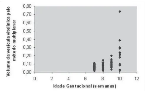
7th and 10th weeks (mean, 0.11 cm3 ± 0.10 cm3). Table 1 shows mean, median, stan-dard deviation, minimum and maximum values for the YS volume at each gesta-tional age evaluated. It was demonstrated that the YS volume is poorly correlated with the gestational age (GA), that is best represented by the quadratic regression corresponding to the following equation: YS volume = 0.9757 – 0.2499 × GA + 0.0172 × GA2, with R2 = 0.234. Figure 2 shows a scatterplot demonstrating YS vol-ume as a function of the gestational age, as well as the poor correlation between both factors.
DISCUSSION
Two-dimensional US at the first gesta-tional trimester provides relevant informa-tion on the embryo feasibility and accu-rately defines the gestational age(10). Some
parameters such as small mean gestational
Figure 2. Scatterplot demonstrating yolk sac volume as a function of the gestational age, with adjust-ment of the quadratic regression.
Table 1 Mean, median, standard deviation, maximum and minimum yolk sac volume between the 7th and 10th gestational weeks.
Multiplanar yolk sac volume (cm3)
Gestational age (weeks)
7 to 7 + 6 8 to 8 + 6 9 to 9 + 6 10 to 10 + 6
n
15 18 22 17
Mean
0.07 0.08 0.12 0.20
Median
0.06 0.08 0.12 0.18
Standard deviation
0.03 0.03 0.03 0.18
Minimum
0.02 0.02 0.04 0.02
Maximum
0.11 0.15 0.20 0.74
362
Rolo LC et al.
Radiol Bras. 2009 Nov/Dez;42(6):359–362 ranged from 0.07 (0.02–0.11) to 0.20 cm3
(0.02–0.74), between the 7th and 10th ges-tational weeks (mean, 0.11 cm3). It is im-portant to note that the values found in the present study are overestimated in relation to the ones found by Kupesic et al.(8) and
Cosmi et al.(17), who have utilized the same
volumetric technique adopted in the present study. In the first study, the authors have observed that the mean YS volume ranged from 0.037 cm3 to 0.058 cm3, while in the second study, the mean YS volume in healthy pregnant women ranged from 0.020 cm3 to 0.082 cm3.
In a previous study developed by our group with this same population group, but utilizing the VOCAL method (with 30° ro-tation angle) as a volumetric technique, they observed that the mean volume ranged from 0.06 to 0.16 cm3, proving that both techniques are in agreement(18). In a study
developed for evaluating the first trimester placental volume, it has been demonstrated that the multiplanar method (1.0 mm inter-vals), VOCAL 30° (six consecutive planes) and VOCAL 12° (15 consecutive planes) methods are in agreement(17). The main
advantage of the VOCAL 30° method is the quickness and practicality, but, considering that the YS is a small and relatively regu-lar structure, it is believed that both tech-niques are feasible for measuring the YS volume.
The authors observed a poor correlation between gestational age and YS volume that is best represented by quadratic regres-sion (R2 = 0.234). In a previous study uti-lizing the VOCAL method, the authors have also observed a poor correlation
be-tween YS volume and gestational age, best represented by the quadratic equation (R2 = 0.188). Such correlation level was al-ready expected because, as the YS reaches its maximum volume, approximately at the 10th gestational week, the degeneration process resulting from the decrease in vas-cularization has already been initiated(8).
CONCLUSION
The YS volume measured by means of 3DUS presented a poor correlation with the gestational age between the 7th and 10th weeks of gestation.
REFERENCES
1. Jurkovic D, Gruboeck K, Campbell S. Ultrasound features of normal early pregnancy development. Curr Opin Obstet Gynecol. 1995;7:493–504. 2. Moore KL. The placenta. In: Moore KL, Persaud
TVN, editors. The developing human: clinically oriented embryology. 7th ed. Philadelphia: WB Saunders; 1993. p. 110–8.
3. Trahair JF, Harding R. Ultrastructural anomalies in the fetal small intestine indicate that fetal swal-lowing is important for normal development: an experimental study. Virchows Arch A Pathol Anat Histophatol. 1992;420:305–12.
4. Cho FN, Chen SN, Tai MH, et al. The quality and size of yolk sac in early pregnancy loss. Aust N Z J Obstet Gynaecol. 2006;46:413–8.
5. Jauniaux E, Jurkovic D, Henriet Y, et al. Devel-opment of the secondary human yolk sac: corre-lation of sonographic and anatomical features. Hum Reprod. 1991;6:1160–6.
6. Küçük T, Duru NK, Yenen MC, et al. Yolk sac size and shape as predictors of poor pregnancy out-come. J Perinat Med. 1999;27:316–20. 7. Cepni I, Bese T, Ocal P, et al. Significance of yolk
sac measurements with vaginal sonography in the first trimester in the prediction of pregnancy out-come. Acta Obstet Gynecol Scand. 1997;76:969– 72.
8. Kupesic S, Kurjak A, Ivanciƒ-Kosuta M. Volume
and vascularity of the yolk sac studied by three-dimensional ultrasound and color Doppler. J Perinat Med. 1999;27:91–6.
9. Figueras F, Torrents M, Muñoz A, et al. Three-di-mensional yolk and gestational sac volume. A pro-spective study of prognostic value. J Reprod Med. 2003;48:252–6.
10. Babinszki A, Nyari T, Jordan S, et al. Three-di-mensional measurement of gestational and yolk sac volumes as predictors of pregnancy outcome in the first trimester. Am J Perinatol. 2001;18: 203–11.
11. Raine-Fenning NJ, Clewes JS, Kendall NR, et al. The interobserver reliability and validity of vol-ume calculation from three-dimensional ultra-sound datasets in the in vitro setting. Ultraultra-sound Obstet Gynecol. 2003;21:283–91.
12. Riccabona M, Nelson TR, Pretorius DH. Three-dimensional ultrasound: accuracy of distance and volume measurements. Ultrasound Obstet Gynecol. 1996;7:429–34.
13. Hösli IM, Tercanli S, Herman A, et al. In vitro volume measurement by three-dimensional ultra-sound: comparison of two different systems. Ul-trasound Obstet Gynecol. 1998;11:17–22. 14. Blaas HG, Eik-Nes SH, Berg S, et al. In-vivo
three-dimensional ultrasound reconstructions of em-bryos and early fetuses. Lancet. 1998;352:1182– 6.
15. Blaas HG, Taipale P, Torp H, et al. Three-dimen-sional ultrasound volume calculations of human embryos and young fetuses: a study on the volumetry of compound structures and its repro-ducibility. Ultrasound Obstet Gynecol. 2006;27: 640–6.
16. Nowak PM, Nardozza LMM, Araujo Júnior E, et al. Comparison of placental volume in early preg-nancy using multiplanar and VOCAL methods. Placenta. 2008;29:241–5.
17. Cosmi E, Piazze JJ, Ruozi A, et al. Structural-tri-dimensional study of yolk sac in pregnancies complicated by diabetes. J Perinat Med. 2005;33: 132–6.