Reproducibility of ultrasonography as a method
to measure abdominal and visceral fat*
Avaliação da reprodutibilidade ultrassonográfica como método para medida da gordura abdominal e visceral
Angélica Lemos Debs Diniz1
, Raphael Alves Ferreira Tomé2
, Cecília Lemos Debs3
, Renata Carraro4 , Leonardo Borges Roever5
, Rogério de Melo Costa Pinto6
OBJECTIVE: The purpose of this study was to evaluate the interobserver variability of ultrasound in the measure-ment of subcutaneous, visceral and perirenal fat through standard technique. MATERIALS AND METHODS: From November 2006 to January 2007, 50 patients were evaluated. The subcutaneous fat thickness was measured with a 7.5 MHz linear transducer transversely positioned 1 cm above the umbilical scar. For the visceral fat, a 3.5 MHz transducer was also positioned 1 cm above the umbilical scar, considering the distance between the internal surface of the abdominal rectus muscle and the posterior aortic wall in the abdominal midline. The perirenal fat was measured in the middle third of the right kidney, with the transducer positioned at the axillary midline. RESULTS: The t-Student test was utilized to analyze the interobserver reproducibility
with significance level of 95%. No statistically significant difference was observed among mean values for
subcutaneous, visceral and perirenal fat (p = 0.7141, 0.7286 and 0.6368, respectively). Mean and standard
deviation corresponded to 2.64 ± 1.37 for subcutaneous thickness, 6.84 ± 2.38 for visceral fat, and 4.89 ± 2.6 for perirenal fat. CONCLUSION: Ultrasound presented a good interobserver reproducibility in the evalu-ation of abdominal fat based on measurement of subcutaneous, visceral and perirenal fat as parameters.
Keywords: Abdominal fat; Ultrasound; Metabolic syndrome.
OBJETIVO: O objetivo deste estudo é avaliar a variabilidade interobservador do método ultrassonográfico para medida da gordura subcutânea, visceral e perirrenal por meio de técnica padronizada. MATERIAIS E MÉTODOS: Foram avaliados 50 pacientes entre novembro de 2006 e janeiro de 2007. A medida da espes-sura subcutânea foi realizada com transdutor linear de 7,5 MHz posicionado transversalmente a 1 cm acima da cicatriz umbilical. Para a gordura visceral foi utilizado transdutor de 3,5 MHz posicionado 1 cm acima da cicatriz umbilical, considerando-se a medida entre a face interna do músculo reto abdominal e a parede pos-terior da aorta na linha média do abdome. A gordura perirrenal foi medida no terço médio do rim direito, com transdutor posicionado na linha axilar média. RESULTADOS: A reprodutibilidade interobservador foi ana-lisada por meio do teste t de Student, com significância de 95%. Não houve diferença significativa entre as
médias das medidas das gorduras subcutânea, visceral e perirrenal, com p = 0,7141, 0,7286 e 0,6368,
respectivamente. As médias encontradas, com seus respectivos desvios-padrão, foram: 2,64 ± 1,37 para a espessura subcutânea, 6,84 ± 2,38 para a espessura visceral e 4,89 ± 2,6 para a espessura perirrenal. CONCLUSÃO: A ultrassonografia apresentou boa reprodutibilidade interobservador para avaliação da gor-dura abdominal por meio das medidas das espessuras subcutânea, visceral e perirrenal.
Unitermos: Gordura abdominal; Ultrassom; Síndrome metabólica.
Abstract
Resumo
* Study developed at Unit of Ultrasonography, Hospital de Clí-nicas da Universidade Federal de Uberlândia (UFU), Uberlândia, MG, Brazil.
1. PhD, Permanent Professor, Post-graduation of Health Sci-ences at Universidade Federal de Uberlândia (UFU), Uberlândia, MG, Brazil.
2. MD, Resident at Department of Radiology and Imaging Diagnosis, Hospital de Clínicas da Universidade Federal de Uber-lândia (UFU), UberUber-lândia, MG, Brazil.
3. MD, Specialist in Radiology and Imaging Diagnosis, spe-cialized in Breast Imaging Diagnosis, Universidade Federal de Uberlândia (UFU), Uberlândia, MG, Brazil.
4. MD, Specialist in Radiology and Imaging Diagnosis, Uni-versidade Federal de Uberlândia (UFU), Uberlândia, MG, Brazil. 5. Post-graduation Student of Health Sciences at Universidade de Uberlândia (UFU), Uberlândia, MG, Brazil.
6. PhD, Professor, Faculdade de Matemática da Universidade Federal de Uberlândia (UFU), Uberlândia, MG, Brazil.
Several studies demonstrate the closed relation between abdominal adiposity and glucose intolerance, hyperinsulinemia, hypertriglyceridemia and arterial hyperten-sion. Recently, it has been believed that, more than a simple association, abdominal fat, particularly the visceral one, plays a significant role in the metabolic syndrome physiopathology(1–4).
Therefore, the quantification of visceral fat is important to identify those individu-als with higher risk for developing meta-bolic syndrome and thus eligible for being
Diniz ALD, Tomé RAF, Debs CL, Carraro R, Roever LB, Pinto RMC. Reproducibility of ultrasonography as a method to measure abdominal and visceral fat. Radiol Bras. 2009;42(6):353–357.
Mailing address: Dra. Angélica Lemos Debs Diniz. Alameda João César de Souza, 110. Uberlândia, MG, Brazil, 38411-154. E-mail: [email protected]
Received May 12, 2009. Accepted after revision September 24, 2009.
INTRODUCTION
submitted to earlier interventions in an at-tempt to reduce the impact of metabolic al-terations on the cardiovascular morbimor-tality in such individuals(3,4).
Computed tomography is the method of choice for quantifying visceral fat, but it has not been solely utilized in the diagnos-tic routine of patients with metabolic syn-drome(5,6). Waist size and waist-hip ratio, although indirectly, constitute other meth-ods for visceral fat evaluation(7). Magnetic resonance imaging has been proposed as an alternative method free from ionizing ra-diation, but the literature reports restric-tions for patients with morbid obesity, claustrophobia, or metal prosthesis and pacemaker(8–10).
Ultrasonography is a low cost and use-ful method besides not requiring radiation for evaluating visceral fat tissue(5,6,8,10). Ad-ditionally, recent publications have demon-strated similar effectiveness of both ultra-sonography and computed tomography in the quantification of visceral fat(5–7). How-ever, ultrasonography is operator-depen-dent, besides requiring the definition of specific anatomic landmarks and a stan-dardized scanning technique.
The present study is aimed at evaluat-ing the interobserver variability of the sonographic method in the measurement of subcutaneous, visceral and perirenal fat by means of a standardized technique.
MATERIALS AND METHODS
Observational study analysing abdomi-nal and visceral fat in 50 patients by means of ultrasonography performed by two ob-servers with a same scanning technique. The abdominal fat was quantified through the following sonographic measurements: abdominal subcutaneous, visceral and peri-renal fat.
The present study was developed in the Division of Ultrasonography at Hospital de Clínicas of Universidade Federal de Uberlândia, Minas Gerais, Brazil, with pre-vious approval by the Committee for Eth-ics in Research of the institution. All the pa-tients signed a term of free and informed consent.
The study group included both male and female patients consecutively and ran-domly selected, independently from their
body mass índex. The mean age of such patients was 42 years ± 12 years.
The ultrasonography scans were per-formed by two radiologists who utilized a same, previously standardized scanning technique. Each of the patients was evalu-ated on the same day by two investigators who were not aware of the results of each other, in order to avoid samples contami-nation.
A Versa Pro (Siemens; Erlangen, Ger-many) equipment was utilized with elec-tronic, linear 7.5 MHz and convex, 3.5 MHz transducers. All the scans were per-formed with the patient under 12-hour fast-ing, lying in dorsal decubitus and maxi-mum abduction of the right arm.
The measurement of subcutaneous fat was performed with a linear 7.5 MHz trans-ducer transversely positioned 1 cm above the umbilical scar, without exerting any pressure over the abdomen in order to avoid underestimation of the measurement. The subcutaneous thickness corresponded to the distance in centimeters between the skin and the outer surface of the fascia of the abdominal muscles.
For the visceral fat, a 3.5 MHz trans-ducer was also transversely positioned 1 cm above the umbilical scar on the abdominal midline, without exerting any pressure over the abdomen. The visceral fat thickness corresponded to the measurement in cen-timeters between the internal surface of the abdominal rectus muscle and the posterior aortic wall in the abdominal midline, dur-ing expiration. The perirenal fat was mea-sured with a convex 3.5 MHz transducer longitudinally positioned on the axillary midline, with identification of the right kidney image. The perirenal fat thickness corresponded to the distance in millimeters between the lateral border of the kidney and the iliopsoas muscle surface adjacent to the middle third of the right kidney (Figure 1).
Statistical analyses
Descriptive statistical analysis was uti-lized for sociodemographic characteriza-tion and for calculating mean and standard deviation of the values found for the three measurements performed by the two inves-tigators.
The data normality was evaluated by the Lilliefors test. The Student t-test was
uti-lized to compare the measurements per-formed by the two investigators for the subcutaneous and visceral fat variables, and the Mann-Whitney test, for perirenal fat.
The intraclass correlation coefficient and respective confidence interval(11) were utilized for determining the reproducibil-ity of the measurements obtained by the two investigators. Additionally, the Bland-Altman plot(12) was utilized for evaluating the interobserver agreement.
The level of statistical significance was defined as p < 0.05.
RESULTS
Mean values and respective standard deviations were 2.64 cm ± 1.37 for subcu-taneous fat thickness, 6.84 cm ± 2.38 for visceral fat thickness, and 4.89 mm ± 2.6 for the perirenal fat thickness.
No statistically significant difference was observed among the measurements of subcutaneous, visceral and perirenal fat performed by the investigators 1 and 2 (p = 0.7141, 0.7286 and 0.6368, respectively), indicating a good level of interobserver agreement (Figure 2). Such good interob-server agreement can be confirmed on Fig-ures 3, 4 and 5 regarding subcutaneous, visceral and perirenal fat, respectively.
Figure 3 (subcutaneous fat) demon-strates that most of times, the values re-mained between mean ± one standard de-viation, and in only seven patients the val-ues were > one standard deviation. The mean difference was very low, i.e., –0.10 cm and standard deviation, 0.31 cm, show-ing excellent interobserver agreement. Ad-ditionally, the maximum and minimum values for the differences were, respec-tively, 0.9 and –0.9.
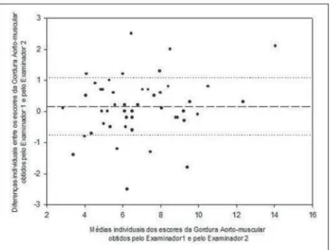
As regards the interobserver agreement for the visceral fat variable (Figure 4), an excellent level was observed, with the ma-jority of values remaining between the mean and one standard deviation. The dif-ferences were a little higher as compared with the subcutaneous fat, i.e., the mean difference was 0.16 cm and standard devia-tion, 0.93 cm. Measurements for eleven patients remained above this threshold.
pre-Figure 1. A: Measurement of muscular aortic thickness. B: Measurement of subcutaneous fat. C: Measurement of perirenal fat.
A B
C
sented a good interobserver agreement, with the majority of values remaining be-tween the mean and one standard deviation (Figure 5). The mean difference was –0.14 mm and standard deviation, 1.39 mm.
Measurements for twelve patients re-mained above this threshold.
As regards the interobserver reproduc-ibility, intraclass correlation coefficient of 0.97 was observed (confidence interval
[CI] 95%: 0.96–0.99, p < 0.01) for subcu-taneous fat, 0.91 (CI 95%: 0.86–0.95, p < 0.01) for visceral fat, and 0.63 (CI 95%: 0.44–0.78, p < 0.01) for perirenal fat. An excellent interobserver reproducibility is
Figure 2. Means and standard deviations of measurements obtained by the investigators 1 and 2, ESC corresponding to subcutaneous thickness, EMA to muscular aortic thickness, and EPR to perirenal thickness. No significant difference was observed among the means for the three measurements ob-tained by the two investigators.
observed for subcutaneous and visceral fat, and satisfactory correlation for perirenal fat.
DISCUSSION
Considering the current worldwide epi-demics of metabolic syndrome, and the knowledge on the obesity’s impact on the morbidity and mortality resulting from car-diovascular events, it is increasingly nec-essary to develop diagnostic methods ca-pable of evaluating and quantifying the distribution of the body fat, particularly the visceral fat(2–4,13,14). It is known that fat may accumulate in several abdominal compart-ments, including the epiploon, viscera such as the liver, as well as in the retroperitoneal region, including the perirenal region.
Ultrasonography has shown to be a practical, effective and low-cost method besides not requiring ionizing radia-tion(5,7,9,10,14,15). Such factors, in association with its relevant role in the identification of individuals with increased central adi-posity, may represent an important step towards the future possibility of selecting patients at high risk for developing meta-bolic syndrome, aiming at allowing an early intervention, thus minimizing the impact resulting from the complications of this condition(3,8,13,16).
In the present study, ultrasonography has demonstrated to be highly reproducible and capable of measuring the thickness of subcutaneous, visceral and perirenal fat.
Among the several advantages of ultra-sonography, it is important to observe that high availability, low cost and reproducibil-ity are factors that meet the needs of devel-oping countries which may utilize this method in public health measures to reduce social costs(7,9,14,15,17,18).
The study of abdominal fat by means of ultrasonography was first undertaken early in the nineties by Armellini et al. by com-parison of sonographic and computed to-mography findings in a group of 50 obese women. The sonographic findings pre-sented a good correlation with the com-puted tomography findings (r = 0.66, p < 0.001), corroborating the hypothesis that ultrasonography could be useful in the as-sessment of abdominal fat(15). Later stud-ies have demonstrated a poor correlation between sonographic and tomographic findings for measurement of visceral fat(7,14,17,18). However, study developed by Stolk et al. concluded that the low reliabil-ity of the measurements of visceral fat by ultrasonography would be a result from failures in a strict standardization of vis-ceral measurements such as inappropriate transducer positioning, pressure on the abdomen and measurements at different phases of the respiratory cycle(17). Such factors may impair the quantification of abdominal fat, leading to poorly reproduc-ible results. This fact has not been observed in the present study that has been developed in compliance with strict standardization for measurement of abdominal fat.
Abdominal computed tomography is considered as the gold standard in the de-termination of visceral fat, since this method can differentiate subcutaneous from visceral fat, besides being highly re-producible(5–7,14). The advantage of com-puted tomography and magnetic resonance imaging in the evaluation of abdominal fat is the fact that these methods do not depend on the operator ability to identify the struc-tures during the examination, besides not being influenced by the pressure exerted by the transducer over the patient’s abdomen during the measurements. However, the ionizing radiation present in computed to-mography and the high cost and long ac-quisition time of magnetic resonance im-aging represent unfavorable aspects in the utilization of such methods in the routine for quantifying abdominal fat, considering that ultrasonography presents an optimum reproducibility(6,7,10).
The present study has demonstrated a high interobserver agreement and thus the ultrasonography reproducibility in the quantification of abdominal fat by means of measurements of subcutaneous, visceral and perirenal fat, with excellent CI for the first two types of fat, and a reasonable CI for perirenal fat – an already expected re-sult because of the higher technical diffi-culty in the evaluation of the perirenal fat. Additionally, the perirenal fat is measured in millimeters, becoming more susceptible to biometric errors. The development of the present study only was possible because the
Figure 4. Means for measurements of muscular aortic fat obtained by the two investigators, demonstrating excellent interobserver agreement.
description and utilization of a strict pro-tocol for measurement of abdominal fat.
It is important to mention some techni-cal limitations, since they may pose some difficulty to the performance of the US scan, limiting its reliability. Among these limitation, one may observe non-coopera-tion of the patient in respiratory maneuvers, utilization of inappropriate transducers and, particularly, inappropriate operator training and technique(5,17,18).
Therefore, ultrasonography presented a good interobserver reproducibility in the evaluation of abdominal and visceral fat by means of measurements of the thickness of subcutaneous, visceral and perirenal fat.
REFERENCES
1. Radominski RB, Vezozzo DP, Cerri GG, et al. O uso da ultra-sonografia na avaliação da distribui-ção de gordura abdominal. Arq Bras Endocrinol Metab. 2000;44:5–12.
2. Timar O, Sestier F, Levy E. Metabolic syndrome X: a review. Can J Cardiol. 2000;16:779–89. 3. World Health Organization. Obesity: preventing
and managing the global epidemic. Report of a WHO consultation. World Health Organ Tech Rep Ser. 2000;894:i–xii, 1–253.
4. van der Kooy K, Seidell JC. Techniques for the measurement of visceral fat: a practical guide. Int J Obes Relat Metab Disord. 1993;17:187–96. 5. Hirooka M, Kumagi T, Kurose K, et al. A
tech-nique for the measurement of visceral fat by ul-trasonography: comparison of measurements by ultrasonography and computed tomography. In-tern Med. 2005;44:794–9.
6. Armellini F, Zamboni M, Robbi R, et al. Total and intra-abdominal fat measurements by ultrasound and computerized tomography. Int J Obes Relat Metab Disord. 1993;17:209–14.
7. Pineau JC, Guihard-Costa AM, Bocquet M. Vali-dation of ultrasound techniques applied to body fat measurement. A comparison between ultra-sound techniques, air displacement plethysmog-raphy and bioelectrical impedance vs. dual-en-ergy X-ray absorptiometry. Ann Nutr Metab. 2007;51:421–7.
8. Pozzato C, Radaelli G, Dall’Asta C, et al. MRI in identifying hepatic steatosis in obese children and relation to ultrasonography and metabolic findings. J Pediatr Gastroenterol Nutr. 2008;47:493–9. 9. Liu KH, Chan YL, Chan WB, et al. Sonographic
measurement of mesenteric fat thickness is a good correlate with cardiovascular risk factors: com-parison with subcutaneous and preperitoneal fat thickness, magnetic resonance imaging and an-thropometric indexes. Int J Obes Relat Metab Disord. 2003;27:1267–73.
10. Gong W, Ren H, Tong H, et al. A comparison of ultrasound and magnetic resonance imaging to assess visceral fat in the metabolic syndrome.
Asia Pac J Clin Nutr. 2007;16 Suppl 1:339–45.
11. Weir JP. Quantifying test-retest reliability using the intraclass correlation coefficient and the SEM. J Strength Cond Res. 2005;19:231–40.
12. Bland JM, Altman DG. Statistical methods for assessing agreement between two methods of clinical measurement. Lancet. 1986;1(8476): 307–10.
13. Sturm W, Sandhofer A, Engl J, et al. Influence of visceral obesity and liver fat on vascular structure and function in obese subjects. Obesity (Silver Spring). 2009;17:1783–8.
14. Ribeiro-Filho FF, Faria AN, Azjen S, et al. Meth-ods of estimation of visceral fat: advantages of ultrasonography. Obes Res. 2003;11:1488–94.
15. Armellini F, Zamboni M, Rigo L, et al. The con-tribution of sonography to the measurement of intra-abdominal fat. J Clin Ultrasound. 1990;18: 563–7.
16. Sabir N, Pakdemirli E, Sermez Y, et al. Sono-graphic assessment of changes in thickness of different abdominal fat layers in response to diet in obese women. J Clin Ultrasound. 2003;31:26– 30.
17. Stolk RP, Wink O, Zelissen PM, et al. Validity and reproducibility of ultrasonography for the mea-surement of intra-abdominal adipose tissue. Int J Obes Relat Metab Disord. 2001;25:1346–51. 18. Tornaghi G, Raiteri R, Pozzato C, et al.