Prevalence of Chlamydial Infections in
Fattening Pigs and Their Influencing Factors
Karolin Hoffmann1,2, Franziska Schott3, Manuela Donati4, Antonietta Di Francesco5, Michael Hässig6, Sabrina Wanninger1,2, Xaver Sidler3, Nicole Borel1*
1Institute of Veterinary Pathology, Vetsuisse Faculty, University of Zurich, Zurich, Switzerland,2Center for Clinical Studies, Vetsuisse Faculty, University of Zurich, Zurich, Switzerland,3Department of Farm Animals, Division of Swine Medicine, Vetsuisse Faculty, University of Zurich, Zurich, Switzerland,4DIMES,
Microbiology, University of Bologna, Bologna, Italy,5Department of Veterinary Medical Sciences, University of Bologna, Ozzano Emilia (BO), Italy,6Department for Farm Animals, Section for Herd Health, Vetsuisse Faculty, University of Zurich, Zurich, Switzerland
*n.borel@access.uzh.ch
Abstract
Chlamydial infections in pigs are associated with respiratory disease, diarrhea, conjunctivi-tis and other pathologies. The aim of this study was to define the prevalence of Chlamydia-ceaein Swiss fattening pigs by applying sensitive and specific detection methods and to correlate prior antibiotic treatment and farm related factors with differences in prevalence. Conjunctival and fecal swabs were collected from 636 pigs in 29 Swiss fattening pig farms with and without antibiotic treatment, at the beginning and the end of the fattening period. The swabs were screened by real-time PCR forChlamydiaceae. For the chlamydial detec-tion and species-identificadetec-tion, a DNA-microarray analysis was performed. All farms were positive forChlamydiaceaewith 94.3 and 92.0% prevalence in fecal swabs as well as 45.9 and 32.6% in conjunctival swabs at the first and second time points, respectively. Antibiotic treatment could not clear the infection on herd level. Potential contact with wild boars was a significant risk factor, while hygiene criteria did not influence chlamydial prevalence. A cor-relation of chlamydial positivity to diarrhea, but not to conjunctivitis was evident.Chlamydia suiswas the predominant species. Mixed infections withC.suisandC.pecorumwere com-mon, with a substantial increase inC.pecorumpositivity at the end of the fattening period, and this finding was associated with ruminant contact.C.abortuswas detected in one con-junctival swab. In this study,C.suisinhabited the intestinal tract of nearly all examined pigs, implying a long-term infection.C.pecorumwas also common and might be transmitted to pigs by ruminants.
Introduction
Members of theChlamydiaceaeare known to cause a broad spectrum of diseases in numerous vertebrate host species worldwide. Chlamydiae are obligate intracellular bacteria sharing a characteristic biphasic lifecycle. Extracellular elementary bodies (EBs) infect the host cell and transform into the replicating reticulate bodies (RBs), which then re-differentiate into EBs.
OPEN ACCESS
Citation:Hoffmann K, Schott F, Donati M, Di Francesco A, Hässig M, Wanninger S, et al. (2015) Prevalence of Chlamydial Infections in Fattening Pigs and Their Influencing Factors. PLoS ONE 10(11): e0143576. doi:10.1371/journal.pone.0143576
Editor:David M. Ojcius, University of the Pacific, UNITED STATES
Received:September 18, 2015
Accepted:November 8, 2015
Published:November 30, 2015
Copyright:© 2015 Hoffmann et al. This is an open access article distributed under the terms of the
Creative Commons Attribution License, which permits unrestricted use, distribution, and reproduction in any medium, provided the original author and source are credited.
Data Availability Statement:All relevant data are within the paper and its Supporting Information files.
Funding:This study was funded by the Swiss Federal Food Safety and Veterinary Office (grant 1.13.03). The funders had no role in study design, data collection and analysis, decision to publish, or preparation of the manuscript.
Replication occurs primarily in epithelial cells of the respiratory, gastrointestinal and urogenital tract as well as in the conjunctival epithelium [1].
To date, there are eleven species included in the GenusChlamydia, four of which have been documented in pigs:Chlamydia suis,Chlamydia pecorum,Chlamydia abortusandChlamydia psittaci, withC.suisbeing the most important.Chlamydiaceaein pigs are associated with disor-ders like conjunctivitis, pneumonia, pericarditis, polyarthritis, polyserositis, enteritis and reproductive problems of sows and boars; however, infections are often asymptomatic and rou-tine diagnostics usually do not include chlamydiae [1]. The seropositivity forChlamydiaceaein pigs from European countries is high and ranges up to 96.5% [2–7]. Not only domestic pigs but also wild boars (Sus scrofa) harborChlamydiaceae, as reported in Germany, Spain and Italy [8–12]. In fecal samples from pigs,C.suisis the most frequently found chlamydial species [7,13]. AlthoughC.suisis most often found in pigs, many other animal species like cattle, horses, frogs and cats can become naturally infected [14–17]. Evidence for the zoonotic poten-tial ofC.suisis emerging, as previously demonstrated in trachoma patients from Nepal, where
C.suiswas found as a single or mixed infection withC.trachomatis[18]. Moreover,C.suis
DNA was found in conjunctival swabs of employees in a Belgian pig slaughterhouse [19,20]. In Switzerland, prophylactic and metaphylactic antimicrobial treatment of fattening pigs is often applied, mostly with sulfonamide and trimethoprim combinations or tetracyclines [21,22]. For decades, tetracyclines, as broad-spectrum antibiotics, have been extensively used in the pig industry for both prophylactic and therapeutic treatment. Over the past years, there have been accumulating reports on the occurrence of tetracycline-resistantC.suisstrains in America, Europe and Asia [23–27]. A rapid selection for tetracycline-resistantC.suiswas detected in a Swiss farm after oral tetracycline medication, while elimination of the organism was not achieved [26].
In Switzerland, pigs are the most important source of meat for human consumption [28] and therefore the pig producing industry plays an important role in human nutrition and the agricultural economy. Consequently, pig pathogens potentially play an important role in human public health.
To date, no large-scale prevalence studies onChlamydiaceaein pigs exist. The available data were obtained by serological methods and included mostly breeding farms or breeding sows [1]. The aim of this study was to collect comprehensive data by applying sensitive and specific direct detection methods to determine the prevalence of infection with all currently known
Chlamydiaceaespecies in the Swiss fattening pig population. Results were correlated with med-ical treatment, hygiene and housing conditions, as well as contact with other farm and/or wild animal species.
Material and Methods
The prevalence ofChlamydiaceaein Swiss fattening pigs was investigated by sampling con-junctival and fecal swabs from 29 different pig herds at the beginning and end of the fattening period.
Farms and animals
In total, 1,359,513 pigs, excluding breeding sows, lived in Switzerland as of May 1st, 2013 [29] and 2,689,327 pigs were slaughtered in the year 2013 [28].
n = 1, Thurgau: n = 2, Waadt: n = 2, Zürich: n = 3). The cantons examined harbor the following proportions of the Swiss pig population [29]: Aargau: 6.5%, Bern: 17.1%, Freiburg: 5.5%, Luzern: 27.6%, St. Gallen: 12.2%, Schaffhausen: 1.3%, Thurgau: 12.6%, Waadt: 2.9%, Zürich: 2.6%, thus, the most important pig producing regions were covered by the sampling. Farm par-ticipation was confirmed upon the request of the farm veterinarians (n = 12), the two main Swiss pig-trading companies (n = 10), the Swiss Pig Health Service (n = 2) or directly by the study investigator (n = 5). The study was approved by the Veterinary Office of Canton Luzern (authorization no. LU03/14) and all efforts were made to minimize the discomfort of the ani-mals during sampling. The pig housings and land that was accessed are privately owned and all owners gave their permission to conduct the study on these sites.
The farms were divided into two groups: farrow-to-finish farms (n = 10) and fattening farms without breeding on farm (n = 19). An all-in/all-out-production system was used on seven farms, while nine out of ten farrow-to-finish farms and 13 fattening farms were rearing the pigs in a continuous system. Regarding the housing system, there were farms with (n = 19) and without (n = 10) outdoor access combined with indoor batch pens. The floor of the indoor pens was covered with bedding material in most farms (n = 19). Outdoor access and presence of bedding material in indoor pens was often combined on the same farm (n = 16). Eight farms routinely applied prophylactic oral antibiotic treatment of the whole herd (combination of tri-methoprime, sulfadimidin and sulfathiazole (TSS) on farms 6, 23 and 26; combination of chlortetracycline, tylosin and sulfadimidin on farms 9, 22, 27 and 29; chlortetracycline in farm 17), whereas one farm administered amoxicillin (A) therapeutic group treatment at the begin-ning of the fattebegin-ning period (farm 7). On one farm, a combination of chlortetracycline, sulfadi-midine and tylosin was known to be administered to the pigs before their entry into the fattening period (farm 28). All group treatments were administered orally for five to twelve days. Individual medical treatment was carried out on 15 farms, nine farms did not administer any medication during the whole sampling period and on farm 8 no medication data was avail-able (S1 Table).
In farm nos. 1, 5 and 9, all newly restocked fattening pigs in the herd were sampled, whereas in the remaining farms (n = 26), 20 randomly selected pigs per newly restocked pig batch were sampled regardless of herd size. In total, 636 pigs were included in this study. Twenty-seven farms reared male and female fattening pigs, two farms (farm nos. 18 and 25) were farrow-to-finish pig farms that only fattened female pigs. Overall, 54.7% (n = 348) of the pigs were female and 45.3% (n = 288) were male. Identical pigs were sampled twice (n = 589): i) at the beginning (first sampling, 1st) of the fattening period (at the age of approximately 12 weeks), and ii) at the end (second sampling, 2nd) of the fattening period (at the age of approximately six months). The first sampling was performed as early as possible after the pigs were introduced into their new pens and before any medical treatment was started, which was between 0 and 72 hours after their arrival. The second sampling was performed approximately one to three weeks prior to slaughtering. In 47 pigs, a second sampling was not possible. In one farm (no. 10), porcine reproductive and respiratory syndrome virus was detected after the first sampling procedure and led to the subsequent eradication of all pigs on this farm and a second farm (no. 8) was lost to follow-up. In another six farms, one animal each (farm nos. 5, 6, 11, 12, 13) and two animals (no. 14), respectively, could not be sampled a second time, because they had been slaughtered or died spontaneously during the fattening period.
Swab sampling
September 2013 and December 2014. Both eyes of each individual pig were sampled using one conjunctival swab. Fecal swabs were taken by rectal insertion of flocked swabs. Additionally, conjunctival swabs from voluntarily participating farmers (n = 9) were obtained with their written informed consent of seven different farms, and swabs (n = 2) from dust in the housing environment of farm no. 29 were collected. A total of 2,461 swabs were obtained and stored at -20°C until processing.
Questionnaire and health assessment
A detailed questionnaire was obtained from the farmers and a summary of the parameters is shown inS2 Table. The cleanliness of the sampled animals and the facilities (housing in gen-eral, indoor pens, outdoor area when existent) were recorded by the study investigators at both time points and classified as clean, moderately dirty or dirty. Hygiene management was scored by giving one point each for the following hygiene criteria as reported by the farmer: Existence of hygiene gate to pig housings, cleaning of pens before introducing new pigs, water tempera-ture in high-pressure cleaner at least 60°C, use of cleaning agents and use of disinfectant on a regular basis. With five points as the maximal score, four to five points were regarded as most hygienic, two to three points as medium and zero to one point as least hygienic. Recruitment of animals was scored as follows: 0 equals recruitment exclusively from own farm (n = 9), 1 equals purchase from one farm (n = 9), 2 equals purchase from 2 to 4 farms (n = 8) and 3 equals pur-chase from more than 4 farms (n = 3). All pig herds had at least one ruminant herd in their sur-rounding area of up to 1000 meters, belonging to the same or neighboring farms. The distance to ruminants was scored as follows: 1 equals direct snout contact is possible (n = 5,S1 Table), 2 equals one to ten meters distance (n = 12), 3 equals 11 to 100 meters distance (n = 7) and 4 equals 150 to 1000 meters distance (n = 5). Indirect contact with ruminants was defined as potential transmission routes by personnel and non-living vectors, such as instruments used in ruminant and pig housings, and occurred in 17 farms. Poultry was present within a 1000 m radius of 19 farms and in eight of these the birds belonged to the same farm. In none of the 19 farms was direct contact between poultry and fattening pigs possible. Direct contact with wild boars and other wild mammals like roe deer was possible due to the absence of double enclo-sures in 9 farms, and 25 herds had potential contact with wild birds. Insect and rodent infesta-tion was estimated by the farmers and graded as low, moderate or high. The farmers were asked to record clinical symptoms and any treatment of the sampled pigs during the fattening period. The health status of the sampled pigs was assessed on the occurrence of eye lesions, change in fecal consistency, respiratory signs, wasting and lameness by the study investigators. Eye changes were assessed in accordance with Englund et al. [30], Polkinghorne et al. [31] and Becker et al. [32] and included reddening (hyperemia) and swelling (edema) of the conjunctiva and/or sclera, epiphora, as well as mucous or purulent discharge.
DNA extraction
DNA from all conjunctival and fecal swabs was extracted using the Maxwell116 Buccal Swab LEV DNA Purification Kit (Promega, Madison, WI, USA) according to the manufacturer’s instructions with an elution volume of 100μl.
Screening for
Chlamydiaceae
threshold (Ct value) of<38 was considered positive. The correspondingChlamydiaceaecopy number perμl was automatically calculated by the PCR instrument for each tested sample. In cases of an inhibited amplification of the internal control DNA, the run was repeated with a 1:10 dilution of the sample. A sevenfold dilution series ofC.abortusDNA constituting the standard curve served as positive control and a reaction mixture with water instead of the tem-plate DNA was used as negative control.
Chlamydia
species identification by Arraymate microarray
All samples classified as positive by real-time PCR (mean Ct value of<38) were further exam-ined by the species-specific 23S Arraymate microarray assay (Alere, Jena, Germany), as described by Borel et al. [34]. The present version [35] carries 34 probes for eleven species of
Chlamydiaceae(four probes each forC.suis,C.trachomatis,C.pneumoniaeandC.psittaci, three probes forC.avium,C.caviae,C.muridarumandC.pecorum, two forC.abortus,C.felis
andC.gallinacea), three genus-specific probes, four family markers and 15 probes for Chla-mydia-like organisms, as well as four probes for the internal control DNA and an internal staining control (biotin marker). Sample DNA was amplified and biotin labeled prior to hybridization according to Borel et al. [34], with the following temperature-time profile: 96°C 10 min, 40 cycles of 94°C 30 s, 50°C 30 s, 72°C 30 s, additionally an internal control DNA was included as recommended by the manufacturer (Intype IC-DNA, Qiagen Labor, Leipzig). In the first step, 8μl of amplification product was loaded on the chip. TheChlamydiaspecies could not be determined when only genus-specific probes were positive and the species-specific probes produced no or only a weak signal.
Data analysis
Data editing and all statistical analyses were done using Stata Software (StataCorp., 2011; Stata Statistical Software: Release 12; College Station, TX, USA: StataCorp LP). Firstly, a quality con-trol of the data and the descriptive analysis was carried out using<codebook varx1 varxn>, where varx1 to varxn represents the variables from x1to xn. The dependence of a positive result in the conjunctival and rectal swab in pigs at one time point and between the two time points was analysed using the chi-square-test. To identify the influencing factors on positivity in con-junctival and fecal swabs at either time point forChlamydiaceae,C.suisandC.pecorum, a logistic regression analysis for the independent variables sex, type of farm (farrow-to-finish or fattening), all-in/all-out production or continuous production, number of purchase farms, total number of fattening pigs on farm, outdoor access, existence of bedding material, existence of own or foreign ruminant species, possible direct or indirect contact and distance to rumi-nants, possible contact to wild boars or wild birds, clinical symptoms (conjunctivitis score; diarrhea, coughing, lameness of the group as reported by farmer), hygiene score, infestation with flies and rodents, cleanliness of animals and facilities at first and second time point and the use and type of antibiotics was carried out using<logistic vary varx1 varxn>, where vary represents the dependent variable, varx1 to varxn represents the dependent variable from x1to xn. The significantly influencing variables were tested for their correlation using the<pwcorr varx1 varxn>command. If correlations of>0.6 occurred, one of the variables was eliminated. The remaining variables were entered into a full regression model for step back procedure [36]. The final model included again only significant dependent variables. The meanChlamydiaceae
regard of anatomical site, time point and antibiotic group treatment. In all analyses, a p-value0.05 was considered statistically significant.
Results
Clinical symptoms
Six farmers stated that they had observed diarrhea during the fattening period. The following infectious agents could be detected by further laboratory investigations initiated by the respec-tive farm veterinarians:Brachyspira pilosicoliandLawsonia intracellularisin farm 4, Brachy-spira hyodysenteriaein farm 9 andLawsonia sp. in farm 19. Farms 11, 17 and 26 had only rare cases of diarrhea, of which infectious agents were not further investigated. In one additional farm (no. 18), the farmer did not report diarrhea, but clinical symptoms were visible in the majority of pig pens at both time points during farm visits by the study investigators.
Sixteen farmers observed lameness in one or more pigs during the fattening period. A diag-nostic workup for infectious diseases was not performed in any of these cases, most farmers suspectedHaemophilus parasuisand/or tail biting (cannibalism) as the cause, one farm (no. 12) assumed a broken leg, and another (no. 4) claw problems (panaritia).
In eleven farms cases of respiratory disease were observed by the farmers, but except for two farms (farm nos. 4 and 7), in which influenza was diagnosed, no causative agents were
investigated.
One farm was known to have conjunctivitis problems in its finishing pigs. In another farm unclassified eye symptoms were observed during the fattening period; all other farmers reported no eye problems. The results of the conjunctivitis assessment in combination with conjunctival chlamydial positivity are displayed inTable 1. While the conjunctival lesions were significantly increasing, the chlamydial detection in conjunctival swabs significantly decreased between the first and second time point.
None of the farmers reported herd problems of wasting, however, there were a few pigs (n = 7) at the first sampling and one pig at the second sampling that were considerably retarded in growth.
Chlamydiaceae
screening
Pigs from all farms were positive forChlamydiaceaeat both samplings (S3 Table,Table 2). In the first sampling, 45.9% (n = 292) and in the second sampling 32.6% (n = 192) of the pigs were positive in the conjunctival swab. The herd-based prevalence on conjunctival swabs ran-ged from 0 to 100% with a mean value of 38.7%. In contrast, 94.3% (n = 600) of the fecal swabs were positive in the first sampling and 92.0% (n = 542) in the second sampling. The herd prev-alence ranged from 55 to 100% with a mean value of 93.0%. Only one pig from farm 18 was negative in all samples, it had diarrhea of unknown origin at both samplings and mild conjunc-tival reddening at the second sampling. Another six pigs were questionably positive in at least
Table 1. Presence of eye lesions and chlamydial positivity byChlamydiaceaespecific real-time PCR.1st= sampling at the beginning of the fattening
period; 2nd= sampling at the end of the fattening period.
Eye lesions No. animals 1st(%) No. chl.-pos. animalsa)(%) No. animals 2nd(%) No. chl.-pos. animalsa)(%)
No 572 (89.9) 274 (93.8) 369 (62.6) 126 (65.6)
Yes 64 (10.1) 18 (6.2) 220 (37.4) 66 (34.4)
Total 636 (100) 292 (100) 589 (100) 192 (100)
apositive byChlamydiaceaespeci
fic real-time PCR.
one of the samples taken and negative in the remaining samples, resulting in a total of 98.9% (n = 629)Chlamydiaceaepositive pigs in at least one sample. On the other hand, 12.7% of the pigs (n = 81) were positive in all samples taken. In one farm (no. 29), two dust swabs taken from the housing environment were both positive forChlamydiaceae.
Regardless of the sampling time point, individual pigs were most frequently positive in fecal and negative in conjunctival swabs (312/636 pigs at first sampling, 356/589 pigs at second sam-pling). The second most common finding was a positive conjunctival and fecal swab in individ-ual pigs (288/636 pigs at the first sampling and 186/589 at the second sampling).
A majority of pigs (n = 513) were positive at both samplings in fecal swabs. In addition, pigs (n = 36) negative in fecal swabs at the first sampling showed a significant change to positive at the second sampling; specifically, 80.6% (n = 29) became positive at the second sampling and 19.4% (n = 7) remained negative. A small proportion of the positive fecal samples from the first sampling became negative in the second (7.2%, n = 40). Comparing the first and second sampling,Chlamydiaceaeprevalence rates in the conjunctival swabs were not significantly cor-related to the sampling time point.
The meanChlamydiaceaecopy numbers perμl of extracted DNA for positive conjunctival swabs were 108 and 437 at the first and second time point, respectively. In fecal swabs, the mean of the positives were 3913 at the first, and 668 copies perμl at the second time point. Regarding all swab samples, the anatomical site and the time points were significant influenc-ing factors, whereas antibiotic group treatment was not.
Chlamydia suis
All farms were positive forC.suisat both sampling time points, as shown inTable 3. This chla-mydial species accounted for the largest proportion of theChlamydiaceaepositive samples: 94.2% (n = 275) and 76.0% (n = 146) in the conjunctival swabs as well as 90.8% (n = 545) and 80.4% (n = 436) in the fecal swabs at both time points, respectively. Both dust swabs from farm no. 29 wereC.suispositive. Mixed infections withC.pecorum(n = 85) andC.abortus(n = 1) were present (Table 3).
Chlamydia pecorum
C.pecorumwas detected in 15 farms at the second sampling and in three of these also at the first sampling (Table 3). In one of the farms (no. 29),C.pecorumwas found in one of two dust
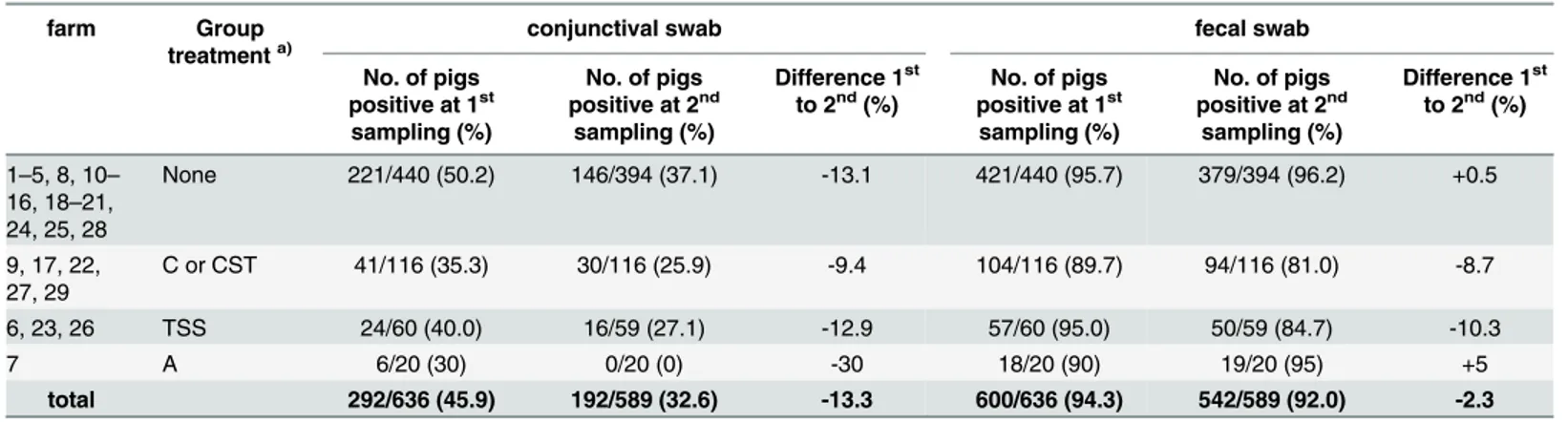
Table 2. Results ofChlamydiaceaescreening according to antibiotic group treatment.
farm Group
treatmenta)
conjunctival swab fecal swab
No. of pigs positive at 1st
sampling (%)
No. of pigs positive at 2nd
sampling (%)
Difference 1st to 2nd(%)
No. of pigs positive at 1st sampling (%)
No. of pigs positive at 2nd
sampling (%)
Difference 1st to 2nd(%)
1–5, 8, 10–
16, 18–21, 24, 25, 28
None 221/440 (50.2) 146/394 (37.1) -13.1 421/440 (95.7) 379/394 (96.2) +0.5
9, 17, 22, 27, 29
C or CST 41/116 (35.3) 30/116 (25.9) -9.4 104/116 (89.7) 94/116 (81.0) -8.7
6, 23, 26 TSS 24/60 (40.0) 16/59 (27.1) -12.9 57/60 (95.0) 50/59 (84.7) -10.3
7 A 6/20 (30) 0/20 (0) -30 18/20 (90) 19/20 (95) +5
total 292/636 (45.9) 192/589 (32.6) -13.3 600/636 (94.3) 542/589 (92.0) -2.3
a)
pro-/metaphylactic group treatment after 1stsampling, type of antimicrobial substance: A = amoxicillin; C = chlortetracycline; CST = chlortetracycline, sulfadimidin, tylosin; TSS = trimethoprime, sulfadimidin, sulfathiazole. 1st= sampling at the beginning of the fattening period; 2nd= sampling at the end of the fattening period.
swabs from the housing environment but not in swabs from tested pigs. In total, 14.5% (n = 92) of the pigs were infected withC.pecorumat least at one site and one sampling time point. Of nine animals (1.4%) positive in at least one site at the first sampling, four were females and five were males. Later on, of 86 animals (14.6%) positive at the second sampling, 59 were females and 27 were males. Three pigs were positive at least at one site at both time points, all others were only positive at one sampling time point (n = 89). The total number of conjunctival specimens positive at both time points did not change (n = 9) but was found in different pigs. In contrast, the positive fecal samples underwent a considerable increase between the first and second time point from one to 81 animals on 14 different farms (0.2% to 13.8%). Few pigs were positive forC.pecorumat both sampling sites, one pig at the first sam-pling and four pigs at the second samsam-pling.C.pecoruminfections were mostly mixed infections withC.suis.
Chlamydia abortus
In farm 15, one animal at the first sampling was positive forC.abortusin the conjunctival swab (Table 3) and it was also positive forC.suisin the same sample. At the second sampling, the conjunctival swab of this animal was negative by means of PCR. The conjunctivae of this pig showed mild reddening at the first and moderate reddening at the second sampling, other-wise the pig appeared clinically healthy. The fecal swabs of this pig were positive at both time points, however, the Arraymate could not identify the chlamydial species from the fecal swab of the first sampling, but revealedC.pecorumin the second swab. These results indicate a potential mixed infection of this pig withC.abortus,C.pecorumandC.suis. On this farm no prophylactic, metaphylactic, therapeutic antibiotics or other medications were administered. Strikingly, it was one of two farms with direct contact to sheep in the outdoor area. The first sampling was performed three days after the pigs’arrival at the farm.
Human samples
All conjunctival samples obtained from farmers (n = 9) were negative by theChlamydiaceae -specific PCR.
Clinical symptoms and influencing factors
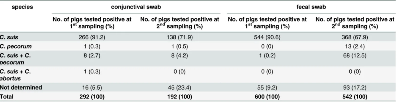
There was no correlation between the presence of conjunctivitis and chlamydial positivity in the conjunctival swabs. In farms with diarrhea observed by the farmers, the likelihood of the Table 3. Chlamydial species differentiation inChlamydiaceaepositive samples.1stsampling = sampling at the beginning of the fattening period; 2nd sampling = sampling at the end of the fattening period.
species conjunctival swab fecal swab
No. of pigs tested positive at 1stsampling (%)
No. of pigs tested positive at 2ndsampling (%)
No. of pigs tested positive at 1stsampling (%)
No. of pigs tested positive at 2ndsampling (%)
C.suis 266 (91.2) 138 (71.9) 544 (90.6) 368 (67.9)
C.pecorum 1 (0.3) 1 (0.5) 0 (0) 13 (2.4)
C.suis + C.
pecorum
8 (2.7) 8 (4.2) 1 (0.2) 68 (12.5)
C.suis + C.
abortus
1 (0.3) 0 (0) 0 (0) 0 (0)
Not determined 16 (5.5) 45 (23.4) 55 (9.2) 93 (17.2)
Total 292 (100) 192 (100) 600 (100) 542 (100)
individual animals having a chlamydial infection was higher compared to that of other farms (Table 4). Lameness observed by the farmers was also positively correlated toChlamydiaceae
positivity at both time points (Table 4). In contrast, there was no association between respira-tory signs, wasting and chlamydial prevalence.
In farrow-to-finish farms, the risk of chlamydial infection at the second sampling was lower than in fattening farms and a continuous production system had a lower risk of chlamydial infection than an all-in/all-out production system (Table 4). The risk of chlamydial infection at the second time point increased depending on the number of purchase farms. Direct or indirect contact to ruminants was a risk factor for a higher rate of chlamydial infections at the second sampling time point. Moreover, the distance to surrounding ruminants significantly influenced the positivity rate at the second sampling. An increasing distance between pigs and ruminants resulted in a decreased chlamydial infection risk (Table 4). InC.pecorum, outdoor access and Table 4. Results of the univariate logistic regression for clinical symptoms and influencing factors.
Independent variable Dependent variablea) Time point OR CI (95%)
Diarrhea Chlamydiaceae* 1st
11.1 1.5, 81.9
Diarrhea C.suis* 1st 1.8 1.1, 2.8
Diarrhea C.suis* 2nd 1.6 1.03, 2.6
Diarrhea C.pecorum* 2nd 2.7 1.7, 4.5
Lameness Chlamydiaceae 1st 2.9 1.4, 6.0
Lameness Chlamydiaceae 2nd 1.9 1.04, 3.4
Farrow-to-finish farms Chlamydiaceae 2nd 0.3 0.2, 0.5
Farrow-to-finish farms C.suis 2nd 0.4 0.3, 0.5
Continuous production Chlamydiaceae 2nd 0.3 0.2, 0.6
Continuous production C.suis* 2nd 0.7 0.4, 0.96
No. of purchase farms Chlamydiaceae 2nd 1.4 1.1, 1.8
No. of purchase farms C.suis 2nd 1.3 1.1, 1.6
Direct contact with ruminants Chlamydiaceae 2nd 3.5 1.4, 8.8
Direct contact with ruminants C.pecorum 2nd 7.1 4.3, 11.7
Indirect contact with ruminants Chlamydiaceae 2nd 2.9 1.7, 5.2
Indirect contact with ruminants C.suis 2nd 2.8 1.7, 4.5
Indirect contact with ruminants C.pecorum 2nd 3.9 1.4, 10.9
Distance to ruminants Chlamydiaceae 2nd 0.6 0.4, 0.7
Distance to ruminants C.suis 2nd 0.7 0.6, 0.9
Distance to ruminants C.pecorum 2nd 0.5 0.4, 0.6
Outdoor access C.pecorum 2nd 15.4 5.6, 42.6
Bedding material C.pecorum 2nd 3.9 2.1, 7.0
Potential direct contact with wild boars Chlamydiaceae 2nd 2.1 1.2, 3.8
Potential direct contact with wild boars C.suis 2nd 1.9 1.2, 2.9
Potential direct contact with wild boars C.pecorum 2nd 6.0 3.7, 9.8
Cleanliness of housing facilities atfirst sampling Chlamydiaceae 2nd 2.5 1.3, 4.7
Cleanliness of housing facilities atfirst sampling C.suis 2nd 2.0 1.3, 3.0
Cleanliness of housing facilities atfirst sampling C.pecorum 2nd 11.3 3.0, 42.0
Prophylactic antibiotic group treatment Chlamydiaceae* 1stpos!2ndneg 4.7 2.4, 9.1 Prophylactic antibiotic group treatment Chlamydiaceae* 1stneg!2ndneg 13.9 1.7, 115.9 a)animals positive in conjunctival and/or fecal swab.
*: fecal swab considered.
1st= sampling at the beginning of the fattening period; 2nd= sampling at the end of the fattening period; OR: odds ratio; CI: con
fidence interval.
the presence of bedding material had a significant influence on chlamydial positivity. Pigs reared in farms with potential direct contact to wild boars had a higher risk of being infected at the second sampling time point. The observed cleanliness of the housing facilities at the first sampling had an impact on the positivity at that time point: the dirtier the facilities were evalu-ated, the higher was the chlamydial prevalence (Table 4).
No clear association was found between chlamydial infection (Chlamydiaceae,C.suis) and the sex of the pig, the total number of fattening pigs on the farm, outdoor access and existence of bedding material (Chlamydiaceae,C.suis), contact to wild birds, the hygiene score and the level of infestation with insects or rodents, the observed cleanliness of the housing facilities at the second sampling and the observed cleanliness of the individual pigs at both sampling time points.
Individual pigs that had received a prophylactic antibiotic group treatment had a higher likelihood of changing fromChlamydiaceaepositive to negative or of remaining negative in their fecal swabs between the first and second sampling (Tables2and4). Amoxicillin treatment did not reduce the chlamydial prevalence in fecal swabs, while treatment with C/CST as well as TSS had a reducing effect (Table 2). On herd level, however, the outcome was variable
(Table 5): five herds showed an increase and four herds a decrease of chlamydial prevalence in conjunctival swabs; in fecal swabs, two herds showed an increase, five herds a decrease and in two herds the chlamydial prevalence remained the same. In farms without antibiotic group treatment (n = 18; farm nos. 1–5, 10–16, 18–21, 24, 25, 28), conjunctival positivity decreased in 10 farms, increased in seven and was unaltered in one farm. Fecal positivity was reduced in four farms, was increased in six farms and was unaltered in eight farms (S3 Table). Overall, the herd-based conjunctival prevalence was reduced by 6.5% in antibiotic group treated farms compared to 13.3% reduction in the untreated group. The fecal prevalence was reduced by 7.1% in the first group compared to a 0.5% rise in the untreated farms. The maximum reduc-tion of 30% of fecal chlamydial burden was reached in farm 27 with tetracycline group treat-ment (Table 5).
Pigs in farms with single-pig antibiotic treatment during the fattening period showed no decrease in positivity.
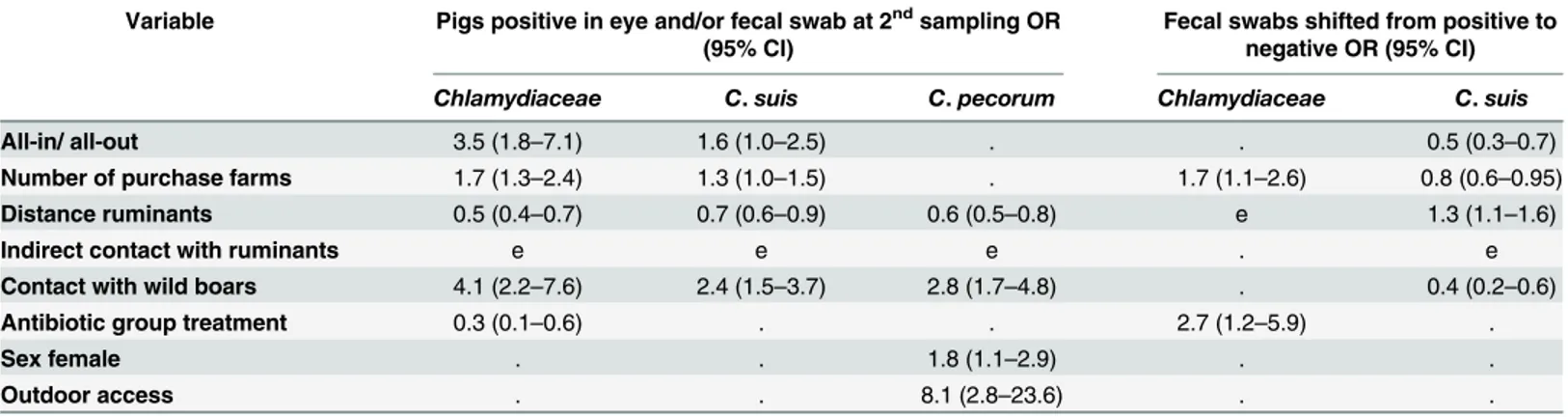
The non-correlated significant influencing factors were included in a full regression model and are summarized inTable 6. Briefly, an independent association of the following factors Table 5. Difference ofChlamydiaceaeprevalence in farms with antibiotic group treatment between the first and second sampling.
farm group treatmenta) conjunctival swab difference 1stto 2nd(%)§ rectal swab difference 1stto 2nd(%)§
9 CST -80.5 -13.9
17 C +20 ±0
22 CST +55 ±0
27 CST -10 -30
29 CST +25 +5
6 TSS -68.4 -5.3
23 TSS +10 -15
26 TSS +20 -10
7 A -30 +5
a)pro-/metaphylactic group treatment after 1st sampling, type of antimicrobial substance: A = amoxicillin; C = chlortetracycline; CST = chlortetracycline,
sulfadimidin, tylosin; TSS = trimethoprime, sulfadimidin, sulfathiazole; 1st= sampling at the beginning of the fattening period; 2nd= sampling at the end of the fattening period
§= signi
ficant difference between the individual farms.
andChlamydiaceaepositivity was found at the second sampling: all-in/all-out production, num-ber of purchase farms, distance to ruminants, possible contact with wild boars and prophylactic antibiotic group treatment. The number of purchase farms and a prophylactic antibiotic group treatment were independently associated with fecal swabs that were positive forChlamydiaceae
at the first and negative at the second sampling. InC.suis, all-in/all-out production, number of purchase farms, distance to ruminants and possible contact with wild boars were independent influencing factors for positivity at the second time point. All-in/all-out production, number of purchase farms, distance to ruminants and possible contact with wild boars were independently associated with fecal swabs that were positive forC.suisat the first and negative at the second sampling. InC.pecorum, the results of the second sampling were independently influenced by distance to ruminants, possible contact with wild boars, sex and outdoor access.
Discussion
Chlamydiaceae
and
Chlamydia suis
This is the first large-scale study to investigateChlamydiaceaeinfections of fattening pigs at two time points in Switzerland. With a prevalence of 98.9%, almost all pigs in this study were positive forChlamydiaceae. In the first sampling, 94.3% and in the second sampling 92.0% of the fecal swabs were positive forChlamydiaceae, as well as 45.9 and 32.6% of the conjunctival swabs, respectively. Of those,C.suiswas identified in 90.6% (first sampling) and 67.9% (second sampling) of fecal swabs and in 91.2% and 71.9% of the conjunctival samples. Thus,C.suiswas by far the most common chlamydial species found in both rectal and conjunctival swabs in this study.C.suishas been previously found to be the most commonChlamydiasp. in pigs [37], being often found in the pig intestine [13,38,39], pig conjunctivae [32,40] and at a variety of other sites, such as in the male and female genital tract [7,41], nasal swabs [42], lung tissue [43] and the liver of aborted fetuses [44]. The high prevalence ofChlamydiaceaeandC.suisin fecal swabs at both sampling time points in the present study was surprising. Englund et al. [30] found that 100% of the pigs (n = 36) from herds with diarrhea problems (n = 6) and 83% of the healthy control pigs (n = 12) from good performance herds (n = 4) tested positive for Chlamy-diaceae; moreover, sequencing of selected ileal tissues revealedC.suis. In contrast, in the study of De Puysseleyr et al. [19], only 52% of rectal swabs from slaughter pigs in a Belgian slaughter-house were tested positive with aC.suisspecies-specific PCR. Kauffold et al. [7] found 37.9% and 8.3%C.suispositive fecal samples in two different boar studs. Pollmann [39] tested 22 breeding sows three times with aC.suis-specific PCR and, compared to a single time sampling, Table 6. Results of multivariate analysis with step back procedure for infection risk.e: entered in full model (p0.05 univariate) but eliminated in the final model;.: not entered in full model (p>0.05 univariate); OR: odds ratio; CI: confidence interval.
Variable Pigs positive in eye and/or fecal swab at 2ndsampling OR (95% CI)
Fecal swabs shifted from positive to negative OR (95% CI)
Chlamydiaceae C.suis C.pecorum Chlamydiaceae C.suis
All-in/ all-out 3.5 (1.8–7.1) 1.6 (1.0–2.5) . . 0.5 (0.3–0.7)
Number of purchase farms 1.7 (1.3–2.4) 1.3 (1.0–1.5) . 1.7 (1.1–2.6) 0.8 (0.6–0.95) Distance ruminants 0.5 (0.4–0.7) 0.7 (0.6–0.9) 0.6 (0.5–0.8) e 1.3 (1.1–1.6)
Indirect contact with ruminants e e e . e
Contact with wild boars 4.1 (2.2–7.6) 2.4 (1.5–3.7) 2.8 (1.7–4.8) . 0.4 (0.2–0.6)
Antibiotic group treatment 0.3 (0.1–0.6) . . 2.7 (1.2–5.9) .
Sex female . . 1.8 (1.1–2.9) . .
Outdoor access . . 8.1 (2.8–23.6) . .
the prevalence rose from 27% (single sampling) to 73% (three samplings), suggesting the possi-bility of intermittent fecal shedding. In the present study, the fecal swabs of most pigs remained positive during the study period and only a small proportion became negative or changed from negative to positive in both treated and untreated farms, which might also be explained by intermittent fecal shedding. In different hosts,Chlamydiasp. is able to inhabit the gastrointesti-nal tract over a long period of time, as recently reviewed by Rank and Yeruva [45]. The results of our study indicate that a similar long-term intestinal infection is common in pigs. Moreover, Pospischil et al. [46] showed that aberrant bodies ofC.suis, the cryptic chlamydial form associ-ated with persistence, also called the chlamydial stress response, can occur in the intestine of naturally and experimentally infected pigs.
In all herds, the prevalence at the first sampling time point was very high, which indicates that the pigs were already infected earlier or acquired the infection on the new farms after mix-ing with infected pigs. In the farrow-to-finish farms, mixmix-ing with foreign animals was not pos-sible, although, the prevalence in these farms was as high as in other farms. The incubation period after chlamydial infection was determined by Guscetti et al. [47] in an intra-gastric inoc-ulation model of 2–3 day-old gnotobiotic piglets, and first fecal shedding occurred as early as the second day after inoculation. Hence, early infection ofChlamydia-negative pigs by fecal-oral transmission through mixing with infected animals could have been possible before the first sampling time point, in particular in those herds where the study investigators performed the first sampling 48–72 hours after arrival of the pigs. Alternatively, piglets could have become infected prior to the fattening period.
The conjunctival prevalence ofChlamydiaceae(45.9% at the first and 32.6% at the second time point) was far below the fecal prevalence, but comparable to findings of a study by Becker et al. [32]. They found 42% positive Swiss pigs, but 89% positive pigs in Germany. Englund et al. [30] detectedChlamydiaceaein conjunctival swabs in Swedish pigs with conjunctivitis (82.8%) and without conjunctivitis (72.4%). Differences might be explained by different hous-ing and management systems in these countries, which might influence predisposhous-ing factors and transmission routes.Chlamydiaceaeshed through conjunctival discharge and feces can easily lead to horizontal transmission, auto- or reinfections. Moreover, contaminated dust can be the source of chlamydial infection, as indicated in our study, whereC.suis and C.pecorum
were detected in the dust of one farm and viableC.suiswere also found in air samples of a Bel-gian pig slaughterhouse [19].
RegardingChlamydiaceaecopy numbers, fecal shedding was significantly higher than con-junctival shedding. This may be due to the fact that the DNA we found in the eyes does not nec-essarily represent the presence of viableChlamydiabut rather an eye contamination with chlamydial DNA fragments. This might also explain the short-term detection of some eye infec-tions. The reasons for the rise in the mean copy number of positive conjunctival swabs at the sec-ond time point remained unclear. Nevertheless, it was still lower than fecal copy numbers indicating that the gut might represent a true site of active chlamydial replication. An explanation for the lower mean of copy numbers in positive fecal swabs at the second time point could be due to stable living conditions of the animals for at least three months, in contrast to the situation at the first sampling time point after introduction into a new farm. The hypothetical time point of the initial infection is thereby also months ago and in the meantime the host organism may have adapted itself leading to a more balanced gut microbiota and reduced replication.
Other
Chlamydia
spp.
detected in conjunctival swabs [26,32,48] and rarely in semen and fecal samples of boars [7] or as a mixed infection withC.suisin aborted material [44]. Ruminants are the main host forC.
pecorumand subclinical infections are common [49]. All pigs in our study were surrounded by ruminants at different distances and contact opportunities. We were able to show a high corre-lation between potential direct or indirect contact between ruminants and pigs andC.pecorum
positive samples. The high likelihood of contact with ruminants in our study compared to other pig husbandry systems could explain the relatively high prevalences. Interestingly, all eye infections at the first time point were no longer present at the second time point in the same individuals. This may be due to intermittent shedding or fast clearance of conjunctival infections.
As it was only detected once in a conjunctival swab,C.abortuswas not an important Chla-mydiasp. in this study. Comparable toC.pecorum, theC.abortusinfection was no longer pres-ent at the second sampling. A possible infection source in the single positive case was a sheep flock with potential direct contact with the pig herd.C.abortuswas found by other investiga-tors in the cervical swabs of breeding sows [4], conjunctival swabs of sows and semen of boars [48], lung and intestine of healthy slaughter pigs and pigs with respiratory disease [50], and in an aborted fetus [44].
C.psittaciwas not detected in any of the pigs, despite possible contact with wild birds and poultry in the surroundings of most pig herds. This contrasts with the report of Vanrompay et al. [5], where a chlamydial strain was found in a Belgian pig, which was highly related to pigeonC.psittaciserovar B strains; thus a potential transmission from birds to pigs was assumed. WhileC.abortusandC.psittaciare known zoonotic pathogens, the zoonotic poten-tial ofC.suisandC.pecorumis still a matter of debate [18–20,51]. All tested conjunctival swabs of farmers in this study were negative, thus a zoonotic transmission could not be detected. However, the human population tested was very small and, apart from the eyes, no other body sites were tested.
Clinical symptoms and influencing factors
It has been shown thatC.suisis not a primary pathogen when colonizing pig intestines during natural infection [2,52] but could cause intestinal lesions and diarrhea after experimental oral or intragastric infection of piglets [47,53].C.pecorumhas been associated with pneumonia, polyarthritis, pleuritis, pericarditis and abortion in pigs [54]. In this study, a correlation between diarrhea and fecal swab positivity forC.suisandC.pecorumcould be found. In some farms, typical diarrhea-causative agents were identified, but a bacteriological examination was not performed in all affected farms. It can be assumed thatC.suiswas acting as a facultative pathogen, as reported previously in co-infections with primary pathogenic agents like Salmo-nella[48,55]. On the other hand, there was no clear association between pneumonia and chla-mydial infection, which is in accordance with the studies of Reinhold et al. [56,57]. These authors showed that in contrast to experimental infection, naturally acquiredChlamydiaceae
infections do not cause respiratory symptoms in pigs.
were only visible by histological examination of the conjunctiva and nictitating membrane. In the present study, conjunctival positivity declined from the first to the second time point, maybe due to increasing immunity, while the conjunctivitis symptoms increased. Hence, it can be assumed that the clinical symptoms were due to other, most probably environmentally related, factors such as a high concentration of dust, ammonia, hydrogen sulfide, and other decomposition gases [59]. The occurrence of other conjunctivitis-causing infectious agents is also conceivable, e.g.Mycoplasmaspp., porcine cytomegalovirus and swine influenza virus, but they were not examined. Although chlamydiae do not seem to cause the clinical symptoms, they may predispose to conjunctivitis. Older literature even reports that the conjunctivae of pigs are moderately reddened physiologically [60]. Apart from that, the study investigators often had the impression that iatrogenic stress during sampling led to reddening of the con-junctivae due to elevated blood pressure. Becker et al. [32] reported a rate of conjunctivitis in Swiss pigs (37.3%) similar to that in this study at the second sampling time point.
When comparing single pig results at the second sampling, a few management factors influ-enced the rate. First of all, farrow-to-finish farms and those with continuous production had a lower risk of being infected than others. However, an increasing number of different pig origins raised the risk ofChlamydiaceaeinfection. In farms where self recruitment is not carried out and fattening pigs are moved in at approximately three months of age, the pigs have to cope with many environmental changes in the new housing facility as well as the microorganisms present in the new housings and in the pigs from foreign farms. Therefore the risk of being infected with any bacteria is increased. In addition, the transport and unfamiliar situation in a new pen with unknown group members puts stress on the pigs, which, in turn, usually enhances the intestinal growth of pathogens or the likelihood of their shedding due to immune suppression [61,62]. Based on the concept of persistence, stress could also reactivate a chla-mydial infection [48].
Interestingly, increased hygiene management (as reported by the farmer) could not lower
Chlamydiapositivity. This means that the type and frequency of the cleaning procedure and use of disinfectants did not influence chlamydial infection. However, the information on hygiene management provided by the farmer was possibly not entirely reliable because of his subjective evaluation. The actual cleanliness of the housing was also evaluated by the study investigators. At the first sampling time point, good housing hygiene had a reducing influence on the detection rate ofChlamydiaceae. On the other hand, this connection could not be made at the second sampling time point or for individual animal-surface cleanliness. In summary, only two individual time points could be assessed limiting the significance of this observation.
Swiss pig farms are usually not double-fenced and are situated in rural areas by law; there-fore contact with wild boars or other wild mammals is possible in farms with outdoor areas. We found a clear association between farms with potential wild boar contact and the risk of infection. This is an important finding, because other more fatal agents could potentially be transmitted in this way, as wild boars represent a reservoir for several pathogens [63]. Having unprotected outdoor areas also enables contact with pastured ruminants and wild birds. Free-ranging ruminants in Switzerland have been shown to sporadically harborChlamydiaceae[64].
InC.pecoruminfections, there was a link between the presence of bedding material and positivity at the second sampling time point. This may be a confounding factor, because these farms also mostly had outdoor access, and this also increased the infectivity rate. However, this association was not present in infections withChlamydiaceaeandC.suis. EBs ofChlamydiasp. are reported to survive in dry feces, dust or litter for several months [49] and contaminated bedding material could consequently represent a source of infection.
intestinal tract and respiratory diseases, and to improve growth performance. A therapeutic effect on chlamydiae can be expected from tetracyclines as well as sulfonamide and trimetho-prim combinations [65]. These and amoxicillin were used for group treatments in the investi-gated farms. Amoxicilllin is a derivate of penicillin, which is known to induce chlamydial persistencein vitroinstead of killing the RBs [45]. In this study, we could not retrieve informa-tion about any treatment before the beginning of the fattening period. Only in one farrow-to-finish farm (no. 28), did we know about the administration of chlortetracycline, sulfadimidine and tylosin after weaning and prior to our first sampling. The rate of positive conjunctival (50%) and fecal samples (90%) in this particular farm at the first sampling time point was com-parable to that in the untreated farms. Five farms in this study treated the pig herd after the first sampling with an oral medication containing chlortetracycline. Four of them used a com-bination product with tylosin, which belongs to the protein synthesis inhibiting macrolides. Azithromycin also belongs to this group and is usually the drug of choice to treat human chla-mydial infections. Nevertheless, theChlamydiaceaeprevalence in conjunctival and fecal sam-ples only decreased in two farms, whereas in the other farms, the prevalences remained unchanged or even increased (Table 5). In the TSS treated farms, the reduction of positivity in conjunctival and fecal swabs was higher than in the untreated farms and also higher than in the tetracycline/tylosin-group. One farm (no. 7) administered Amoxicillin as an oral group treat-ment, because the pigs developed respiratory symptoms. All conjunctival samples that were positive at the beginning were negative at the second time point, but fecal positivity increased slightly. None of the individually antimicrobial treated pigs became negative forChlamydiaceae
at the second time point.
In summary, none of the antibiotic treatments in this study was able to clear the chlamydial infections on herd level despite individual pigs becoming negative by the end of the fattening period. The tetracycline resistance ofC.suisis well known, while resistance to sulfonamide and trimethoprim combinations was described once [23]. Our observations match those of a study of Reinhold et al. [42], in which the short-term treatment with enrofloxacin, a fluoroquinolone, resulted in a recurrence and increased quantity ofChlamydiaspp. in fecal and nasal swabs after initial reduction. It is possible that the treatment duration in the farms of this study was too short to clear the infection; it was administered for five to twelve days. Moreover, Yeruva et al. [66,67] showed that the gut is a site of persistence and source of possible reinfection in a mouse model. This is facilitated by an immune down-regulating effect in the intestine, resulting in the inability of the host to resolve the infection. The same group also proved a reduced sus-ceptibility of the gut compared to the genital tract to azithromycin treatment.
In conclusions, the recommendation of antibiotic treatment in pig chlamydiosis should be reconsidered regarding its necessity and effectivity. Apart from that, the efficacy of prophylac-tic or metaphylacprophylac-tic use of oral group treatment with antibioprophylac-tics in pig farms in light of the highly prevalent intestinal chlamydial infections should be critically reviewed. The clinical impact of chlamydial infection seems to be of low significance in regard to conjunctivitis but they may contribute to diarrhea. Therefore, routine diagnostics of herd-based diarrhea prob-lems should includeChlamydiaceaetesting. A follow-up examination will investigate the path-ogenicity and antibiotic susceptibility ofC.suisfound in this study.
Supporting Information
S1 Table. Details of farms investigated in this study. (DOCX)
S3 Table. Results ofChlamydiaceaescreening of fattening pigs from 29 farms. (DOCX)
S1 Dataset. Listing of all samples with relevant herd-based information, PCR and Array-mate microarray results.
(XLSX)
Acknowledgments
The authors are grateful to Carmen Kaiser and Regula Güttinger, Institute of Veterinary Pathology, Vetsuisse Faculty, University of Zurich, for the excellent technical assistance. We thank all students and assistents of the Institute for Veterinary Pathology Zurich who kindly helped during the sampling procedure and the Swiss pig health service, external veterinarians and the Swiss pig trading companies agrifera and anicom for their help in the acquisition of farms. The laboratory work was partly performed using the logistics of the Center for Clinical Studies at the Vetsuisse Faculty of the University of Zurich. Special thanks to the participating farmers and pigs, whose collaboration made this study possible in the first place.
Author Contributions
Conceived and designed the experiments: NB XS KH FS MD AF. Performed the experiments: KH FS SW. Analyzed the data: KH MH. Wrote the paper: KH NB.
References
1. Schautteet K, Vanrompay D.Chlamydiaceaeinfections in pigs. Vet Res. 2011; 42: 29. doi:10.1186/ 1297-9716-42-29PMID:21314912
2. Szeredi L, Schiller I, Sydler T, Guscetti F, Heinen E, Corboz L, et al. IntestinalChlamydiain Finishing Pigs. Vet Pathol. 1996; 33(4): 369–374. doi:10.1177/030098589603300401PMID:8817833
3. Eggemann G, Wendt M, Hoelzle LE, Jäger C, Weiss R, Failing K. [Prevalence ofChlamydiainfections in breeding sows and their importance in reproductive failure]. Dtsch Tierarztl Wochenschr. 2000; 107 (1): 3–10. German. PMID:10689792
4. Camenisch U, Lu ZH, Vaughan L, Corboz L, Zimmermann DR, Wittenbrink MM, et al. Diagnostic inves-tigation into the role of Chlamydiae in cases of increased rates of return to oestrus in pigs. Vet Rec. 2004; 155(19): 593–596. doi:10.1136/vr.155.19.593PMID:15573952
5. Vanrompay D, Geens T, Desplanques A, Hoang TQT, De Vos L, Van Loock M, et al. Immunoblotting, ELISA and culture evidence forChlamydiaceaein sows on 258 Belgian farms. Vet Microbiol. 2004; 99 (1): 59–66. doi:10.1016/j.vetmic.2003.08.014PMID:15019112
6. Di Francesco A, Baldelli R, Cevenini R, Magnino S, Pignanelli S, Salvatore D, et al. Seroprevalence to chlamydiae in pigs in Italy. Vet Rec. 2006; 159(25): 849–850. doi:10.1136/vr.159.25.849PMID:
17172480
7. Kauffold J, Melzer F, Henning K, Schulze K, Leiding C, Sachse K. Prevalence of chlamydiae in boars and semen used for artificial insemination. Theriogenology. 2006; 65(9): 1750–1758. doi:10.1016/j. theriogenology.2005.10.010PMID:16280160
8. Hotzel H, Berndt A, Melzer F, Sachse K. Ocurrence ofChlamydiaceaespp. in a wild boar (Sus scrofa
L.) population in Thuringia (Germany). Vet Microbiol. 2004; 103(1–2): 121–126. doi:10.1016/j.vetmic. 2004.06.009PMID:15381275
9. Salinas J, Caro MR, Vicente J, Cuello F, Reyes-Garcia AR, Buendía AJ, et al. High prevalence of anti-bodies againstChlamydiaceaeandChlamydophila abortusin wild ungulates using two "in house" blocking-ELISA tests. Vet Microbiol. 2009; 135(1–2): 46–53. doi:10.1016/j.vetmic.2008.10.001PMID:
19010612
10. Di Francesco A, Donati M, Morandi F, Renzi M, Masia MA, Ostanello F, et al. Seroepidemiologic Survey forChlamydia suisin Wild Boar (Sus scrofa) Populations in Italy. J Wildl Dis. 2011; 47(3): 709–712. doi:
http://dx.doi.org/10.7589/0090-3558-47.3.709PMID:21719838
spain. J Zoo Wildl Med. 2013; 44(1): 159–162. doi:http://dx.doi.org/10.1638/1042-7260-44.1.159
PMID:23505718
12. Di Francesco A, Baldelli R, Donati M, Cotti C, Bassi P, Delogu M. Evidence forChlamydiaceaeand
Parachlamydiaceaein a wild boar (Sus scrofa) population in Italy. Vet Ital. 2013; 49(1): 119–122. PMID:
23564593
13. Zahn I, Szeredi L, Schiller I, Straumann Kunz U, Bürgi E, Guscetti F, et al. [Immunohistochemical deter-mination ofChlamydia psittaci/pecorumandC.trachomatisin the pig gut]. Zentralbl Veterinarmed B. 1995; 42(5): 266–276. German. PMID:8592901
14. Taenkum K, Pospischil A, Janett F, Brugnera E, Hoelzle LE, Hoelzle K, et al. Prevalence of chlamydiae in semen and genital tracts of bulls, rams and bucks. Theriogenology. 2007; 67(2): 303–310. doi:10. 1016/j.theriogenology.2006.07.012PMID:16935325
15. Blumer C, Zimmermann DR, Weilenmann R, Vaughan L, Pospischil A. Chlamydiae in Free-Ranging and Captive Frogs in Switzerland. Vet Pathol. 2007; 44(2): 144–150. doi:10.1354/vp.44-2-144PMID:
17317791
16. Pantchev A, Sting R, Bauerfeind R, Tyczka J, Sachse K. Detection of allChlamydophilaandChlamydia
spp. of veterinary interest using species-specific real-time PCR assays. Comp Immunol Microb Infect Dis. 2010; 33(6): 473–484. doi:10.1016/j.cimid.2009.08.002
17. Szymańska-Czerwińska M, Niemczuk K, Galińska EM. Serological and nested PCR survey to deter-mine the occurence of chlamydia infections in the Polish cattle population. Ann Agric Environ Med. 2013; 20(4): 682–686. PMID:24364434
18. Dean D, Rothschild J, Ruettger A, Kandel RP, Sachse K. ZoonoticChlamydiaceaeSpecies Associated with Trachoma, Nepal. Emerg Infect Dis. 2013; 19(12): 1948–1955. doi:http://dx.doi.org/10.3201/ eid1912.130656doi:10.3201/eid1912.130656PMID:24274654
19. De Puysseleyr K, De Puysseleyr L, Dhondt H, Geens T, Braeckmann L, Morré SA, et al. Evaluation of the presence and zoonotic transmission ofChlamydia suisin a pig slaughterhouse. BMC Infect Dis. 2014; 14: 560. doi:10.1186/s12879-014-0560-xPMID:25358497
20. De Puysseleyr K, De Puysseleyr L, Geldhof J, Cox E, Vanrompay D. Development and Validation of a real-time PCR forChlamydia suisdiagnosis in swine and humans. PLoS One. 2014; 9(5): e96704. doi:
10.1371/journal.pone.0096704PMID:24816542
21. Regula G, Torriani K, Gassner B, Stucki F, Müntener CR. Prescription patterns of antimicrobials in vet-erinary practices in Switzerland. J Antimicrob Chemother. 2009; 63(4): 805–811. doi:10.1093/jac/ dkp009PMID:19218273
22. Müntener CR, Stebler R, Horisberger U, Althaus FR, Gassner B. [Calculation of therapeutic intensity for piglets and fatteners treated with medicated feed]. Schweiz Arch Tierheilkd. 2013; 155(6): 365–72. German. doi:10.1024/0036-7281/a000472PMID:23732383
23. Lenart J, Andersen AA, Rockey DD. Growth and development of tetracycline-resistantChlamydia suis. Antimicrob Agents Chemother. 2001; 45(8): 2198–2203. doi:10.1128/AAC.45.8.2198–2203.2001
PMID:11451674
24. Dugan J, Rockey DD, Jones L, Andersen AA. Tetracycline resistance inChlamydia suismediated by genomic islands inserted into the chlamydialinv-like gene. Antimicrob Agents Chemother. 2004; 48 (10): 3989–3995. doi:10.1128/AAC.48.10.3989–3995.2004PMID:15388463
25. Di Francesco A, Donati M, Rossi M, Pignanelli S, Shurdhi A, Baldelli R, et al. Tetracycline-resistant
Chlamydia suisisolates in Italy. Vet Rec. 2008; 163(8): 251–252. doi:10.1136/vr.163.8.251PMID:
18723867
26. Borel N, Regenscheit N, Di Francesco A, Donati M, Markov J, Masserey Y, et al. Selection for tetracy-cline-resistantChlamydia suisin treated pigs. Vet Microbiol. 2012; 156(1–2): 143–146. doi:10.1016/j. vetmic.2011.10.011PMID:22036200
27. Schautteet K, De Clercq E, Miry C, Van Groenweghe F, Delava P, Kalmar I, et al. Tetracycline-resistant
Chlamydia suisin cases of reproductive failure on Belgian, Cypriote and Israeli pig production farms. J Med Microbiol. 2013; 62(Pt 2): 331–334. doi:10.1099/jmm.0.042861–0PMID:23105027
28. Swiss Farmers’Union, Agristat: Fleischbilanz; je-d-07.02.04.05 [updated 2014 Sept 29; cited 2015 Sept 9]. Available:http://www.bfs.admin.ch/bfs/portal/de/index/themen/07/03/blank/data/01/04.html
29. Swiss Federal Statistical Office, Landwirtschaftliche Betriebsstrukturerhebung: Nutztierbestand der Landwirtschaftsbetriebe; je-d-07.02.03.03–3 [cited 2015 Sept 9]. Available:http://www.bfs.admin.ch/ bfs/portal/de/index/themen/07/03/blank/data/01/03.html
30. Englund S, Segerstad CH, Arnlund F, Westergren E, Jacobson M. The occurrence ofChlamydiaspp. in pigs with and without clinical disease. BMC Vet Res. 2012; 8: 9. doi:10.1186/1746-6148-8-9PMID:
31. Polkinghorne A, Borel N, Becker A, Lu ZH, Zimmermann DR, Brugnera E, et al. Molecular evidence for chlamydial infections in the eyes of sheep. Vet Microbiol. 2009; 135(1–2): 142–146. doi:10.1016/j. vetmic.2008.09.034PMID:18945556
32. Becker A, Lutz-Wohlgroth L, Brugnera E, Lu ZH, Zimmermann DR, Grimm F, et al. Intensively Kept Pigs Pre-disposed to Chlamydial Associated Conjunctivitis. J Vet Med A Physiol Pathol Clin Med. 2007; 54(6): 307–313. doi:10.1111/j.1439-0442.2007.00963.xPMID:17650151
33. Blumer S, Greub G, Waldvogel A, Hässig M, Thoma R, Tschuor A, et al.Waddlia,Parachlamydiaand
Chlamydiaceaein bovine abortion. Vet Microbiol. 2011; 152(3–4): 385–93. doi:10.1016/j.vetmic.2011. 05.024PMID:21658867
34. Borel N, Kempf E, Hotzel H, Schubert E, Torgerson P, Slickers P, et al. Direct identification of chlamyd-iae from clinical samples using a DNA microarray assay–A validation study. Mol Cell Probes. 2008; 22 (1): 55–64. doi:10.1016/j.mcp.2007.06.003PMID:17714911
35. Schnee C, Sachse K. DNA microarray-based detection of multiple pathogens:Mycoplasmaspp. and
Chlamydiaspp. Methods Mol Biol. 2015; 1247: 193–208. doi:10.1007/978-1-4939-2004-4_15PMID:
25399098
36. Altmann DG. Practical statistics for medical research. 1st ed. London: Chapman & Hall; 1994. PMID:
25144107
37. Longbottom D. Chlamydial infections of domestic ruminants and swine: new nomenclature and new knowledge. Vet J. 2004; 168(1): 9–11. doi:10.1016/S1090-0233(03)00106-0PMID:15158203
38. Schiller I, Koesters R, Weilenmann R, Kaltenboeck B, Pospischil A. PCR detection of porcine Chla-mydia trachomatisand ruminantChlamydia psittaciserovar 1 DNA in formalin fixed intestinal speci-mens from swine. Zentralbl Veterinarmed B. 1997; 44(3): 185–91. PMID:9197211
39. Pollmann M. [Effects of a probiotic strain ofEnterococcus faeciumon natural infection rate of Chlamyd-iae in swine.] Doctoral Thesis, Freie Universität Berlin. 2006. German. Available: http://www.diss.fu-berlin.de/diss/receive/FUDISS_thesis_000000002542
40. Davidson HJ, Rogers DP, Yeary TJ, Stone GG, Schoneweis DA, Chengappa MM. Conjunctival micro-bial flora of clinically normal pigs. Am J Vet Res. 1994; 55(7): 949–51. PMID:7978633
41. Kauffold J, Melzer F, Berndt A, Hoffmann G, Hotzel H, Sachse K. Chlamydiae in oviducts and uteri of repeat breeder pigs. Theriogenology. 2006; 66(8): 1816–1823. doi:10.1016/j.theriogenology.2006.04. 042PMID:16837032
42. Reinhold P, Liebler-Tenorio E, Sattler S, Sachse K. Recurrence ofChlamydia suisinfection in pigs after short-term antimicrobial treatment. Vet J. 2011; 187(3): 405–407. doi:10.1016/j.tvjl.2010.01.008PMID:
20800518
43. Sachse K, Hotzel H, Slickers P, Ellinger T, Ehricht R. DNA microarray-based detection and identifica-tion ofChlamydiaandChlamydophilaspp. Mol Cell Probes. 2005; 19(1): 41–50. doi:10.1016/j.mcp. 2004.09.005PMID:15652219
44. Schiller I, Koesters R, Weilenmann R, Thoma R, Kaltenboeck B, Heitz P, et al. Mixed infections with porcineChlamydia trachomatis/pecorumand infections with ruminantChlamydia psittaciserovar 1 associated with abortion in swine. Vet Microbiol. 1997; 58(2–4): 251–260. doi:10.1016/S0378-1135 (97)00154-5PMID:9453135
45. Rank RG, Yeruva L. Hidden in Plain Sight: Chlamydial Gastrointestinal Infection and Its Relevance to Persistence in Human Genital Infection. Infect Immun. 2014; 82(4): 1362–1371. doi:10.1128/IAI. 01244-13PMID:24421044
46. Pospischil A, Borel N, Chowdhury EH, Guscetti F. Aberrant chlamydial developmental forms in the gas-trointestinal tract of pigs spontaneously and experimentally infected withChlamydia suis. Vet Microbiol. 2009; 135(1–2): 147–156. doi:10.1016/j.vetmic.2008.09.035PMID:18950970
47. Guscetti F, Schiller I, Sydler T, Heinen E, Pospischil A. Experimental enteric infection of gnotobiotic pig-lets withChlamydia suisstrain S45. Vet Microbiol. 2009; 135(1–2): 157–168. doi:10.1016/j.vetmic. 2008.09.038PMID:18950966
48. Schautteet K, Beeckmann DSA, Delava PP, Vanrompay D. Possible pathogenic interplay between
Chlamydia suis,Chlamydophila abortusand PCV-2 on a pig production farm. Vet Rec. 2010; 166(11): 329–333. doi:10.1136/vr.b4714PMID:20228367
49. Reinhold P, Sachse K, Kaltenboeck B.Chlamydiaceaein cattle: Commensals, trigger organisms, or pathogens? Vet J. 2011; 189(3): 257–267. doi:10.1016/j.tvjl.2010.09.003PMID:20980178
50. Hoelzle LE, Steinhausen G, Wittenbrink MM. PCR-based detection of chlamydial infection in swine and subsequent PCR-coupled genotyping of chlamydialomp1-gene amplicons by DNA-hybridization, RFLP-analysis, and nucleotide sequence analysis. Epidemiol Infect. 2000; 125(2): 427–439. PMID:
51. Rodolakis A, Mohamad KY. Zoonotic potential ofChlamydophila. Vet Microbiol. 2010; 140(3–4): 382–
391. doi:10.1016/j.vetmic.2009.03.014PMID:19345022
52. Nietfeld JC, Leslie-Steen P, Zeman DH, Nelson D. Prevalence of intestinal chlamydial infection in pigs in the midwest, as determined by immunoperoxidase staining. Am J Vet Res. 1997; 58(3): 260–264. PMID:9055971
53. Rogers DG, Andersen AA. Intestinal lesions caused by two swine chlamydial isolates in gnotobiotic pigs. J Vet Diagn Invest. 1996; 8(4): 433–440. doi:10.1177/104063879600800405PMID:8953527
54. Mohamad KY, Rodolakis A. Recent advances in the understanding ofChlamydophila pecorum infec-tions, sixteen years after it was named as the fourth species of theChlamydiaceaefamily. Vet Res. 2010; 41(3): 27. doi:10.1051/vetres/2009075PMID:19995513
55. Pospischil A, Wood RL. IntestinalChlamydiain Pigs. Vet Pathol. 1987; 24(6): 568–570. PMID:
3331859
56. Reinhold P, Jaeger J, Melzer F, Sachse K. Evaluation of Lung Function in Pigs Either Experimentally or Naturally Infected WithChlamydiaceae. Vet Res Commun. 2005; 29(Suppl.1): 125–150. PMID:
15943072
57. Reinhold P, Kirschvink N, Theegarten D, Berndt A. An experimentally inducedChlamydia suisinfection in pigs results in severe lung function disorders and pulmonary inflammation. Vet Res. 2008; 39(3): 35. doi:10.1051/vetres:2008012PMID:18298931
58. Rogers DG, Andersen AA. Conjunctivitis caused by a swineChlamydia-trachomatis-like organism in gnotobiotic pigs. J Vet Diagn Invest. 1999; 11(4): 341–344. doi:10.1177/104063879901100408PMID:
10424650
59. Done S, Williamson SM, Strugnell BW. Nervous and Locomotor Systems. In: Zimmermann JJ, Karriker LA, Ramirez A, Schwartz KK, Stevenson GW, editors. Diseases of Swine. 10th ed. West Sussex: John Wiley & Sons, Inc.; 2012. pp. 301–302.
60. Baumgartner W, Hess M, Ketz-Riley CJ, Kölle P, Schuh M, Schusser G, et al. [Eye and Conjunctiva]. In: Baumgartner W, editor. [Clinical propaedeutics of the internal diseases and skin diseases of domes-tic and companion animals]. German. 6th edition. Stuttgart: Parey; 2005. pp. 76–77.
61. Callaway TR, Morrow JL, Edrington TS, Genovese KJ, Dowd S, Carrol J, et al. Social stress increases fecal shedding of Salmonella typhimurium by early weaned piglets. Curr Issues Intest Microbiol. 2006; 7(2): 65–71. PMID:16875421
62. Khafipour E, Munyaka PM, Nyachoti CM, Krause DO, Rodriguez-Lecompte JC. Effect of crowding stress andEscherichia coliK88+ challenge in nursery pigs supplemented with anti-Escherchia coliK88 + probiotics. J Anim Sci. 2014; 92(5): 2017–2029. doi:10.2527/jas.2013-7043PMID:24663172
63. Montagnaro S, Sasso S, De Martino L, Longo M, Iovane V, Ghiurmino G, et al. Prevalence of Antibod-ies to Selected Viral and Bacterial Pathogens in Wild Boar (Sus scrofa) in Campania Region, Italy. J Wildl Dis. 2010; 46(1): 313–319. doi:http://dx.doi.org/10.7589/0090-3558-46.1.316
64. Regenscheit N, Holzwarth N, Greub G, Aeby S, Pospischil A, Borel N. Deer as a potential wildlife reser-voir forParachlamydiaspecies. Vet J. 2012; 193(2): 589–592. doi:10.1016/j.tvjl.2012.02.016PMID:
22460045
65. Sandoz KM, Rockey DD. Antibiotic resistance in Chlamydiae. Future Microbiol. 2010; 5(9): 1424–1442. doi:10.2217/fmb.10.96
66. Yeruva L, Spencer N, Bowlin AK, Wang Y, Rank RG. Chlamydial infection of the gastrointestinal tract: a reservoir for persistent infection. Pathog Dis. 2013; 68(3): 88–95. doi:10.111/2049-632XPMID:
23843274