Surface-plasmon-enhanced frequency upconversion in Pr3+ doped
tellurium-oxide glasses containing silver nanoparticles
Vineet Kumar Rai, Leonardo de S. Menezes, Cid B. de Araújo, Luciana R. P. Kassab, Davinson M. da Silva et al.
Citation: J. Appl. Phys. 103, 093526 (2008); doi: 10.1063/1.2919566
View online: http://dx.doi.org/10.1063/1.2919566
View Table of Contents: http://jap.aip.org/resource/1/JAPIAU/v103/i9
Published by the AIP Publishing LLC.
Additional information on J. Appl. Phys.
Journal Homepage: http://jap.aip.org/
Journal Information: http://jap.aip.org/about/about_the_journal
Top downloads: http://jap.aip.org/features/most_downloaded
Surface-plasmon-enhanced frequency upconversion in Pr
3+doped
tellurium-oxide glasses containing silver nanoparticles
Vineet Kumar Rai,1Leonardo de S. Menezes,1Cid B. de Araújo,1,a兲Luciana R. P. Kassab,2 Davinson M. da Silva,3and Renata A. Kobayashi3
1
Departamento de Física, Universidade Federal de Pernambuco, 50670-901 Recife, Pernambuco, Brazil 2
Faculdade de Tecnologia de São Paulo, CEETEPS/UNESP, São Paulo 01124-060, Brazil 3
Departamento de Engenharia de Sistemas Eletrônicos, Escola Politécnica da USP, São Paulo 05508-900, Brazil
共Received 9 January 2008; accepted 6 March 2008; published online 9 May 2008兲
A frequency upconversion process in Pr3+doped TeO2– ZnO glasses containing silver nanoparticles is studied under excitation with a nanosecond laser operating at 590 nm, in resonance with the 3
H4→1D2 transition. The excited Pr3+ ions exchange energy in the presence of the nanoparticles, originating efficient conversion from orange to blue. The enhancement in the intensity of the luminescence at⬃482 nm, corresponding to the3P0→3H4transition, is due to the influence of the
large local field on the Pr3+ ions, which are located near the metallic nanoparticles. © 2008
American Institute of Physics.关DOI:10.1063/1.2919566兴
I. INTRODUCTION
The study of glasses doped with rare earth ions are of significant interest due to their potential applications in many optical devices.1–3 Specifically, heavy metal oxide 共HMO兲
glasses doped with trivalent rare earth ions are very impor-tant because they show strong luminescence due to their small cutoff phonon frequencies.1However, in order to make devices with enhanced optical characteristics, the concentra-tion of rare earth ions has to be low enough so that lumines-cence quenching is minimized. It is also possible to prevent the quenching effect by modifying the environment felt by the luminescent ions.4–6Therefore, glasses containing metal-lic nanoparticles doped with low concentration of rare earth ions are of particular interest because the large local field acting on the ions positioned near the nanoparticles may in-crease the luminescence efficiency when the optical quency of the excitation beam and/or the luminescence fre-quency are near resonance with the surface plasmon frequency of the nanoparticles.7
Recently, the luminescence properties of HMO glasses doped with rare earth ions and containing metallic nanostruc-tures have been investigated. Initially, optical absorption and
Stokes luminescence were investigated in Pr3+ doped
PbO– GeO2 glasses containing silver nanoparticles.
8
The
lu-minescence efficiency of Pr3+ ions was influenced by the
nanoparticle concentration, which was controlled by the ap-propriate heat treatment of the samples. The influence of
silver nanoparticles on the luminescence of Pr3+ doped
TeO2– PbO– GeO2was investigated in Ref. 9. Enhancement
of Stokes emissions and frequency upconversion共UC兲 inten-sity were characterized for different nanoparticle concentra-tions. In another work, the luminescence properties of Eu3+
doped TeO2– PbO– GeO2 glasses containing gold
nanopar-ticles were also investigated.10 The emission spectra of the
samples exhibited an enhancement of Eu3+luminescence due
to the presence of gold nanoparticles. The emission at 614 nm due to the Eu3+ hypersensitive transition 5D0-7F2 was influenced by the gold nanoparticles and increases by
⬇100% for samples heat treated at 350 ° C for 41 h. In the works referred above, detailed measurements of the Stokes luminescence were performed to characterize the influence of the isolated metallic nanoparticles and their ag-gregates on the optical properties of the systems. On the other hand, the influence of metallic nanoparticles on the UC luminescence of HMO doped with rare earth ions was not
much exploited. The infrared-to-visible UC of Er3+ doped
PbO– GeO2was reported in Ref. 11. The experiments were
made, wherein the samples were excited with a cw diode laser共980 nm兲and the red and green luminescence due to the
Er3+ ions were recorded. The excitation mechanism for UC
generation was found to be the excited state absorption in
isolated ions. UC in Pr3+ doped TeO
2– PbO– GeO2 glasses, excited at 520 nm 共off resonance兲, was also reported.9 The generation of the signal was attributed to a phonon-assisted optical transition from the Pr3+ground state共3H
4兲to the关 3P
J
共J= 0 , 1 , 2兲,1I6兴manifold simultaneously with phonon anni-hilation, which was corroborated by the fact that the depen-dence on the incident light intensity of the UC intensity was linear.
This paper reports a study of UC processes in Pr3+doped TeO2– ZnO 共Pr3+: TZO兲 containing silver nanoparticles. In
this case, the Pr3+ ions are excited in resonance with the
3H 4→
1D
2transition, and the main goal is to investigate the influence of the metallic nanoparticles on the UC emission that corresponds to the3P0→3H4transition. The UC process studied here is different from the ones reported in Refs.9and
11 because the interaction between two Pr3+ ions plays the
essential role in the process and its efficiency depends on the strength of the interaction between Pr3+ ions. The metallic nanoparticles contribute for a luminescence enhancement of
⬇120% with respect to the UC emission observed for
samples without metallic nanostructures. a兲
Author to whom correspondence should be addressed. Electronic mail: [email protected].
II. EXPERIMENTAL
The samples were fabricated by the melt-quenching
method with the following composition: 85.5 TeO2– 14.5
ZnO 共in wt %兲. The doping components were Pr2O3
共1.0 wt%兲 and AgNO3 共1.0 wt%兲. The reagents were
melted in a platinum crucible at 800 ° C, quenched inside a brass mold, annealed at 350 ° C, and then cooled to room
temperature. Sample A 共with Pr3+ and without Ag兲 and
sample B 共containing Pr3+ and Ag兲 were annealed for 1 h.
Three samples were submitted to heat treatment at 350 ° C for 10 h共sample C兲, 20 h共sample D兲, and 40 h共sample E兲to thermally reduce the Ag+ions into Ag0, which is needed to nucleate metallic nanoparticles.
A 200 kV transmission electron microscope共TEM兲was
used to investigate the nucleation of metallic nanoparticles. Optical absorption spectra were measured with a double-beam spectrometer operating in the visible range. For
exci-tation within the 1D2 band of the Pr3+ ions, a dye laser
共⬇590 nm, pulses of 8 ns, peak power of ⬃20 kW, and
linewidth of⬃0.5 cm−1兲pumped by the second harmonic of
a Nd:YAG 共yttrium aluminum garnet兲 laser was used. The
excitation beam was focused onto the sample with a 5 cm focal length lens, and the fluorescence was collected in a direction perpendicular to the incident beam; the signal was
analyzed by a 0.5 m spectrophotometer 共resolution of 0.5
nm兲 attached to a photomultiplier tube. The signals were
recorded by using a digital oscilloscope connected to a com-puter. The experiments were made with the samples at room temperature.
III. RESULTS AND DISCUSSION
Figure 1共a兲 shows one representative TEM image of a
sample that contains metallic nanoparticles, while the corre-sponding histogram of their sizes is shown in Fig.1共b兲. The
average diameter of the nanostructures is ⬇6 nm. The
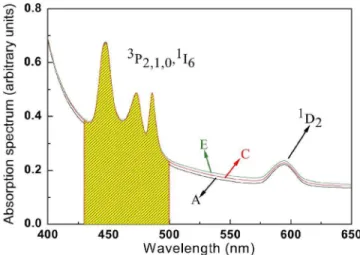
ab-sorption spectra of three Pr3+: TZO samples are shown in
Fig.2. The bands are due to Pr3+transitions starting from the ground state 共3H
4兲 to final excited states, as indicated. The surface plasmon band is not seen because the nanoparticle concentration is small and, as a consequence, the amplitude of the Pr3+bands does not change for different samples. The spectra of the other samples are similar. The shaded area in Fig.2indicates the expected location of the surface plasmon band based on the results of Refs.8 and11.
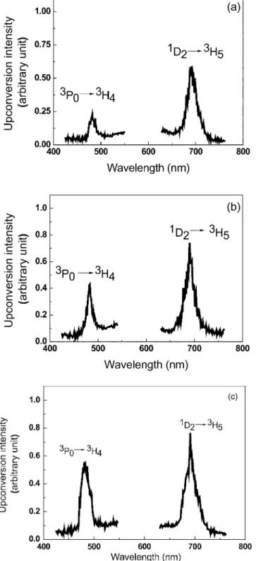
Figures 3共a兲–3共c兲 show the luminescence spectra for samples A, C, and E. The dye laser wavelength at 590 nm
was in resonance with the 3H4→1D2 transition. The two
bands are observed to peak at⬃486 and⬃692 nm and are
attributed to the 3P0→3H4 and 1D2→3H5 transitions, re-spectively. The behavior of the UC luminescence intensity at
⬃486 nm versus the laser intensity is quadratic, showing
that the excitation of the3P0state is due to the absorption of two photons; the temporal behavior of the UC signal shows
FIG. 1. TEM image of the sample annealed for 20 h共sample D兲.共b兲 His-togram of the size distribution of the metallic NPs. Average diameter: 6 nm.
FIG. 2. 共Color online兲Absorption spectra of samples A, C, and E. The features are due to transitions in the Pr3+ions. The shaded area indicates the
expected location of the surface plasmon band.
rise and decay times of ⬇10 and ⬇100 s, respectively.
This process was already observed in other glassy systems and vitreous ceramics, which was attributed to an energy exchange process involving a pair of Pr3+ions.12–18
Accord-ingly, two ions initially excited to level1D2exchange energy
in such a way that one ion is promoted to the 关3P
J 共J = 0 , 1 , 2兲, 1I6兴 manifold, while the other nonradiatively de-cays to lower energy levels. On the other hand, the intensity of the emission corresponding to the 1D2→3H5 transition linearly depends on the laser intensity.
The dependence of the ratio R between the integrated
intensities of transitions3P0→3H4and1D2→3H5as a func-tion of the heat-treatment time was determined. The results are shown in TableI, where it can be seen thatRincreases by
more than⬃120%for sample E in comparison to sample A.
The UC enhancement is due to the proximity between the surface plasmon band and the关3P
J共J= 0 , 1 , 2兲;1I6兴manifold. The intensity of the emission centered at 692 nm was not affected by the presence of the metallic nanoparticles be-cause the1D2multiplet is located far from the surface
plas-mon band. Pr3+: TZO samples 共without silver兲 were heat
treated for different time intervals, but no variation in the luminescence intensity was observed. This result confirms that the enhancement of the UC intensity in the other samples is due to the presence of the metallic nanoparticles and not due to changes in the glass structure.
In conclusion, we remark that a comparison between the optical absorption spectra and the luminescence results illus-trates the larger sensitivity of the UC process to changes in the environment of the rare earth ions hosted in glasses with metallic particles. This is clearer when energy levels closer to the surface plasmon resonance participate in the UC pro-cess. Finally, the present results suggest that the presence of silver nanoparticles may enhance UC processes of high or-der, such as the ones involving triads and quartets of Pr3+,19,20
by a factor much larger than is shown here for ion pairs.
ACKNOWLEDGMENTS
We acknowledge financial support from the Conselho Nacional de Desenvolvimento Científico e Tecnológico
共CNPq兲and the Fundação de Amparo à Ciência e Tecnologia
do Estado de Pernambuco 共FACEPE兲. The Laboratório de
Microscopia Eletrônica 共IFUSP兲 is also acknowledged for
the TEM images. This work was performed under the
Mille-nium Institute 共Nonlinear Optics, Photonics and
Bio-Photonics兲Project and the Nanophotonics Network Program.
1
M. Yamane and Y. Asahara,Glasses for Photonics共Cambridge University Press, Cambridge, England, 2000兲.
2
A. J. Kenyon, Prog. Quantum Electron.26, 225共2002兲, and references therein.
3
F. Auzel,Chem. Rev.共Washington, D.C.兲104, 139共2004兲. 4
E. Snoeks, A. Lagendijk, and A. Polman, Phys. Rev. Lett. 74, 2459 共1995兲.
5
G. M. Kumar, D. N. Rao, and G. S. Agarwal,Opt. Lett.30, 732共2005兲.
6
G. M. Kumar, D. N. Rao, and G. S. Agarwal,Phys. Rev. Lett.91, 203903 共2003兲.
7
P. N. Prasad,Nanophotonics共Wiley, New York, 2004兲.
8
L. P. Naranjo, C. B. de Araújo, O. L. Malta, P. A. S. Cruz, and L. R. P. TABLE I. RatioRbetween the integrated intensities corresponding to3P0 →3H4and1D2→3H5transitions for samples heat treated at different times.
Sample
Heat-treatment time
共h兲 R
A 1 0.29
B 1 0.42
C 10 0.47
D 20 0.52
E 40 0.64
Kassab,Appl. Phys. Lett.87, 241914共2005兲. 9
L. R. P. Kassab, C. B. de Araújo, R. A. Kobayashi, R. A. Pinto, and D. M. da Silva,J. Appl. Phys.102, 103515共2007兲.
10
R. de Almeida, D. M. da Silva, L. R. P. Kassab, and C. B. de Araújo, Opt. Commun.281, 108共2008兲.
11
D. M. da Silva, L. R. P. Kassab, S. R. Lüthi, C. B. de Araújo, A. S. L. Gomes, and M. J. V. Bell,Appl. Phys. Lett.90, 081913共2007兲.
12
E. M. Pacheco and C. B. de Araújo,Chem. Phys. Lett.148, 334共1988兲.
13
L. E. E. de Araújo, A. S. L. Gomes, C. B. de Araújo, Y. Messaddeq, A. Florez, and M. A. Aegerter,Phys. Rev. B50, 16219共1994兲.
14
V. K. Rai, L. de S. Menezes, and C. B. de Araújo,J. Appl. Phys.101,
123514共2007兲.
15
H. Zheng, X. Wang, S.-X. Qu, M. J. Dejneka, and R. S. Meltzer, J. Lumin.
119-120, 153共2006兲.
16
H. Zheng, X. Wang, M. J. Dejneka, W. M. Yen, and R. S. Meltzer, J. Lumin.108, 395共2004兲.
17
J. Fernández, R. Balda, A. Mendioroz, and A. J. García-Adeva,J. Phys.: Condens. Matter13, 10347共2001兲.
18
R. Rolli, S. Ronchin, M. Montagna, E. Moser, C. Duverger, V. T. Tikhomirov, A. Jha, and M. Ferrari, J. Non-Cryst. Solids280, 266共2001兲.
19
A. Lezama, J. R. Rios Leite, and C. B. de Araújo,Phys. Rev. B32, 7139 共1985兲.
20
E. L. Falcão-Filho, C. B. de Araújo, and Y. Messaddeq,J. Appl. Phys.92,
3065共2006兲.