A Protein Microarray for the Rapid Screening of Patients
Suspected of Infection with Various Food-Borne
Helminthiases
Jia-Xu Chen1., Mu-Xin Chen1., Lin Ai1,2
, Jun-Hu Chen1, Shao-Hong Chen1, Yong-Nian Zhang1, Yu-Chun Cai1, Xing-Quan Zhu2, Xiao-Nong Zhou1*
1National Institute of Parasitic Diseases, Chinese Center for Disease Control and Prevention, WHO Collaborating Center for Malaria, Schistosomiasis and Filariasis, Key Laboratory of Parasite and Vector Biology, Ministry of Health, Shanghai, People’s Republic of China,2State Key Laboratory of Veterinary Etiological Biology, Key Laboratory of Veterinary Parasitology of Gansu Province, Lanzhou Veterinary Research Institute, CAAS, Lanzhou, People’s Republic of China
Abstract
Background: Food-borne helminthiases (FBHs) have become increasingly important due to frequent occurrence and worldwide distribution. There is increasing demand for developing more sensitive, high-throughput techniques for the simultaneous detection of multiple parasitic diseases due to limitations in differential clinical diagnosis of FBHs with similar symptoms. These infections are difficult to diagnose correctly by conventional diagnostic approaches including serological approaches.
Methodology/Principal Findings:In this study, antigens obtained from 5 parasite species, namelyCysticercus cellulosae, Angiostrongylus cantonensis, Paragonimus westermani, Trichinella spiralis and Spirometra sp., were semi-purified after immunoblotting. Sera from 365 human cases of helminthiasis and 80 healthy individuals were assayed with semi-purified antigens by both a protein microarray and the enzyme-linked immunosorbent assay (ELISA). The sensitivity, specificity and simplicity of each test for the end-user were evaluated. The specificity of the tests ranged from 97.0% (95% confidence interval (CI): 95.3–98.7%) to 100.0% (95% CI: 100.0%) in the protein microarray and from 97.7% (95% CI: 96.2–99.2%) to 100.0% (95% CI: 100.0%) in ELISA. The sensitivity varied from 85.7% (95% CI: 75.1–96.3%) to 92.1% (95% CI: 83.5–100.0%) in the protein microarray, while the corresponding values for ELISA were 82.0% (95% CI: 71.4–92.6%) to 92.1% (95% CI: 83.5– 100.0%). Furthermore, the Youden index spanned from 0.83 to 0.92 in the protein microarray and from 0.80 to 0.92 in ELISA. For each parasite, the Youden index from the protein microarray was often slightly higher than the one from ELISA even though the same antigen was used.
Conclusions/Significance:The protein microarray platform is a convenient, versatile, high-throughput method that can easily be adapted to massive FBH screening.
Citation:Chen J-X, Chen M-X, Ai L, Chen J-H, Chen S-H, et al. (2012) A Protein Microarray for the Rapid Screening of Patients Suspected of Infection with Various
Food-Borne Helminthiases. PLoS Negl Trop Dis 6(11): e1899. doi:10.1371/journal.pntd.0001899
Editor:Patrick J. Lammie, Centers for Disease Control and Prevention, United States of America
ReceivedDecember 18, 2011;AcceptedSeptember 26, 2012;PublishedNovember 29, 2012
Copyright:ß2012 Chen et al. This is an open-access article distributed under the terms of the Creative Commons Attribution License, which permits
unrestricted use, distribution, and reproduction in any medium, provided the original author and source are credited.
Funding:This work was supported in part by the Program for National S & T Major Program (Grant No. 2012ZX10004220, 2008ZX10004-011, 2009ZX10004-302,
2009ZX10004-201), and National Key Technology R & D Program (Grant No. 2008BAI56B03). The funders had no role in study design, data collection and analysis, decision to publish, or preparation of the manuscript.
Competing Interests:The authors have declared that no competing interests exist.
* E-mail: [email protected]
.These authors contributed equally to this work.
Introduction
Food-borne helminthiases (FBHs) are considered neglected tropical diseases (NTDs) by the World Health Organization (WHO). These infections are increasingly found to cause public health problems and pose socio-economic concerns worldwide. FBHs are caused by infections by helminths through consumption of infected or contaminated water and food [1–8]. The five most common FBHs, namely cysticercosis, trichinellosis, paragonimia-sis, sparganosis and angiostrongyliaparagonimia-sis, are not only the most important FBHs in the People’s Republic of China (P.R. China) but have also a worldwide distribution. Multiple infections are common and co-endemicity areas have been found with more than two different FBHs overlapping geographically.
Numerous reports describing the adverse impact of FBHs on human health have appeared, some of which reporting particu-larly serious morbidity and commonly causing outbreaks in Southeast Asia. For instance, neurocysticercosis is an infection caused by Cysticercus cellulosae, the metacestode larvae of Taenia solium, that can result in raised intracranial pressure, meningoen-cephalitis, focal neurological deficit and psychiatric symptoms [9]. Human trichinellosis has been reported in quite a number of countries around the world [10], with symptoms of malaise, anorexia, nausea, vomiting, abdominal pain, fever, diarrhea, and even death [11]. Paragonimiasis can lead to systemic infections with brain injuries that include hypertensive hemorrhage, coagulopathy, aneurysm, and arteriovenous malformation,
times even lead to death due to intracranial hemorrhage [12]. Spirometraspp. infections can frequently cause migrating granulo-matous lesions in the subcutaneous tissue or pathological changes in the central nervous system [13]. Systemic and/or local eosinophilia is frequently observed in angiostrongyliasis. Several such outbreaks occurred recently in P.R. China and Thailand [14,15]. Helminth infections, in particular, are associated with low socio-economic status and specific behavior (e.g., nutritional habits).
Given that multi-parasitic helminth infections are very com-mon, we conducted the present study in order to establish a protein chip technique for the simultaneous detection of five major FBHs, namely cysticercosis, trichinellosis, paragonimiasis, sparga-nosis and angiostrongyliasis. There are three major reasons to do so. Firstly, similar symptoms presented in most of FBHs patients when they were admitted to the hospital could not simply be identified by conventional diagnostic approaches [16–29]. How-ever, several kinds of parasites may be present in the same organs or the same symptoms may be caused by different parasites, which increase the difficulties of clinical differential diagnosis. For example, Angiostrongylus cantonensis, C. cellulosae, Paragonimus wester-maniandSpirometraspp., can parasitize human brains which cause dizziness, headache, aphasia, even epilepsy [12–15,29,30]. Low prevalence and minimal parasite burdens in some FBHs make it more difficult to detect the parasitic infection by conventional approach.
Secondly, population-based epidemiological studies require fast and high-throughput approach. Immunoassays for antibody and molecular detection have proven useful for epidemiological studies of FBHs for a long time [16–18,21,28,29,31,32]. However, each of these methods and techniques has limitations in that they could not detect multiple parasitic infections by the application of just one test.
Thirdly, bio-chip technology has proven advantageous com-pared to traditional methods. It is a versatile, miniaturized, convenient approach that provides high-throughput diagnosis that can be adapted for a variety of screening uses [33–35]. A protein microarray is included in the bio-chip, which used to track the
interactions and activities of proteins, and to determine their function, and determining function on a large scale [36]. There are a support surface such as a glass slide, nitrocellulose membrane, bead, or microtitre plate consisted of, to which an array of capture proteins is bound [37]. Probe molecules, typically labeled with a fluorescent dye, are added to the array. Reactions between the probe and the immobilized protein, which have been labeled with fluorescent dye, emit a fluorescent signal read by a laser scanner. These kinds of chips are rapid, automated, economical, and highly sensitive, consuming small quantities of samples and reagents [38].
Hence, the study is expected to produce a platform with higher sensitivity and specificity in population-based screening of multiple FBHs in comparison with traditional ELISA-based immunoassay.
Materials and Methods
Ethics statement
Ethical clearance for the retrospective use of human sera for test development and quality control was obtained from the Ethics Committee of the National Institute of Parasitic Diseases (NIPD), Chinese Center for Disease Control and Prevention (China CDC). The objectives, procedures and potential risks were orally explained to all participants. Collection of serum specimens was also conducted with approval from the Ethics Committee of NIPD, China CDC. All participants were given written informed consent to sign, and all child participants had a parent/guardian signing for them.
All animals were handled in strict accordance with good animal practice according to the Animal Ethics Procedures and Guide-lines of the People’s Republic of China, and the study was approved by the Laboratory Animal Welfare & Ethics Committee of NIPD, China CDC (Permit No: IPD2008-4).
Human sera
Human sera were collected from both patients and healthy people. All of those positive sera harvested from patients who were confirmed either by ‘‘gold standard assays’’ that was pathological/ parasitological examination or serum testing by immunological method (Table S1), or by combination of specific clinical symptoms and routine serological methods in accordance with the national criteria for clinical diagnosis of parasitic diseases. A total of 55 serum samples of patients infected withC. cellulosaewere collected from Yunnan province, P.R. China. Among them, 21 patients had been confirmed microscopically by pathological anatomical diagnosis (PAD). The others were diagnosed by serological methods (using an ELISA kit produced by Combined Biotech Company, Shenzhen, Guangdong, P.R. China). All these patients presented with symptom of dizziness, raised intracranial pressure or emotional disturbance and a history of consuming raw pork. A total of 38 serum samples were obtained from patients with angiostrongyliasis. Among them, three patients were confirmed parasitologically (presence of larvae in the cerebrospinal fluid) and 35 patients were identified clinically along with a previous history of eating raw or undercooked food contaminated with parasites or intermediate hosts or transport hosts of A. cantonensis.. The diagnosis was arrived at Shenzhen Center for Diseases Control and Prevention, Shenzhen, P.R. China in accordance with the National Diagnosis Criteria of Angiostrongy-liasis issued by Ministry of Health of P.R. China [39], in Shenzhen Center for Diseases Control and Prevention. In this group, eosinophilia (with at least 7% of the white cell count in the peripheral blood) and evidence of brain image abnormality had been observed. There was also high specificity in ELISA to the
Author Summary
wholeA. cantonensisantigen [14,39,40]. Furthermore, there were 45 serum samples from patients infected withP. westermaniconfirmed parasitologically, i.e. the eggs were detected in sputum or in the pleural fluid [25]. Another 42 serum samples of trichinellosis patients were collected from Yunnan province, P.R. China, including five cases confirmed pathologically or by finding T. spiralis larvae in muscle sections. The remaining 37 cases were diagnosed serologically by using an ELISA kit (Combined Biotech Company). Moreover, a total of 50 serum samples were collected from patients infected with Spirometra spp., in which 23 patients were confirmed by surgery (larvae of subcutaneousSpirometraspp. cysticerci)The others were diagnosed serologically by using an ELISA kit (Combined Biotech Company) (Table 1 and Table S1). All serum samples obtained from Chinese patients infected with Clonorchis sinensis, Schistosoma japonicum, Ascaris lumbricoides, Trichuris trichiura, andAncylostoma duodenalewere diagnosed by parasitological examination, or the eggs were detected in feces [41]. A total of nine serum samples were collected from patients infected with Taenia saginata orT. asiatica in Guizhou province. Five patients infected with T. saginata and four infected with T. asiatica were diagnosed by identification of adult worms from their feaces after orally taken a concoction of areca and pumpkin seeds [42]. Seven patients infected withEchinococcus granulosuslarvae were confirmed parasitologically after surgery, while the patients infected with Toxoplasma gondiiand Filariaspp. were confirmed serologically by using ELISA kits (Combined Biotech Company). Sera of healthy adult people were collected from the serum bank stored at280uC temperature at NIPD, China CDC and used as negative controls (Table 1 and Table S1). Information on the other patient groups including control groups can be found in the same place (Table S1).
Parasites
Four parasite species are available in our laboratory, and one species was isolated from infected animals naturally. Thus, A. cantonensis, originally coming from an isolate collected from Fujian province, P.R. China, has been maintained in rats and the snail Pomacea canaliculata in our laboratory [43]. Rats were orally infected with the third-stage larvae (L3), and then adult worms were harvested from the lungs of the rat on day 33 post-infection [43]. Likewise,P. westermaniadult worms were obtained from dog lungs 70 days post-infection with P. westermani metacercaria and metacercaria were collected from freshwater crab muscles in Fujian province, P.R. China [31].T. spiralisinfective larvae were collected directly from the skeletal muscle of naturally infected pigs in Henan province, P.R. China, by artificial digestion using pepsin according to standard procedures [44]. Plerocercoids ofSpirometra spp., originally isolated from frogs in Guangdong province, P.R. China, were stored at280uC in our laboratory.
Antigens
Scolice antigens ofC. cellulosaewere kindly provided by Dr. R.L. Zhang of Shenzhen Center for Diseases Control and Prevention, Shenzhen, P.R. China. Whole worm antigens ofA. cantonensis,P. westermani, T. spiralis and plerocercoids of Spirometra spp. were prepared following the standard protocol according to Ishida et al. [45] and Chen et al [43]. Briefly, parasites were washed in phosphate-buffered saline (PBS), pH7.4 3 times, then homoge-nized in PBS with a tissue homogenizer (Bio-Gen PRO200 Homogenizer, PRO Scientific Inc., Oxford, CT, USA) for 3 minutes. After tissue homogenization, all samples were sonicated at 13 Hg for 10 seconds and 25 cycles with an Ultrasonic Processor XL (Heat Systems Inc., Farmingdale, NY, USA). After keeping the sonicated homogenate at 4uC overnight, it was
centrifuged at 10,000 rpm for 30 min at 4uC. The clear supernatant was gathered, the protein concentration measured by the Lowry method [46] and the liquid was stored at280uC for further use.
Electrophoresis
The crude protein preparations were separated according to molecular weight on 12% SDS-PAGE gel [47] and finally stained with 0.25% Coomassie blue G250 solution from Merck (http:// www. merck.com).
Immunoblot
Immunoblotting was performed as described previously with a slight modification [48]. The antigens of the different parasites were separated by electrophoresis on 12% SDS-PAGE, and blotted onto a nitrocellulose membrane (Bio-Rad, Hercules, CA, USA) using a semi-dry horizontal electro-transfer system (ATTO, Tokyo, Japan). After washing the membrane with PBS containing 0.05% tween-20 (PBST), blocking was done with 1% (v/v) bovine serum albumin (BSA) in PBST (BSA-PBST) at 4uC overnight. The strips were subsequently probed with sera from infected patients harboring parasites or normal sera for 1 h at room temperature (RT), and then incubated again for 1 h at RT with horseradish-peroxidase (HRP)-conjugated goat anti-human immunoglobulin G (IgG) (Sigma, St. Louis, MO, USA), diluted at 1/1000 in BSA-PBST. After incubation, the membrane was washed in PBST, followed by adding DAB+(3,39-diaminobenzidine tetrahydrochlor-ide) (Sigma) and the color bands monitored (Washed in PBST+DAB).
Antigen purification
Semi-purification of antigens was performed following a protocol reported previously with some modifications [49]. Briefly, SDS-PAGE gel electrophoresis was performed to separate the various parasite antigens. After staining, the target bands were viewed and cut with a scalpel. Each band was broken into pieces of approximately 161 mm size and transferred into centrifuge tubes.
PBS was subsequently added in sufficient quantity to cover the gel pieces. The gel pieces were centrifuged at 10, 000 rpm for 30 min at 4-uC and the homogenates kept at 4uC over night. The supernatants were gathered into dialysis membranes and subjected to dialysis in 2 liters of PBS at 4uC for 12 h with buffer solution changes and two repetitions of the procedure. The protein concentrations of the semi-purified antigens were measured by the Lowry method [46] after which the semi-purified antigens were placed in microcentrifuge tubes and stored at280-uC until use.
ELISA assays
Each well of polystyrene 96-well plates (BBI, Shanghai, P.R. China) were sensitized using a standardized quantity (1mg) of semi-purified antigen, diluted in 100ml 0.05 M bicarbonate buffer (pH 9.6) and then incubated overnight at 4-uC. After the incubation, the plates were manually washed three times with PBST after which the wells were blocked with BSA-PBST by incubating them at RT for 2 h. After removing the fluid, the different human sera (in 1:100 dilution with BSA-PBST) were added to each well (100ml/well) and incubated at 37uC for 1 h, followed by three washings with PBST. After this procedure, 100ml of HRP-conjugated goat anti-human IgG (diluted 1/3000 in BSA-PBST) were put into the wells. The plates were incubated at 37uC for 45 min and washed three times in PBST after which the substrate solution (0.02% H2O2), 2 mg/ml o-phenylenedi-A Protein Microarray for Food-Borne Helminthiases
Table 1.Detection of food-borne helminthiases (FBHs) in various human sera using ELISA and protein microarrays with semi-purified antigens.
Serum samples No. examined
No. positive ofC. cellulosaesAg*
No. positive ofA. cantonensissAg*
No. positive ofP. westermanisAg*
No. positive of
T. spiralissAg*
No. positive ofSpirometra
plerocercoids sAg*
ELISA Arrays ELISA Arrays ELISA Arrays ELISA Arrays ELISA Arrays
Patients withC. cellulosae 55 47 49 0 0 0 0 0 0 2 3
Patients withA. cantonensis 38 0 0 35 35 0 0 0 0 0 0
Patients withP. westermani 45 0 0 0 0 39 40 0 0 4 5
Patients withT. spiralis 42 0 0 0 0 0 0 35 36 0 0
Patients withSpirometraplerocercoids 50 0 0 0 0 0 0 0 0 41 43
Patients withT. gondii 20 0 0 0 0 0 0 0 0 0 0
Patients withC. sinensis 20 0 0 0 0 3 3 0 0 0 1
Patients withS. japonicum 20 0 0 0 0 0 0 0 0 0 0
Patients withA. lumbricoides 20 0 0 0 0 0 0 0 1 0 0
Patients withT. trichiura 20 0 0 0 0 0 0 0 0 0 0
Patients withA. duodenale 15 0 0 0 0 0 0 0 0 0 0
Patients withE. granulosus 7 2 3 0 0 0 0 0 0 1 1
Patients withT. saginata 5 1 1 0 0 0 0 0 0 2 2
Patients withT. asiatica 4 1 1 0 0 0 0 0 0 0 0
Patients withFilaria 4 0 0 0 0 0 0 0 0 0 0
Healthy adult 80 0 0 0 0 0 1 0 0 0 0
*sAg: semi-purified antigens. doi:10.1371/journal.pntd.0001899.t001
A
Protein
Microarray
for
Food-Borne
Helminth
iases
Neglected
Tropical
Diseases
|
www.plosntds
.org
4
November
2012
|
Volume
6
|
Issue
11
|
amine (www.Sigmaaldrich.com/) and 0.1 M citrate-phosphate buffer (pH 5.8) were added to each well (100ml/well) followed by 5 min incubation at 37uC [43]. Finally, the reaction was stopped with 2 M H2SO4(50ml/well). The optical density (OD value) of
the reaction product was read at 492 nm in an ELISA reader (Thermo Scientific, Waltham, MA, USA) [43]. The positive cutoff value was calculated as the mean OD value of the normal controls plus 2 standard deviations (SD).
Protein microarray
The preparation of the protein microarray followed previous descriptions [38,50]. Briefly, the semi-purified parasite proteins at a concentration of 0.2 mg/ml in spotting solution (9.3 mM phosphate buffer, pH 7.4) was spotted to each well of the microarrays (CapitalBio, Beijing, P.R. China) and incubated for 2 hrs at 37uC. The microarray contained an area spotted with cyanine dye 3 (Cy3)-labeled goat anti-human IgG (Sigma) as quality control, human IgG (Sigma-Aldrich) for a positive control and spotting solution only for a blank control.
A total of 365 human sera collected from patients infected with different parasites and 80 healthy individuals were examined by the protein microarray following previous descriptions [38,50]. The microarray slides were blocked by adding BSA-PBST for 1 h at 37uC. After washing for 365 min with PBST, individual human sera were diluted 1/100 in BSA-PBST and then incubated for 1 h at 37uC. The slides were washed for 365 min in PBST and incubated for 1 h at 37uC with a 1:1000 dilution of Cy3-labeled goat anti-human IgG. They were then washed for 365 min in PBST and scanned at 532 and 635 nm using a microarray scanner (CapitalBio). If the relative ratio of signal intensity (SI) was.2.0 (test relative to negative control .2.0), the response was considered as positive overall and the response statistically significant (p,0.05) compared to the negative control SI.
Statistical analysis
All data were processed and analyzed with Microsoft Office Excel 2007 for Windows. The sensitivity, specificity, positive predictive value (PPV) and negative predictive value (NPV) were calculated relative to the results of ‘‘gold standard assays’’ described as above, or if parasitic eggs had been detected parasitologically in the feces from the patient from whom the serum came (Table S1). The 95% confidence intervals (CI) were
determined as described previously [51,52]. The Youden index, defined as sensitivity plus specificity minus 1, was used as a summary measure of the ability of a test to discriminate true positives from true negative results [53].
Results
Antigen purification and immunoblot
After electrophoresis of the semi-purified antigens from each parasite investigated, immunoblotting with sera from patients infected withC. cellulosae,A. cantonensis,P. westermani,T. spiralisand Spirometraspp., respectively, was carried out to show the presence, place and relative purity of each antigen. The antigens were demonstrated as the 95 kDa band of C. cellulosae, the approxi-mately 98 kDa band of A. cantonensis, the 35 kDa band of P. westermani, the 55 kDa band ofT. spiralisand the 90 kDa band of Spirometraspp. plerocercoids (Figure 1).
Immunoblotting profiles obtained with sera from patients infected with C. cellulosae, A. cantonensis, P. westermani, T. spiralis andSpirometraspp., are shown in Figure 2. Each band represents a reaction with the semi-purified antigens of C. cellulosae, A. cantonensis,P. westermani,T. spiralisandSpirometraspp. Plerocercoids, respectively (Figure 2).
ELISA assays
The outcome of the ELISA tests carried out with the 365 sera from patients infected with different parasites, as well as those from the 80 healthy individuals, are summarized in Table 1.
The specificities and sensitivities of the ELISA tests ranged from 97.7% (95% CI: 96.2–99.2%) to 100.0%, and from 82.0% (95% CI: 71.4–92.6%) to 92.1% (95% CI: 83.5–100.0%), respectively. The Youden index varied from 0.80 to 0.92 (Table 2).
Protein microarray
The results of the 365 sera from patients and the 80 sera from the healthy individuals examined by protein microarray using the semi-purified antigens are summarized in Table 1 and also shown in Figure 3.
The specificities and sensitivities of protein microarray ranged from 97.0% (95% confidence interval (CI): 95.3–98.7%) to 100.0%, and from 85.7% (95% CI: 75.1–96.3%) to 92.1% (95% CI: 83.5–100.0%), respectively. The Youden index of protein microarray varied from 0.83 to 0.92 (Table 2). The results show that, under the condition of using the same antigen, most of the
Figure 1. SDS-PAGE profile of semi-purified antigens from five parasites confirmed as food-borne helminthiases.Lanes 1 to 5 represent the semi-purified antigens with the molecular weight of 95, 98, 35, 55 and 90 kDa fromC. cellulosae,A. cantonensis,P. westermani,T. spiralisandSpirometraplerocercoids, respectively.
doi:10.1371/journal.pntd.0001899.g001
Figure 2. Immunoblot profiles of five parasites confirmed as food-borne helminthiases with corresponding sera.Lanes 1, 3, 5, 7 and 9 represent the semi-purified antigens of C. cellulosae, A. cantonensis, P. westermani, T. spiralis and Spirometra plerocercoids recognized by the corresponding sera of the patients, respectively. Lanes 2, 4, 6, 8 and 10 represent the semi-purified antigens of C. cellulosae, A. cantonensis, P. westermani, T. spiralis and Spirometra plerocercoids recognized by the sera of healthy adult, serving as an example.
doi:10.1371/journal.pntd.0001899.g002
A Protein Microarray for Food-Borne Helminthiases
Youden indices of protein microarray results were higher than those obtained from the ELISA tests used for single parasite detection.
Discussion
Recently, considerable efforts have been dedicated to establish-ing useful and convenient methods for sero-diagnosis of FBHs [54– 57]. However, the procedures are commonly limited as they are time-consuming, require intensive labor, or produce low specific-ity. With the development of etiological detection technology, microarrays have rapidly expanded into the area of biological research, including gene expression, signal transection, genome mismatch scanning, and protein trafficking. Among the many types of microarrays, the protein microarray offers an opportunity to study the full spectrum of protein attributes in a parallel, miniaturized, and automated fashion representing a significant shift from the traditional ‘‘one protein at a time’’ methods [58,59]. Furthermore, the method not only aids in improved diagnosis, but also identifies autoantibody signatures that may represent disease subgroups, early diagnostics [60,61] and facilitates the analysis of the outcome of vaccine trials [62].
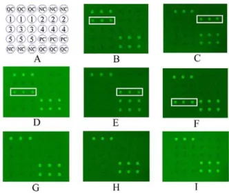
In this study, a protein chip was used as a diagnostic tool for FBHs to detect disease-specific antibodies. Our study results indicate that different parasites have variant diagnostic antigens, therefore they can be used for specific and differential detections. For example, the proteins ofT. soliumcysticerci orC. cellulosaewith molecular weights of 100, 95 or 26 kDa have been used in confirmatory tests for neurocysticercosis-positive cases [49]. Meanwhile, the 98 kDa antigen of A. cantonensis as a candidate antigen can be used for the diagnosis of angiostrongyliasis [43]. The 35 kDa protein spotted into protein chip could be used for the diagnosis of paragonimiasis, although further verification is needed to investigate if the protein is the same as the 35 kDa in P. Figure 3. Protein microarrays probed with human sera with various food-borne helminthiases.Disposition of microarray (A), the semi-purified antigens of the parasites recognized by the sera from patients infected withC. cellulosae(B),A. cantonensis(C),P. westermani (D), T. spiralis (E), Spirometraplerocercoids (F), healthy adult (G), A. lumbricoides (H) and S. japonicum (I) with the method of protein microarrays, respectively. On panel A, QC = quality control; PC = positive control of arrays; NC = negative control of arrays; Spot 1 to 5 = the semi-purified antigens with the molecular weight of 95, 98, 35, 55 and 90 kDa fromC. cellulosae,A. cantonensis,P. westermani,T. spiralisand Spirometraplerocercoids, respectively.
westermaniadult worm soluble antigens [63]. Moreover, it has been reported that the specificities using well-archived serum samples of sera-diagnosis for detection of different FBHs are ideal with positive percentages ranging from 83% to 100%, while the sensitivities ranged from 50 to 98% [16–18,21,28,29,64,65]. In spite of the semi-purified proteins prepared for each parasites in the study, such as the 95 kDa protein ofC. cellulosae, the 98 kDa protein ofA. cantonensis, the 35 kDa protein of P. westermani, the 55 kDa protein ofT. spiralisand the 90 kDa protein ofSpirometra spp. plerocercoids, our results show that the approach of protein microarray display higher specificity in the detection of each infection, ranging from 97.0% to 100.0%.
It was also displayed that the protein microarray had excellent NPV (98.2%–99.3%) and PPV (78.2%–100.0%). In contrast, both of the values varied slightly in ELISA. These data indicate that there is a high probability that the protein microarray negatives are truly uninfected and that the positive results with the protein microarray indeed infected in the population investigated. When it comes to using the same antigen, most of the Youden indices for the protein microarray were a little higher than those produced by the ELISA test used for individual FBH detection. In terms of the advantage of this technology, the protein microarray can be used for epidemiological screening of FBHs on a large scale and play a role in prevention and treatment of parasitic diseases.
Protein microarray technology has played a significant role in the study of protein-protein interactions and been used for the identification of antigenic targets of serum autoantibodies in a variety of diseases. More recently, this technology has been applied in the detection and the identification of autoantibody signatures in five FBHs. The development of 3-dimensional surfaces will support a high-throughput approach which can detect more than one pathogen with only one chip and avoid time-consuming, high costs, and labor required for conventional tests, such as ELISA and colloidal gold testing. The two indices, e.g. predictive values influenced by prevalence of the detected population, and properties of diagnostics including sensitivity and specificity, are
the main indices to assess the yield of screening test when it is used in the field [51]. This research should be seen as a primary attempt in our laboratory, since this method of protein microarray did not generate significant differences with respect to sensitivity. Hence, it is expected that the application of monoclonal antibody might increase the sensitivity of the method in the future. Moreover, it is useful that this chip can be used for clinical detections as well as field screening. In our further study, we aim to produce monoclonal antibodies with specificities for each of the five helminthiases for protein chip testing.
In conclusion, the present study established a protein micro-array for the rapid, sensitive and high-throughput diagnosis of cysticercosis, trichinellosis, paragonimiasis, sparganosis and an-giostrongyliasis. This technique may contribute to better control and prevention of FBHs.
Supporting Information
Table S1 The diagnoses of 365 patients by parasitolog-ical or serologparasitolog-ical methods. Found at: doi:10.1371/ journal.pntd.0000771.s001 (0.14 KB DOC).
(DOCX)
Acknowledgments
We express our special thanks to Dr. Ren-Li Zhang of Shenzhen Center for Diseases Control and Prevention, P.R. China for his kind help in providing the antigens and serum samples. We also would like to thank Dr. Min-Jun Xu, Lanzhou Veterinary Research Institute, P.R. China, for his valuable comments on the draft manuscript. Thanks to Dr Robert Bergquist for critical reviewing of the manuscript.
Author Contributions
Conceived and designed the experiments: JXC MXC LA XNZ. Performed the experiments: JXC MXC LA YCC YNZ SHC. Analyzed the data: JXC MXC LA XNZ. Contributed reagents/materials/analysis tools: JXC MXC LA JHC. Wrote the paper: JXC MXC LA XQZ XNZ.
References
1. Chai JY, Darwin MK, Lymbery AJ (2005) Fish-borne parasitic zoonoses: status and issues. Int J Parasitol 35: 1233–1254.
2. Sripa B (2012) Global burden of food-borne trematodiasis. Lancet Infect Dis 12: 171–172.
3. Dorny P, Praet N, Deckers N, Gabriel S (2009) Emerging food-borne parasites. Vet Parasitol 163: 196–206.
4. Keiser J, Utzinger J (2009) Food-borne trematodiases. Clin Microbiol Rev 22: 466–483.
5. Murrell KD, Cross JH, Looareesuwan S (2001) Food- and water-borne parasitic zoonoses in the 21st century. Trends Parasitol 17: 163–164.
6. Sripa B, Kaewkes S, Intapan PM, Maleewong W, Brindley PJ (2010) Food-borne trematodiases in Southeast Asia epidemiology, pathology, clinical manifestation and control. Adv Parasitol 72: 305–350.
7. Sripa B, Kaewkes S, Intapan PM, Maleewong W, Brindley PJ (2010) Food-borne trematodiases in Southeast Asia epidemiology, pathology, clinical manifestation and control. Adv Parasitol 72: 305–350.
8. Zhou P, Chen N, Zhang RL, Lin RQ, Zhu XQ (2008) Food-borne parasitic zoonoses in China: perspective for control. Trends Parasitol 24: 190–196. 9. Chan EW, Looi A (2010) Cysticercosis of the levator palpebrae superioris. Ann
Acad Med Singapore 39: 932–938.
10. Pozio E (2007) World distribution of Trichinella spp. infections in animals and humans. Vet Parasitol 149: 3–21.
11. Bruschi F, Murrell KD (2002) New aspects of human trichinellosis: the impact of new Trichinella species. Postgrad Med J 78: 15–22.
12. Choo JD, Suh BS, Lee HS, Lee JS, Song CJ, et al. (2003) Chronic cerebral paragonimiasis combined with aneurysmal subarachnoid hemorrhage. Am J Trop Med Hyg 69: 466–469.
13. Yang HJ (2008) Changes of serum adiponectin levels in murine experimental sparganosis. Korean J Parasitol 46: 91–93.
14. Slom TJ, Cortese MM, Gerber SI, Jones RC, Holtz TH, et al. (2002) An outbreak of eosinophilic meningitis caused by Angiostrongylus cantonensis in travelers returning from the Caribbean. N Engl J Med 346: 668–675.
15. Zhang RL, Chen MX, Gao ST, Geng YJ, Huang DN, et al. (2008) Enzootic angiostrongyliasis in Shenzhen, China. Emerg Infect Dis 14: 1955–1956. 16. Agudelo-Florez P, Palacio LG (2009) Dot blot for the diagnosis of porcine
cysticercosis. Rev Sci Tech 28: 1077–1084.
17. Deckers N, Dorny P (2010) Immunodiagnosis of Taenia solium taeniosis/ cysticercosis. Trends Parasitol 26: 137–144.
18. Eamsobhana P, Yong HS (2009) Immunological diagnosis of human angiostrongyliasis due to Angiostrongylus cantonensis (Nematoda: Angiostron-gylidae). Int J Infect Dis 13: 425–431.
19. Gottstein B, Pozio E, Nockler K (2009) Epidemiology, diagnosis, treatment, and control of trichinellosis. Clin Microbiol Rev 22: 127–145.
20. Kliks MM, Kroenke K, Hardman JM (1982) Eosinophilic radiculomyeloence-phalitis: an angiostrongyliasis outbreak in American Samoa related to ingestion of Achatina fulica snails. Am J Trop Med Hyg 31: 1114–1122.
21. Nkouawa A, Sako Y, Itoh S, Kouojip-Mabou A, Nganou CN, et al. (2010) Serological studies of neurologic helminthic infections in rural areas of southwest cameroon: toxocariasis, cysticercosis and paragonimiasis. PLoS Negl Trop Dis 4: e732.
22. Ozeretskovskaia NN, Potekaeva MA, Tumol’Skaia NI, Margulis TD, Vishnevskii VA (1966) The effect of steroid therapy on the acute and convalescent period of trichinellosis. II. The effect of steroids on the symptomatology and morphology of muscle lesions in trichinellosis. Med Parazitol (Mosk) 35: 164–171.
23. Slais J (1968) Morphology and histological diagnosis of the parasite in cysticercosis cerebri. Acta Neuropathol 10: 295–307.
24. Stefanko S, Zebrowski S (1961) The morphology of cerebral paragonimiasis. Acta Med Pol 2: 111–122.
25. Chen MG, Chang ZS, Shao XY, Liu MD, Blair D, et al. (2001) Paragonimiasis in Yongjia County, Zhejiang Province, China: clinical, parasitological and karyotypic studies on Paragonimus westermani. Southeast Asian J Trop Med Public Health 32: 760–769.
26. Wang ZQ, Cui J (2008) Diagnosis and treatment of trichinellosis. Zhongguo Ji Sheng Chong Xue Yu Ji Sheng Chong Bing Za Zhi 26: 53–57.
A Protein Microarray for Food-Borne Helminthiases
27. Zheng SZ, Cui CQ, Shen CH (2005) Subcutaneous sparganosis: biopsy report of four cases. Zhongguo Ji Sheng Chong Xue Yu Ji Sheng Chong Bing Za Zhi 23: 431.
28. Nishiyama T, Ide T, Himes SJ, Ishizaka S, Araki T (1994) Immunodiagnosis of human sparganosis mansoni by micro-chemiluminescence enzyme-linked immunosorbent assay. Trans R Soc Trop Med Hyg 88: 663–665.
29. Sahu PS, Parija SC, Jayachandran S (2010) Antibody specific to 43 kDa excretory-secretory antigenic peptide of Taenia solium metacestode as a potential diagnostic marker in human neurocysticercosis. Acta Trop 115: 257– 261.
30. Mahanty S, Garcia HH (2010) Cysticercosis and neurocysticercosis as pathogens affecting the nervous system. Prog Neurobiol 91: 172–184.
31. Chen MX, Ai L, Zhang RL, Xia JJ, Wang K, et al. (2011) Sensitive and rapid detection of Paragonimus westermani infection in humans and animals by loop-mediated isothermal amplification (LAMP). Parasitol Res 108: 1193–1198. 32. Zhu XQ, Beveridge I, Berger L, Barton D, Gasser RB (2002) Single-strand
conformation polymorphism-based analysis reveals genetic variation within Spirometra erinacei (Cestoda: Pseudophyllidea) from Australia. Mol Cell Probes 16: 159–165.
33. Chen LF, Hu N, Liu N, Guo B, Yao J, et al. (2010) The design and preparation of a flexible bio-chip for use as a visual prosthesis, and evaluation of its biological features. Cell Tissue Res 340: 421–426.
34. Jokerst JV, Chou J, Camp JP, Wong J, Lennart A, et al. (2011) Location of Biomarkers and Reagents within Agarose Beads of a Programmable Nano-bio-chip. Small.
35. Jain T, Muthuswamy J (2007) Bio-chip for spatially controlled transfection of nucleic acid payloads into cells in a culture. Lab Chip 7: 1004–1011. 36. Hartmann M, Roeraade J, Stoll D, Templin MF, Joos TO (2009) Protein
microarrays for diagnostic assays. Anal Bioanal Chem 393: 1407–1416. 37. Kijanka G, Murphy D (2009) Protein arrays as tools for serum autoantibody
marker discovery in cancer. J Proteomics 72: 936–944.
38. Lisa Melton. (2004) Protein arrays: Proteomics in multiplex. Nature 429: 101– 107.
39. Lv S, Zhang Y, Chen SR, Wang LB, Fang W, et al. (2009) Human angiostrongyliasis outbreak in Dali, China. PLoS Negl Trop Dis 3: e520. 40. Chau TT, Thwaites GE, Chuong LV, Sinh DX, Farrar JJ (2003) Headache and
confusion: the dangers of a raw snail supper. Lancet 361: 1866.
41. Satoskar, AR: Medical Parasitology (2009) Available: http://www.ebook3000. com/Medical-Parasitology_51496.html2009.
42. Feng LZ (1956), The research of using composition of pumpkin seed and betel nut to treat taeniasis. Chinese Medical Journal, 42:138–139 (Chinese). 43. Chen MX, Zhang RL, Chen JX, Chen SH, Li XH, et al. (2010) Monoclonal
antibodies against excretory/secretory antigens of Angiostrongylus cantonensis. Hybridoma (Larchmt) 29: 447–452.
44. Crum ED, Despommier DD, McGregor DD (1977) Immunity toTrichinella spiralis. I. Transfer of resistance by two classes of lymphocytes. Immunology 33: 787–795.
45. Ishida K, Yoshimura K (1992) Characterization of monoclonal antibodies against eosinophil chemotactic factors from young adult worms ofAngiostrongylus cantonensis. Parasite Immunol 14: 633–644.
46. Lowry OH, Rosebrough NJ, Farr AL, Randall RJ (1951) Protein measurement with the Folin phenol reagent. J Biol Chem 193: 265–275.
47. Laemmli UK (1970) Cleavage of structural proteins during the assembly of the head of bacteriophage T4. Nature 227: 680–685.
48. Towbin H, Staehelin T, Gordon J (1992) Electrophoretic transfer of proteins from polyacrylamide gels to nitrocellulose sheets: procedure and some applications. 1979. Biotechnology 24: 145–149.
49. Iudici NF, Pianetti-Filho G, Araujo RN, Nascimento E (2007) Immunodiagnosis of human neurocysticercosis by using semi-purified scolex antigens fromTaenia soliumcysticerci. Rev Soc Bras Med Trop 40: 163–169.
50. List C, Qi W, Maag E, Gottstein B, Muller N, et al. (2010) Serodiagnosis of Echinococcusspp. infection: explorative selection of diagnostic antigens by peptide microarray. PLoS Negl Trop Dis 4: e771.
51. Banoo S, Bell D, Bossuyt P, Herring A, Mabey D, et al. (2006) Evaluation of diagnostic tests for infectious diseases: general principles. Nat Rev Microbiol 4: S21–S31.
52. Chen JH, Lu F, Lim CS, Kim JY, Ahn HJ, et al. (2010) Detection ofPlasmodium vivax infection in the Republic of Korea by loop-mediated isothermal amplification (LAMP). Acta Trop 113: 61–65.
53. Youden WJ (1950) Index for rating diagnostic tests. Cancer 3: 32–35. 54. Pin˜a R, Gutie´rrez AH, Gilman RH, Rueda D, Sifuentes C, et al. (2011) A
dot-ELISA using a partially purified cathepsin-L-like protein fraction fromTaenia soliumcysticerci, for the diagnosis of human neurocysticercosis.Ann Trop Med Parasitol 105: 311–318.
55. Ben R, Rodrigues R, Agostini AA, Graeff-Teixeira C (2010) Use of heterologous antigens for the immunodiagnosis of abdominal angiostrongyliasis by an enzyme-linked immunosorbent assay. Mem Inst Oswaldo Cruz 105: 914–917. 56. Cui J, Li N, Wang ZQ, Jiang P, Lin XM (2011) Serodiagnosis of experimental
sparganum infections of mice and human sparganosis by ELISA using ES antigens of Spirometra mansoni spargana. Parasitol Res 108: 1551–1556. 57. Aranzamendi C, Tefsen B, Jansen M, Chiumiento L, Bruschi F, et al. (2011)
Glycan microarray profiling of parasite infection sera identifies the LDNF glycan as a potential antigen for serodiagnosis of trichinellosis. Exp Parasitol 129: 221– 226.
58. Gutjahr C, Murphy D, Lueking A, Koenig A, Janitz M, et al. (2005) Mouse protein arrays from a TH1 cell cDNA library for antibody screening and serum profiling. Genomics 85: 285–296.
59. Horn S, Lueking A, Murphy D, Staudt A, Gutjahr C, et al. (2006) Profiling humoral autoimmune repertoire of dilated cardiomyopathy (DCM) patients and development of a disease-associated protein chip. Proteomics 6: 605–613. 60. Balboni I, Chan SM, Kattah M, Tenenbaum JD, Butte AJ, et al. (2006)
Multiplexedproteinarray platforms for analysis of autoimmune diseases. Annu Rev Immunol 24: 391–418.
61. SartainMJ, Slayden RA, SinghKK, Laal S, Belisle JT (2006) Disease state differentiation and identification of tuberculosis biomarkers via native antigen array profiling. Mol Cell Proteomics 5: 2102–2113.
62. DaviesDH, Liang X,Hernandez JE, Randall A, Hirst S, et al. (2005) Profiling the humoral response to infection using proteome microarrays: high through-put vaccine and diagnostic antigen discovery. Proc Natl Acad Sci USA 102: 547– 552.
63. Kong Y, Chung JY, Yun DH, Kim LS, Kang SY, et al. (1997) Variation of antigenic proteins of eggs and developmental stages ofParagonimus westermani. Korean J Parasitol 35: 197–202.
64. Chung YB, Kong Y, Yang HJ, Cho SY (2000) IgG antibody responses in early experimental sparganosis and IgG subclass responses in human sparganosis. Korean J Parasitol 38: 145–150.