Review
Printed in Brazil - ©2016 Sociedade Brasileira de Química0103 - 5053 $6.00+0.00*e-mail: [email protected]; [email protected]
Nanobiotechnology Solutions against
Aedes aegypti
Nelson Durán,*,a,b,c German A. Islan,d Marcela Durán*,c,e and Guillermo R. Castrod
aLaboratório de Química Biológica and bLaboratório NanoBioss, Instituto de Química,
Universidade Estadual de Campinas (Unicamp), 13083-970 Campinas-SP, Brazil
cLaboratório Nacional de Nanotecnologia (LNNano-CNPEM), 13083-100 Campinas-SP, Brazil
dNanobiomaterials Laboratory, Applied Biotechnology Institute (CINDEFI, UNLP-CONICET CCT
La Plata), School of Sciences, Universidad Nacional de La Plata, La Plata, Argentina
eLaboratório deCarcinogênese Urogenital eImunoterapia, Instituto de Biologia,
Universidade Estadual de Campinas (Unicamp), 13083-970 Campinas-SP, Brazil
United Nations Children’s Fund (UNICEF)/United Nations Development Programme (UNDP)/ World Bank/World Health Organization (WHO) implemented the Training in Tropical Diseases (TDR) program with excellent results; however, due to current challenges, this active program requires new and innovative solutions. Nowadays, Aedes aegyptis-borne diseases can be added among neglected diseases. Surveillance and control must be considered owing to a great risk of infection with dengue, chikungunya and zika viruses. Although investigations on several vaccines are in progress, new insights in term of development of drugs that evade from resistance are of paramount importance. Nanobiotechnology appears as one of the most innovative strategy in the search of new uses for old pharmaceuticals or in the development of innovative and intelligent nanomedicines for neglected diseases. Liposomes, solid lipid nanoparticles, nanoemulsions, polymeric nanoparticles, metallic nanoparticles, quantum dots, carbon dots and carbon nanotubes were the focus of the current advances. In this direction, we have focused this overview on new advances in diagnostic assays as nanobiosensors, antivirus and nanoinsecticides on Aedes aegyptis
control.
Keywords: Aedes aegypti, mosquitoes, nanostructures, nanobiotechnology, antiviral, nanobiosensors, nanoinsecticides
1. Introduction
The Training in Tropical Diseases (TDR) program, approaches and contributions to drug discovery research and development (R&D) and the optimization of known treatments against infectious diseases of poor people were discussed.1,2 The current challenges are completely different from those at TDR’s beginning. Now, there are different players and more funding than before, and the primarily public or nonprofit organization, researching new drugs and diagnostics, but they still requires a strong coordination to optimize resources. The facts from the literature showed that this active environment needs new and innovative solutions.1
In our opinion, the most innovative strategy at this time should be the use of nanotechnology in the search
of new uses for old pharmaceuticals or the development of innovative and intelligent nanomedicines for neglected diseases.2,3
Among neglected diseases, one of most currently importance is the infection through Aedes aegypti and Ae. albopictus-borne and, the surveillance and control must be considered due to a great risk of infection with dengue, chikungunya, zika and also with yellow fever viruses. The principal vectors of dengue (DENV-1, DENV-2, DENV-3 and DENV-4), chikungunya (CHIKV), yellow fever (YFV) and zika (ZIKV) viruses, transmitted by Ae.aegypti and Ae. albopictus, are now under the concern of Center of Diseases Control and Prevention (CDC),4 USA, and by Brazilian Government.5
infection (e.g., influenza, typhoid fever, leptospirosis and measles), or non-specific viral syndrome, so diagnosis of DF, therefore, can be made only by specific laboratory test.6
DV infection by mosquitoes affects 2.5 billion people in tropical and sub-tropical regions of the world. DV is a member of the Flaviviridae family and is transmitted to humans by females of the genus Aedes, especially Ae. aegypti or Ae. albopictus.7
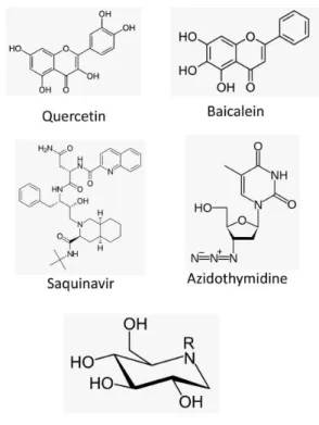
The dengue treatment is preventive and supportive care. The treatment of DV infection with small interfering RNA (siRNA) has been shown to be effective against DV replication.8 However, some studies showed that antivirals could be interesting in the future. Deoxynojirimycin and its N-alkylated derivatives (iminosugars) can be effective against DV infection by targeting host cellular factors that are required for viral morphogenesis.9 Flavonoids, fisetin, quercetin and baicalein, also exhibited anti-dengue virus activities (Figure 1).10
The first major success of the sterile insect technique was achieved against Culex quinquefasciatus in Myanmar in 1967 and efforts were being made against Culex quinquefasciatus and Ae. aegypti in India in 1967 and, recently in Brazil.5,11 Another strategy was used in 2000 by means of bacterium Wolbachia pipientis into Aedes aegypti populations for dengue control with a 50% reduction in the lifespan of adult female mosquito.12
Although several vaccine candidates are in advanced stages of development, no licensed dengue vaccine is
available yet, then, new insights in term of development of drug that evade resistance from resistance are of paramount importance.
2. Nanotechnology Presence on Dengue Virus and Other Viruses
During the last decades, the eradication of viral infections has still been a challenge in the medical field due to not only the problem of the spread, but also to their ability to evolve by genetic mutations, making them a real nightmare in health. In particular, dengue virus possesses four distinct serotypes, but closely related ones that the viral attack may cause a spectrum of illnesses, as discussed above. If subsequent infections by other serotypes are produced, there is an increase in the risk of developing severe dengue infection.6
Once there is no approved vaccine or antiviral agents against dengue virus, this opens the door for the design of new strategies to combat the infection, and nanobiotechnology appears to be a new feasible alternative. Nanobiotechnology is one of the most promising area that could be important in all of these diseases, including cancer13,14 and antibacterial agents.15
2.1. Nanobiosensors
Nowadays, the only therapy against dengue is prevention and palliative care, as mentioned before. For
these reasons, there is a need for the development of new, rapid and specific diagnostic systems to be useful tools for early detection of the infection, at its initial stages. Some nanodiagnostic tools are based on nanomaterials such as liposomes, nanopores and nanowires, which are coupled to conventional methods such as fluorescence, potentiometry and voltammetry.16 Most of them include enzyme-linked immunosorbent assay (ELISA) and reverse transcriptase polymerase chain reaction. Although these systems give rapid diagnostic information and use of inexpensive materials, they are currently unavailable or are scarce for clinical practice.17
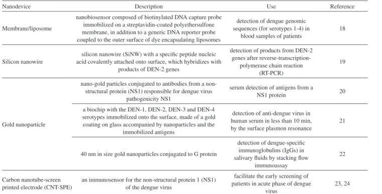
A nanobiosensor was developed by the use of an optical membrane-based DNA/RNA hybridization system utilizing a liposome amplification, which allows the detection of a genomic sequence in patient blood samples (e.g., generic and serotype-specific synthetic dengue sequences, for serotypes 1-4). The method uses a biotinylated DNA capture probe immobilized on a streptavidin-capped polyethersulfone membrane, in addition to a generic DNA reporter probe bound to the outer surface of dye-encapsulating liposomes. While nonspecific RNA molecule generates no signal, generic sequence added to the dengue RNA for the amplification step is hybridized and the signal is turned on. This nanobiosensor showed an excellent correlation in the detection and was mobile, inexpensive and easy to use (Figure 2).18
Another strategy in diagnostics proposes the use of silicon nanowire (SiNW)-based sensor for detection of products from DEN-2 genes after reverse transcription-polymerase chain reaction (RT-PCR) by hybridization with a specific peptide nucleic acid (PNA) attached covalently to the SiNW surface. The positive sign was demonstrated
by measuring the resistance change of the SiNW before and after hybridization.19
More recently, gold nanoparticles (NP) have been used for extremely sensitive and fast detection of dengue infection. A diagnostic device was developed for serum detection of antigens from a non-structural protein (NS1) responsible for dengue virus pathogenicity, using an immunoassay with gold nanoparticles conjugated to antibodies against NS1.20 Jahanshahi et al.21 reported a method based on surface plasmon resonance (SPR) for detection of anti-dengue virus in human serum in less than 10 min. The DEN-1, DEN-2, DEN-3 and DEN-4 serotypes were immobilized onto the biochip surface that consists of a gold coating on glass accompanied by nanoparticles and immobilized antigens. Other work22 has proposed the detection of dengue-specific immunoglobulins (IgGs) in salivary fluids by piling flow immunoassay, in which the liquid conjugate was constructed of G protein bound to 40-nm gold nanoparticles. The important aspect in this assay was that the IgG detection lies in distinguishing primary and secondary dengue infection since IgGs are specifically present in second episodes (Table 1).22 The developed nano-diagnostic systems can be extrapolated for the detection of other viral diseases.
An immunosensor for NS1 of the dengue virus based on carbon nanotube-screen printed electrodes (CNT-SPE) was successfully developed. A homogeneous mixture containing carboxylated carbon nanotubes was dispersed in carbon ink to prepare a screen printed working electrode or also using a poly(allylamine) as support associated to carbon nanotubes. These development and approach represent a great potential value for use in epidemic situations and can facilitate the early screening of patients in acute phase of dengue virus.23,24
2.2. Antiviral drugs
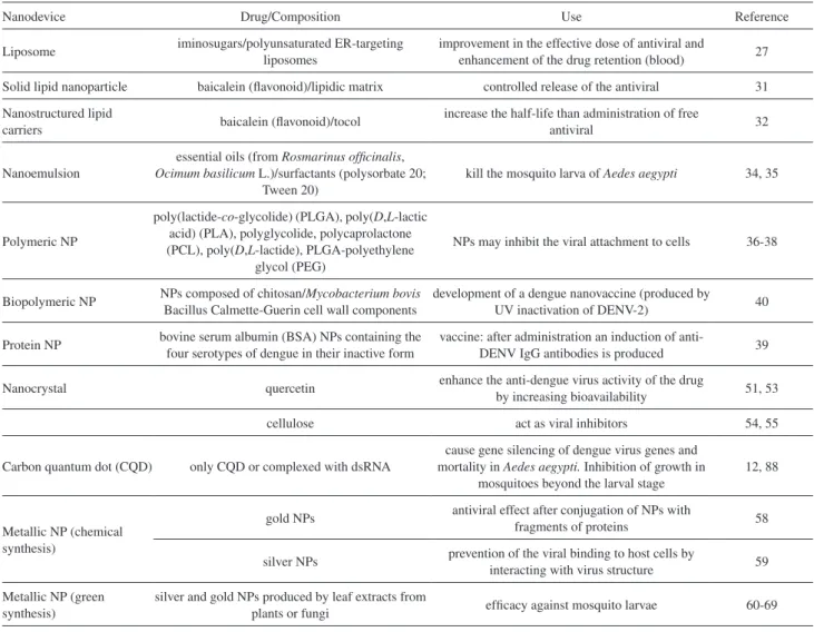
Some studies have shown that antivirals could be an interesting trend in the coming future.9,10,25 Nevertheless, these types of compounds commonly show a low bioavailability, especially for therapeutic uses, the reason why several strategies to overcome this limitation focus on the use of a wide variety of nanocarriers, including liposomes, solid lipid nanoparticles, nanoemulsions and nanocrystals, or polymeric nanoparticles, among others.26
Liposomal formulations have shown interesting properties in terms of the effective antiviral dose, decreasing the toxic concentrations and enhancing the retention time of the drug in blood circulation (due to pegylation). Experiments in vivo demonstrated that the delivery of iminosugars (all deoxynojirimycin derivatives) via
polyunsaturated ER-targeting liposomes (PERLs) increased the survival rate and prevented viral accumulation in organs and serum. In comparison to the administration of free iminosugars, liposomes only needed a 3-log lower dose to achieve survival of the animals. In addition, the formulation exhibited a greater in vitro potency against dengue virus by inhibiting of both the number of infected cells and the release of infectious viral particles induced by primary human monocyte-derived macrophages.27
More interesting is the extrapolation of the technique to treat infections with other related viruses. The ability of PERLs to produce a cholesterol-lowering effect has a positive impact in the inhibition of virus replication, and antiviral activities were observed not only for dengue virus but also hepatitis B virus (HBV), hepatitis C virus (HCV) and human immunodeficiency virus type 1 (HIV-1).28,29 Clayton et al.30 designed pegylated liposomes with the surface covered by labeled ligands from the Fab fragment of anti-HIV-gp120 monoclonal antibody (F105), and assessed the encapsulation of a novel HIV-1 protease inhibitor. They showed that immunoliposomes were effective in targeting and internalization into HIV-1-infected cells, delivering and accumulating the protease inhibitor in the cytoplasm. These results highlight the potential of liposomal carriers as vehicles for antivirals and the possibility to transport other cytotoxic drugs for a wide array of viruses.
Other studies have considered the development of solid lipid nanoparticles (SLNs) and nanostructured lipid
carriers (NLCs) as possible matrices for the encapsulation of antivirals against dengue virus. Baicalein, a well-known flavonoid with antiviral activity, was encapsulated into SLN at 60.73% by the solvent injection method and showed a controlled release profile.31 In a similar way, baicalein was incorporated into 100-nm tocol NLCs for intravenous administration, resulting in higher plasma levels and a longer half-life than with free antiviral.32 Another successful application of SLNs was the improvement in the poor oral availability of lopinavir (antiviral for HIV) by encapsulation in glyceryl behenate-based SLNs for delivery targeted to intestinal lymphatic vessels.33
Nanoemulsions from essential oils are also effective carriers for antiviral substances acting on vector-borne diseases.34,35
Several polymers such as poly(lactide-co-glycolide) (PLGA), poly(D,L-lactic acid) (PLA), polyglycolide, polycaprolactone (PCL), poly(D,L-lactide), chitosan and PLGA-polyethylene glycol (PEG) were developed for passive and ligand-targeted delivery of therapeutic drugs.36,37 In particular, it was observed that carriers for antiviral drugs exert the antiviral activity of the encapsulated molecules, indicating that direct interaction between the nanoparticles and the virus may inhibit viral attachment to cells.38 A tetravalent dengue system, composed of bovine serum albumin (BSA) nanoparticles, was evaluated in a murine model.39 BSA-NPs could absorb the four serotypes of dengue in their inactive form and,
Table 1. Nanodispositives in biosensing
Nanodevice Description Use Reference
Membrane/liposome
nanobiosensor composed of biotinylated DNA capture probe immobilized on a streptavidin-coated polyethersulfone membrane, in addition to a generic DNA reporter probe coupled to the outer surface of dye encapsulating liposomes
detection of dengue genomic sequences (for serotypes 1-4) in
blood samples of patients
18
Silicon nanowire
silicon nanowire (SiNW) with a specific peptide nucleic acid covalently attached onto surface, which hybridizes with
products of DEN-2 genes
detection of products from DEN-2 genes after
reverse-transcription-polymerase chain reaction (RT-PCR)
19
Gold nanoparticle
nano-gold particles conjugated to antibodies from a non-structural protein (NS1) responsible for dengue virus
pathogenicity NS1
serum detection of antigens from a
NS1 protein 20
a biochip with the DEN-1, DEN-2, DEN-3 and DEN-4 serotypes immobilized onto the surface, made of a gold coating on glass accompanied by nanoparticles and the
immobilized antigens
detection of anti-dengue virus in human serum in less than 10 min, by the surface plasmon resonance
21
40 nm in size gold nanoparticles conjugated to G protein
detection of dengue-specific immunoglobulins (IgGs) in salivary fluids by stacking flow
immunoassay
22
Carbon nanotube-screen printed electrode (CNT-SPE)
an immunosensor for the non-structural protein 1 (NS1) of the dengue virus
facilitate the early screening of patients in acute phase of dengue
virus
after administration, an induction of anti-DENV IgG antibodies was produced (Figure 3).
Other nanoparticles composed of chitosan/bacillus Calmette-Guerin (BCG) cell wall components were able to encapsulate a novel dengue nanovaccine (produced by UV inactivation of DENV-2) and showed interesting immunogenic properties in a Swiss albino mouse model by generation of humoral and cellular immune responses.40 Polymeric nanoparticles of polylactic acid (PLA) and methacrylic acid copolymers were designed to entrap the peptidomimetic compound CGP 57813, a potent HIV-1 protease inhibitor. Due to its high lipophilicity, it showed a poor bioavailability. While PLA nanoparticles (PLA NPs) showed an increase of at least two times in the plasma concentration-time curve (area) after intravenous injection in mice, no sufficient plasma level was detected after oral administration, and only the methacrylic acid copolymer nanoparticles provided reasonable values.41 However, in other studies, PLA NPs were demonstrated to be feasible drug delivery carriers to enhance tissue uptake and the targeting of other macromolecules with anti-HIV-1 activity.42 Also, PLGA nanoparticles have shown interesting applications as vaccine-delivery vehicles for treatment of viral infections.43
Polyhexylcyanoacrylate nanoparticles were loaded with saquinavir (another HIV protease inhibitor) or zalcitabine (a nucleoside analog) and tested under in vitro conditions in primary human monocytes/macrophages. A
dose-dependent reduction in HIV type 1 antigen was observed for both types of nanoparticles.44 Hexylcyanoacrylate nanoparticles were developed as a colloidal azidothymidine (AZT) carrier for specific targeting of the antiviral drug to reticuloendothelial cells by oral administration in HIV patients.45 By modifying this kind of nanoparticles through the coating with polysorbate 80, it was possible to change the body distribution of AZT after intravenous injection in rats, with a higher concentration of the drug in the brain. Although the drug was bound to the nanoparticles, its efficacy was not reduced.45 AZT was also encapsulated in polybutylcyanoacrylate (PBCA) and methylmethacrylate-sulfopropylmethacrylate (MMA-SPM) nanoparticles, and the study of their permeability across the blood-brain barrier revealed an enhancement in their permeability with a reduction in nanoparticles size.46 Furthermore, AZT- loaded poly-(isohexylcyanoacrylate) nanospheres displayed promising features to target the antiviral to the epithelium of the intestine and gut-associated lymphoid tissues, which are the main reservoirs of HIV in the gastrointestinal (GI) tract.47 The preparation of pH sensitive NPs was an interesting alternative to improve the bioavailability of HIV-1 protease inhibitor with a very low water-solubility. In this sense, nanoparticles made of the poly(methacrylic acid-co -ethylacrylate) copolymer Eudragit® L100-55 were orally administered to dogs of beagle breed and led to an increase in plasma concentrations due to a specific release of the antiviral near its absorption site.48 Shah and Amiji49 demonstrated that
poly(ethylene oxide)-modified poly(epsilon-caprolactone) (PEO-PCL) nanoparticles were a feasible vehicle for the intracellular delivery of saquinavir. PEO-PCL nanoparticles exhibited spherical shape and uniform size distribution around 200 nm. Their uptake by THP-1 (human leukemic) monocyte/macrophage (Mo/Mac) cells indicated a significantly higher incorporation of the drug in comparison with aqueous solution.
It is important to mention that the surface of the described nanoparticles can be tailored to modify the biodistribution of the antiviral to target specific organs and tissues or, with more precision, to deliver the drug to a particular cell type by attachment of ligands that recognize cell surface receptors.50 All of these facts are easily extrapolated to Ae. aegypti virus.
Although there are currently few reports in the literature, the first products are appearing on the market and a new tendency is indicating that nanocrystals could be a possible delivery system to improve the bioavailability of antiviral drugs.51,52 For example, pure quercetin, a flavonoid with anti-dengue virus activity, showed a limited in vivo efficacy because of its low solubility and reduced absorption at the intestine level. However, the synthesis of four types of co-crystals led to an improvement in physicochemical and pharmacokinetic characteristics in comparison with free quercetin.53 Other reports have described the use of cellulose nanocrystals (CNCs) as viral inhibitors. The first approach was carried out with unmodified CNCs derived from tunicates in a single model, and a decrease in phage infection of host E. coli was observed.54 In the next step, the modification of CNCs by surface attachment of tyrosine sulfate mimetic ligands (multivalent displays) resulted in the inhibition of alphavirus infectivity in Vero cells. Considering these results and the chemical structure of other known polyanionic inhibitors, the possibility to using CNCs for inhibition of other viruses (e.g., HIV and herpes simplex viruses) should be explored.55 A recent work also reported the production of nanocrystals of a reverse transcriptase inhibitor from HIV-1 virus (CSIC) by a three-phase nanoparticle engineering technology for intravaginal delivery.56
During the last decade, investigations have shown the potential of metal nanoparticles for treatment of viral infections. The interaction of silver and gold nanoparticles with proteins is under exploration, and the mechanism of antiviral activity is not well established.57 However, it was observed that gold nanoparticles conjugated to fragments of the HIV inhibitor TAK-779 (named SDC-1721) showed an excellent antiviral effect, while free SDC-1721 had no activity. This means that gold nanoparticles are able to convert inactive molecules into potent antivirals.58 On the
other hand, silver nanoparticles have also shown interesting properties by interacting with HIV virus and thereby preventing their binding to host cells, as long as they are in the range of 1-10 nm in size.59
2.3. Nanoinsecticide against mosquitoes
A nanoinsecticide is defined as a nanostructure containing a formulation that includes nanoscale elements with novel properties associated with some actives acting on insects. The benefits of this formulation are: efficiency due to higher surface area, sometimes higher solubility, higher mobility and a low toxicity, because no organic solvent is used related to the conventionally used in many pesticides.
An important group of nanoinsecticides is found with a nanoemulsion containing Rosmarinus officinalis essential oil (250 ppm), and its larvicidal activity against Ae. aegypti larvae, showed a mortality levels of 80 ± 10 and 90 ± 10% after 24 and 48 h, respectively.35
The green synthesis of metal nanoparticles has been proposed as an alternative to chemical methods, particularly through the use of fungi, since that the benign environment and renewable source of fungi act as reducing agents in the preparation of metal nanoparticles. These kinds of nanoparticles have exhibited efficacy against mosquito larvae (e.g., Aedes, Culex and Anopheles).60-65 In addition, leaf extracts from plants have been used for silver nanoparticle production and have become an eco-friendly alternative for adulticidal activity against filarial, dengue and malaria vector mosquitoes (Table 2).66-69
An interesting review on biogenic silver nanoparticles against mosquitoes was recently reported.70,71
Biogenic silver nanoparticles acting on Aedes aegypti and Culex quinquefasciatus (instars IV) demonstrated median lethal concentrations (LC50) of 0.30 and 0.41 µg mL-1, respectively. Adult longevity (days) in male and female mosquitoes exposed as larvae to 0.1 µg mL-1 silver nanoparticles was reduced by 30%, whereas the number of eggs laid by females exposed as larvae to this concentration decreased by 36%.72
The larval susceptibility to the basil oil (mainly estragole) nanoemulsion against third-instar larvae of Ae. aegypti showed a ten-fold diluted basil oil formulation (standard solution of 6% basil oil), inducing 100% larval mortality in 15 min. Complete loss of larval viability was observed after an exposure period of 90 min.34
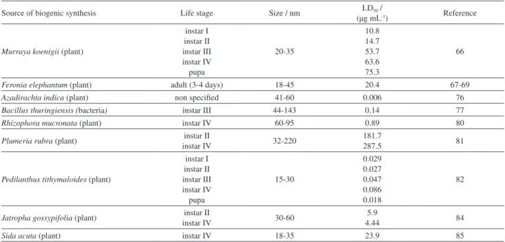
those values cited.70Ae. aegypti showed very sensitivity towards the mortality with biogenic silver nanoparticles (Table 3).66-69,76-82 Besides these actions, silver nanoparticles were effective against Anopheles and Culex.83-85 Against Ae.
Albopictus, biogenic silver nanoparticles exhibited a 100% mortality at 5 µg mL-1 level.86 Probably the large difference between different sources for biogenic synthesis of silver nanoparticles was due to the presence of different kind of capped protein on the nanoparticles or due to the presence of different silver nanostructures (e.g., silver chloride nanoparticles or/and silver oxides).87
A 3 µg mL-1 dose of water soluble carbon nanoparticles (carbon dots) in a stagnant water pool was found as optimum to arrest further growth of mosquitoes beyond the larval stage. At this concentration, the larvae stayed in suspended animation for four weeks and beyond that period of death without any toxicity or disturbing on surrounding ecosystem.12
The composite chitosan, carbon quantum dots and silica complexed with double-stranded RNA (dsRNA), was tested in order to target genes (SNF7 and SRC) to control Ae. aegyptis larvae. The result showed that knockdown efficiency correlated with dsRNA mediated larval mortality, being quantum dots the most efficient one and causing gene silencing and mortality in Ae. aegypti.88
3. Perspectives and Final Remarks
All the information discussed in this overview pointed out to the real potential of nanobiotechnology on virus inactivation and on insect control. Other important aspects in this overview are the safe use of nanomaterials that must be known through their toxicity and the effect in the environment, before its use. We hope this review encourages other researchers to do further research related to nanobiotechnology against Ae. aegyptis.
Table 2. Nanodispositives in treatment of dengue virus and mosquito/larvae of Aedes aegypti
Nanodevice Drug/Composition Use Reference
Liposome iminosugars/polyunsaturated ER-targeting liposomes
improvement in the effective dose of antiviral and
enhancement of the drug retention (blood) 27
Solid lipid nanoparticle baicalein (flavonoid)/lipidic matrix controlled release of the antiviral 31
Nanostructured lipid
carriers baicalein (flavonoid)/tocol
increase the half-life than administration of free
antiviral 32
Nanoemulsion
essential oils (from Rosmarinus officinalis,
Ocimum basilicum L.)/surfactants (polysorbate 20; Tween 20)
kill the mosquito larva of Aedes aegypti 34, 35
Polymeric NP
poly(lactide-co-glycolide) (PLGA), poly(D,L-lactic acid) (PLA), polyglycolide, polycaprolactone (PCL), poly(D,L-lactide), PLGA-polyethylene
glycol (PEG)
NPs may inhibit the viral attachment to cells 36-38
Biopolymeric NP NPs composed of chitosan/Mycobacterium bovis Bacillus Calmette-Guerin cell wall components
development of a dengue nanovaccine (produced by
UV inactivation of DENV-2) 40
Protein NP bovine serum albumin (BSA) NPs containing the four serotypes of dengue in their inactive form
vaccine: after administration an induction of
anti-DENV IgG antibodies is produced 39
Nanocrystal quercetin enhance the anti-dengue virus activity of the drug
by increasing bioavailability 51, 53
cellulose act as viral inhibitors 54, 55
Carbon quantum dot (CQD) only CQD or complexed with dsRNA
cause gene silencing of dengue virus genes and mortality in Aedes aegypti. Inhibition of growth in
mosquitoes beyond the larval stage
12, 88
Metallic NP (chemical synthesis)
gold NPs antiviral effect after conjugation of NPs with
fragments of proteins 58
silver NPs prevention of the viral binding to host cells by
interacting with virus structure 59
Metallic NP (green synthesis)
silver and gold NPs produced by leaf extracts from
plants or fungi efficacy against mosquito larvae 60-69
Acknowledgments
Support from INOMAT (CNPq), Brazilian Network on Nanotechnology (MCTI/CNPq), NanoBioss (MCTI) and FAPESP are acknowledged. The authors also thank Maurício Ramalho Custodio for the design art photography of the cover image.
Nelson Durán is a Professor of
Chemistry at Universidade Estadual de Campinas (Unicamp, Brazil). He received his PhD at University of Porto Rico (USA) working on photolysis and thermolysis of 1,2-dioxolanes (1972). He was Associated Professor at Universidad Catolica de Valparaiso, Chile (1973-1975), and carried out Visiting Professorship at Universidade de São Paulo (USP), Brazil (1975), investigating enzymatic generation of excited states intermediates. In 1978, he joined the Institute of Chemistry at Unicamp working in biological chemistry and biotechnology. His present research interests are nanobiotechnology in cosmetics and in pharmaceuticals, besides metallic nanoparticles as antibiotics and anticancer carriers, and in carbon and silica nanocarriers. He is the Coordinator of the Brazilian Nanotoxicology Network, member of INOMAT (Instituto Nacional de P&D&I em Materiais Complexos Funcionais, MCTI/CNPQ), Vice-Coordinator of NanoBioss Laboratory (MCTI, Instituto de Química, Unicamp) and member of Brazilian-NanoReg (European Community) in vivo nanotoxicology.
Germán Abel Islan is Assistant
Researcher at National Council of Research (CONICET, Argentina) which belongs to the Nanobiomaterial Laboratory at Applied Biotechnology Institute (CINDEFI, La Plata, Argentina). He is graduated from National University of La Plata (UNLP) with Master degree in Biotechnology and Molecular Biology (2004-2009). He finished his PhD studies with honors (2014) under the supervision of Prof Guillermo R. Castro, with focus in the development of micro- and nanodevices for enzymes and antibiotic encapsulation in the treatment of cystic fibrosis disease. He received a postdoctoral scholarship from CONICET for the development of smart lipid-biopolymeric nanoparticles for the controlled release of drugs, co-supervised by Prof Nelson Durán (Institute of Chemistry, Unicamp, Brazil). He also teaches as a Graduate Assistant in Organic Chemistry (2008) and Microbiology courses (2009-up the present) at School of Sciences (UNLP).
Guillermo R. Castro is currently Professor of Department of Chemistry, School of Sciences, Universidad Nacional de La Plata, and Senior Research Scientist at the National Council of Research (CONICET) in Argentina. He got MSc and PhD degrees in Biological Chemistry and Chemistry from Universidad de Buenos Aires, Argentina. He was postdoctoral researcher of the
Table 3. Sensitivity towards the mortality of Aedes aegypti with biogenic silver nanoparticles
Source of biogenic synthesis Life stage Size / nm LD50 /
(µg mL-1) Reference
Murraya koenigii (plant)
instar I instar II instar III instar IV
pupa
20-35
10.8 14.7 53.7 63.6 75.3
66
Feronia elephantum (plant) adult (3-4 days) 18-45 20.4 67-69
Azadirachta indica (plant) non specified 41-60 0.006 76
Bacillus thuringiensis (bacteria) instar III 44-143 0.14 77
Rhizophora mucronata (plant) instar IV 60-95 0.89 80
Plumeria rubra (plant) instar II
instar IV 32-220
181.7
287.5 81
Pedilanthus tithymaloides (plant)
instar I instar II instar III instar IV pupa
15-30
0.029 0.027 0.047 0.086 0.018
82
Jatropha gossypifolia (plant) instar II
instar IV 30-60
5.9
4.44 84
Department of Chemistry at Massachusetts Institute of Technology (MIT, Cambridge, USA) and later of the Department of Biomedical Engineering at Tufts University (Boston, USA), where became as Adjunct Professor. Presently, PhD Castro is the Principal Investigator (P.I.) of the Nanobiomaterial Laboratory at Applied Biotechnology Institute (CINDEFI) with the research focus in the development of biopolymeric matrices for molecular controlled release. His interests are in the field of biotechnology and nanobiotechnology using applied enzymology in aqueous and non-conventional media, natural polymers (e.g., production, purification and modification), gel formation and characterization using biophysical methods, coacervates and molecular controlled release. He published more than 90 papers in peer-reviewed journals, more than 100 meeting presentations, 20 book chapters and 4 patents.
Marcela Durán is a pharmacist,
R e s e a rch e r A s s o c i a t e d i n t h e Carcinogenesis Laboratory Urogenital and Immunotherapy Anatomy, at Institute of Biology at Universidade Estadual de Campinas (Unicamp, Brazil). She is graduated from San Francisco University (USF, Brazil). Currently, she is developing research project related to nanotechnology area, such as cell regeneration and stem cells. She has a Master degree in Food Technology (Unicamp, 2004) and PhD in Medical Sciences (Unicamp, 2014). She is member of Brazilian NanoReg Toxicology group (European Community) working in vivo study of carbon nanotubes by instillation on rats associated to NanoBioss Laboratory (MCTI, Institute of Chemistry, Unicamp).
References
1. Olliaro, P. L.; Kuesel, A. C.; Reeder, J. C.; PLoS Negl. Trop. Dis. 2015, 9, e3379.
2. Rossi-Bergmann, B.; Pacienza-Lima, W.; Marcato, P. D.; De Conti, R.; Durán, N. ; J. Nano Res. 2012, 20, 89.
3. Durán, N.; Marcato, P. D.; Teixeira, Z.; Durán, M.; Costa, F. T. M.; Brocchi, M.; Curr. Nanosci. 2009, 5, 396.
4. Center of Diseases Control and Prevention (CDC); Surveillance and Control of Aedes aegypti and Aedes albopictus in the United States, http://www.cdc.gov/chikungunya/resources/ vector-control.html, accessed on April 14, 2016.
5. Araújo, H. R. C.; Carvalho, D. O.; Ioshino, R. S.; Costa-da-Silva, A. L.; Capurro, M. L.; Insects2015, 6, 576.
6. Mungrue, K.; Adv. Lab. Med. Int. 2014, 4, 1.
7. Beatty, M. E.; Stone, A.; Fitzsimons, D. W.; Hanna, J. N.; Lam,
S. K.; Vong, S.; Gusman, M. G.; Mendez-Galvan, J. F.; Halstead, S. B.; Letson, G. W.; Kuritsky, J.; Mahoney, R.; Margolis, H. S.; PLoS Negl. Trop. Dis. 2010, 4, e890.
8. Idrees, S.; Ashfaq, U. A.; Asian Pac. J. Trop. Biomed. 2013, 3, 232.
9. Sayce, A. C.; Miller, J. L.; Zitzmann, N.; Trends Microbiol.
2010, 18, 323.
10. Zandi, K.; Teoh, B.-T.; Sam, S.-S.; Wong, P.-F.; Mustafa, M. R.; AbuBakar, S.; BMC Complement. Altern. Med.2012, 12, 214.
11. Carvalho, D. O.; Nimmo, D.; Naish, N.; McKemey, A. R.; Gray, P.; Wilke, A. B. B.; Marrelli, M. T.; Virgilio, J. F.; Alphey, L.; Capurro, M. L.; J. Vis. Exp.2014, 83, e3579.
12. Saxena, M.; Sonkar, S. K.; Sarkar, S.; RSC Adv. 2013, 3, 22504.
13. Deda, D. K.; Araki, K.; J. Braz. Chem. Soc. 2015, 26, 2448. 14. Silveira, C. P.; Apolinário, L. M.; Fávaro, W. J.; Paula, A. J.;
Durán, N.; RSC Adv. 2015, 5, 81348.
15. Durán, N.; Durán, M.; de Jesus, M. B.; Fávaro, W. J.; Seabra, A. B.; Nakazato, G.; Nanomedicine: NBM2015, 12, 789. 16. Rai, M.; Gade, A.; Gaikwad, S.; Marcato, P. D.; Durán, N.;
J. Braz. Chem. Soc.2012, 23, 14.
17. Peh, A. E.; Leo, Y. S.; Toh, C. S.; Front. Biosci., Scholar Ed.
2010, 3, 806.
18. Baeumner, A. J.; Schlesinger, N. A.; Slutzki, N. S.; Romano, J.; Lee, E. M.; Montagna, R. A.; Anal. Chem. 2002, 74, 1442.
19. Zhang, G. J.; Zhang, L.; Huang, M. J.; Luo, Z. H. H.; Tay, G. K. I.; Lim, E. J. A.; Kang, T. G.; Chen, Y.; Sens. Actuators B 2010, 146, 138.
20. Hussain, A.; Rehman, S. U.; Aslam, S.; Javed, N.; Abbas, Z.;
J. Anim. Plant Sci.2014, 24, 1110.
21. Jahanshahi, P.; Zalnezhad, E.; Sekaran, S. D.; Adikan, F. R. M.;
Sci. Reports2014, 4, 3851.
22. Zhang, Y.; Bai, J.; Ying, J. Y.; Lab Chip2015, 15, 1465. 23. Dias, A. C.; Gomes-Filho, S. L. R.; Silva, M. M.; Dutra, R. F.;
Biosens. Bioelectron.2013, 44, 216.
24. Silva, M. M. S.; Dias, A. C. M. S.; Silva, B. V. M.; Gomes-Filho, S. L. R.; Kubota, L. K.; Goulart, M. O. F.; Dutra, R. F.; J.Chem. Technol. Biotechnol.2015, 90, 194.
25. Zandi, K.; Teoh, B. T.; Sam, S. S.; Wong, P. F.; Mustafa, M. R.; AbuBakar, S.; J.Virol. 2011, 8, 560.
26. Teles, F. R. R.; Prazeres, D. M. F.; Lima‐Filho, J. L.; Rev. Med. Virol. 2005, 15, 287.
27. Miller, J. L.; Lachica, R.; Sayce, A. C.; Williams, J. P.; Bapat, M.; Dwek, R.; Zitzmann, N.; Antimicrob. Agents Chemother.
2012, 56, 6379.
28. De Mareuil, J.; Mabrouk, K.; Doria, E.; Moulard, M.; De Chasteigner, S.; Oughideni, R.; van Rietschoten, J.; Rochat, H.; De Waard, M.; Sabatier, J.-M.; Antiviral Res. 2002,
29. Pollock, S.; Nichita, N. B.; Böhmer, A.; Radulescu, C.; Dwek, R. A.; Zitzmann, N.; Proc. Natl. Acad. Sci. U. S. A. 2010, 107, 17176.
30. Clayton, R.; Ohagen, A.; Nicol, F.; Del Vecchio, A. M.; Jonckers, T. H.; Goethals, O.; Hertogs, K.; Antiviral Res. 2009, 84, 142. 31. Yumei, L.; Kun, Z.; Haiming, M.; Pract. Pharm. Clin. Remedies
2009, 12, 104.
32. Tsai, M. J.; Wu, P. C.; Huang, Y. B.; Chang, J. S.; Lin, C. L.; Tsai, Y. H.; Fang, J. Y.; Int. J. Pharm.2012, 423, 461. 33. Alex, M. A.; Chacko, A. J.; José, S.; Souto, E. B.; Eur. J. Pharm.
Sci.2011, 42, 11.
34. Ghosh, V.; Mukherjee, A.; Chandrasekaran, N.; Asian J. Chem.
2013, 25, S321.
35. Duarte, J. L.; Amado, J. R.; Oliveira, A. E.; Cruz, R. A.; Ferreira, A. M.; Souto, R. N.; Fernandes, C. P.; Rev. Brasil. Farmacogn.
2015, 25, 189.
36. Prabhu, R. H.; Patravale, V. B.; Joshi, M. D.; Int. J. Nanomed.
2015, 10, 1001.
37. Jhaveri, A.; Torchilin, V.; Expert Opin. Drug Delivery2016,
13, 49.
38. Lembo, D.; Cavalli R.; Antiviral Chem. Chemother. 2010, 21, 53.
39. Silva, E. F.; Orsi, M.; Andrade, A. L.; Domingues, R. Z.; Silva, B. M.; de Araújo, H. R.; Pimenta, P. F. P.; Diamond, M. S.; Rocha, E. S. O.; Kroon, E. G.; Malaquias, L. C. C.; Coelho, L. F.; J. Nanobiotechnol.2012, 10, 13.
40. Hunsawonga, T.; Sunintaboon, P.; Warit, S.; Thaisomboonsuk, B.; Jarmane, R. G.; Yoon, I.-K.; Ubol, S.; Fernandez, S.; Vaccine
2015, 33, 1702.
41. Leroux, J.-C.; Doelker, E.; Gurny, R.; Cozens, R.; Roesel, J. L.; Galli, B.; Kubel, F.; Doelker, E.; Gurny, R.; J. Pharm. Sci. 1995,
84, 1387.
42. Ham, A.; Cost, M.; Sassi, A.; Dezzutti, C.; Rohan, L.; Pharm. Res. 2009, 26, 502.
43. Demento, S. L.; Bonafé, N.; Cui, W.; Kaech, S. M.; Caplan, M. J.; Fikrig, E.; Ledizet, M.; Fahmy, T. M.; J. Immunol. 2010,
185, 2989.
44. Bender, A. R.; von Briesen, H.; Kreuter, J.; Duncan, I. B.; Rübsamen-Waigmann, H.; Antimicrob. Agents Chemother.
1996, 40, 1467.
45. Löbenberg, R.; Araujo, L.; von Briesen, H.; Rodgers, E.; Kreuter, J.; J. Controlled Release1998, 50, 21.
46. Kuo, Y.-C.; Chen, H.-H.; Int. J. Pharm. 2006, 327, 160. 47. Dembri, A.; Montisci, M. J.; Gantier, J. C.; Chacun, H.; Ponchel,
G.; Pharm. Res. 2001, 18, 467.
48. De Jaeghere, F.; Allémann, E.; Kubel, F.; Galli, B.; Cozens, R.; Doelker, E.; Gurny R.; J. Controlled Released2000, 68, 291. 49. Shah, L.; Amiji, M.; Pharm. Res. 2006, 23, 2638.
50. Gunaseelan, S.; Gunaseelan, K.; Deshmukh, M.; Zhang, X.; Sinko, P. J.; Adv. Drug Delivery Rev.2010, 62, 518.
51. Shegokar, R.; Müller, R H.; Int. J. Pharm. 2010, 399, 129.
52. Durán, N.; Durán, M.; Tasic, L.; Marcato, P. D.; Quim. Nova
2010, 33, 151.
53. Smith, A. J.; Kavuru, P.; Wojtas, L.; Zaworotko, M. J.; Shytle, R. D.; Mol. Pharmacol. 2011, 8, 1867.
54. S e r i z awa , T. ; S awa d a , T. ; O k u r a , H . ; Wa d a M . ;
Biomacromolecules2013, 14, 613.
55. Zoppe, J. O.; Ruottinen, V.; Ruotsalainen, J.; Rönkkö, S.; Johansson, L.-S.; Hinkkanen, A.; Jarvinen, K.; Seppala, J.;
Biomacromolecules2014, 15, 1534.
56. Gong, T.; Parniak, M.; Gupta, P.; Rohan, L.; Aids Res. Human Retroviruses2014, 30, A143.
57. Bhattacharya, R.; Mukherjee, P.; Adv. Drug Delivery Rev.2008,
60, 1289.
58. Bowman, M. C.; Ballard, T. E.; Ackerson, C. J.; Feldheim, D. L.; Margolis, D. M.; Melander, C.; J. Am. Chem. Soc. 2008, 130, 6896.
59. Elechiguerra, J. L.; Burt, J. L.; Morones, J. R.; Camacho-Bragado, A.; Gao, X.-X.; Lara, H. H.; Yacaman, M. J.;
J. Nanobiotechnol. 2005, 3, 6.
60. Salunkhe, R. B.; Patil, S. V.; Patil, C. D.; Salunke, B. K.;
Parasitol. Res.2011, 109, 823.
61. Soni, N.; Prakash, S.; Parasitol. Res. 2012a, 110, 175. 62. Soni, N.; Prakash, S.; Rep. Parasitol. 2012b, 2, 1. 63. Soni, N.; Prakash, S.; Parasitol. Res. 2012c, 111, 2091. 64. Soni, N.; Prakash, S.; Asian Pacific J. Trop. Dis. 2012d, 2,
S356.
65. Soni, N.; Prakash, S.; Adv. Nanopart. 2013, 2, 125.
66. Suganya, A.; Murugan, K.; Kovendan, K.; Kumar, P. M.; Hwang, J. S.; Parasitol. Res.2013, 112, 1385.
67. Veerakumar, K.; Govindarajan, M.; Hoti, S. L.; Parasitol. Res.
2014, 113, 4567.
68. Veerakumar, K.; Govindarajan, M.; Rajeswary, M.; Muthukumaran, U.; Parasitol. Res. 2014, 113, 1775. 69. Veerakumar, K.; Govindarajan, M.; Rajeswary, M.;
Muthukumaram, U.; Parasitol. Res. 2014, 113, 2663. 70. Hajra, A.; Mondal, N. K.; Int. J. Sci. Res. Environ. Sci.2015,
3, 47.
71. Rai, M.; Kon, K. In Nanotechnology in Diagnosis, Treatment and Prophylaxis of Infectious Diseases; Rai, M.; Kon, K., eds.; Elsevier: London, USA, 2015, ch. 3.
72. Arjunan, N. K.; Murugan, K.; Rejeeth, C.; Madhiyazhagan, P.; Barnard, D. R.; Vector Borne Zoonotic Dis.2012, 12, 262. 73. Durán, N.; Marcato, P. D.; De Conti, R.; Alves, O. L.; Costa,
F. T. M.; Brocchi M.; J. Braz. Chem. Soc.2010, 21, 949. 74. Gaikwad, S. C.; Birla, S. S.; Ingle, A. P.; Gade, A. K.; Marcato,
P. D.; Rai, M.; Durán, N.; J. Braz. Chem. Soc. 2013, 24, 1974.
75. Mashwani, Z.; Khan, T.; Khan, M. A.; Nadhman, A.; Appl. Microbiol. Biotechnol. 2015, 99, 9923.
77. Banu, A. N.; Balasubramanian, C.; Moorthi, P. V.; Parasitol. Res.2014, 113, 311.
78. Gnanadesigan, M.; Anand, M.; Ravikumar, S.; Maruthupandy, M.; Vijayakumar, V.; Selvam, S.; Dhineshkumar, M.; Kumaraguru, A. K.; Asian Pacific J. Trop. Med.2011, 4, 799. 79. Patil, C. D.; Patil, S. V.; Borase, H. P.; Salunke, B. K.; Salunkhe,
R. B.; Parasitol. Res. 2012, 110, 1815.
80. Sundaravadivelan, C.; Padmanabhan M. N.; Sivaprasath, P.; Kishmu, L.; Parasitol. Res.2013, 112, 303.
81. Borase, P. H.; Patil, D.; Salunkhe, R. B.; Narkhede, C. P.; Salunke, B. K.; Patil, S. V. J.; Nanomed. Biother. Discovery
2013, 3, 111.
82. Veerakumar, K.; Govindarajan, M.; Rajeswary, M.; Parasitol. Res. 2013, 112, 4073.
83. Priyadarshini, K. A.; Murugan, K.; Panneerselvam, C.; Ponarulselvam, S.; Hwang, J. S.; Nicoletti, M.; Parasitol. Res.
2012, 111, 997.
84. Kalimuthu, K.; Panneerselvam, C.; Murugan, K.; Hwang, J. S.;
J. Entomol. Acarol. Res. 2013, 45, 57.
85. Sathishkumar, M.; Sneha, K.; Won, W. S.; Cho, C.-W.; Kim, S.; Yun, Y.-S.; Colloids Surf., B2009, 73, 332.
86. Sareen, S. J.; Pillai, R. K.; Chandramohanakumar, N.; Balagopalan, M.; Res. J. Recent Sci.2012, 1, 52.
87. Durán, N.; Durán, M.; de Jesus, M. B.; Fávaro, W. J.; Nakazato, G.; Seabra, A. B.; Nanomedicine: NBM2016, 12, 789. 88. Das, S.; Debnath, N.; Cui, Y.; Unrine, J.; Palli, S. R.; ACS Appl.
Mater. Interfaces2015, 7, 19530.
Submitted: March 3, 2016
Published: April 20, 2016