Proteins Using Angle-Distance Image-Based Secondary
Structural Matching Techniques
Chia-Han Chu1., Wei-Cheng Lo2,3., Hsin-Wei Wang4, Yen-Chu Hsu4, Jenn-Kang Hwang3, Ping-Chiang Lyu2, Tun-Wen Pai4*, Chuan Yi Tang1,5*
1Department of Computer Science, National Tsing Hua University, Hsinchu, Taiwan, Republic of China,2Institute of Bioinformatics and Structural Biology, National Tsing Hua University, Hsinchu, Taiwan, Republic of China,3Institute of Bioinformatics and Systems Biology, National Chiao Tung University, Hsinchu, Taiwan, Republic of China, 4Department of Computer Science and Engineering, National Taiwan Ocean University, Keelung, Taiwan, Republic of China,5Department of Computer Science and Information Engineering, Providence University, Taichung, Taiwan, Republic of China
Abstract
This work presents a novel detection method for three-dimensional domain swapping (DS), a mechanism for forming protein quaternary structures that can be visualized as if monomers had ‘‘opened’’ their ‘‘closed’’ structures and exchanged the opened portion to form intertwined oligomers. Since the first report of DS in the mid 1990s, an increasing number of identified cases has led to the postulation that DS might occur in a protein with an unconstrained terminus under appropriate conditions. DS may play important roles in the molecular evolution and functional regulation of proteins and the formation of depositions in Alzheimer’s and prion diseases. Moreover, it is promising for designing auto-assembling biomaterials. Despite the increasing interest in DS, related bioinformatics methods are rarely available. Owing to a dramatic conformational difference between the monomeric/closed and oligomeric/open forms, conventional structural comparison methods are inadequate for detecting DS. Hence, there is also a lack of comprehensive datasets for studying DS. Based on angle-distance (A-D) image transformations of secondary structural elements (SSEs), specific patterns within A-D images can be recognized and classified for structural similarities. In this work, a matching algorithm to extract corresponding SSE pairs from A-D images and a novel DS score have been designed and demonstrated to be applicable to the detection of DS relationships. The Matthews correlation coefficient (MCC) and sensitivity of the proposed DS-detecting method were higher than 0.81 even when the sequence identities of the proteins examined were lower than 10%. On average, the alignment percentage and root-mean-square distance (RMSD) computed by the proposed method were 90% and 1.8A˚ for a set of 1,211 DS-related pairs of proteins. The performances of structural alignments remain high and stable for DS-related homologs with less than 10% sequence identities. In addition, the quality of its hinge loop determination is comparable to that of manual inspection. This method has been implemented as a web-based tool, which requires two protein structures as the input and then the type and/or existence of DS relationships between the input structures are determined according to the A-D image-based structural alignments and the DS score. The proposed method is expected to trigger large-scale studies of this interesting structural phenomenon and facilitate related applications.
Citation:Chu C-H, Lo W-C, Wang H-W, Hsu Y-C, Hwang J-K, et al. (2010) Detection and Alignment of 3D Domain Swapping Proteins Using Angle-Distance Image-Based Secondary Structural Matching Techniques. PLoS ONE 5(10): e13361. doi:10.1371/journal.pone.0013361
Editor:Andreas Hofmann, Griffith University, Australia
ReceivedJuly 10, 2010;AcceptedSeptember 13, 2010;PublishedOctober 14, 2010
Copyright:ß2010 Chu et al. This is an open-access article distributed under the terms of the Creative Commons Attribution License, which permits unrestricted use, distribution, and reproduction in any medium, provided the original author and source are credited.
Funding:This work was funded by the National Science Council, Taiwan (http://www.nsc.gov.tw) with NSC grant number 99-2745-B-009-001-ASP (Academic Summit Program) to Jenn-Kang Hwang; 99-2811-M-007-073 and 99-3112-B-007-003 to Ping-Chiang Lyu; 98-2221-E-019-031-MY2 to Tun-Wen Pai; 97-2221-E-007-080-MY3 to Chuan Yi Tang. The Ministry of Education, Taiwan also funded this study with the Development Plan for World Class University and Research Centers of Excellence (MOU ATU Plan). The funders had no role in study design, data collection and analysis, decision to publish, or preparation of the manuscript.
Competing Interests:The authors have declared that no competing interests exist. * E-mail: [email protected] (T-WP); [email protected] (CYT)
.These authors contributed equally to this work.
Introduction
Involved in the formation of quaternary structures from monomers, three-dimensional (3D) domain swapping refers to two or more identical proteins exchanging equivalent parts of their structures to form intertwined oligomers, inclusive of dimers [1,2,3]. The term ‘‘3D domain swapping’’ was first created in 1994 to describe the dimeric structure of diphtheria toxin [4,5]. Subsequently, this led to the discovery of a considerable number of other domain-swapped proteins, such as some ribonucleases [6,7,8], cysteine proteinase inhibitors [9,10,11], SH2 and SH3
A subunit of a 3D domain-swapped oligomer appears to have two conformational states, a monomeric closed-form and an oligomeric open-form. 3D domain swapping (abbreviated as DS) has been accordingly classified into three types [2]. First, in bona fide cases, both the closed monomer and domain-swapped oligomer of a protein exist stably. Second, although capable of forming intertwined and apparently domain-swapped oligomers, some proteins cannot exist as closed monomers. Quasi-domain-swapped cases refer to the domain-Quasi-domain-swapped proteins that have structural homologs known to be closed monomers. Third, DS candidates refer to the opposite situation in which no closed homolog is found for these oligomeric proteins.
DS can originate from environmental changes such as variations in pH values and protein concentrations [1,23]. Additionally, two evolutionary mechanisms have been proposed for DS [1,3]. First, as the hinge loop, the loop connecting the swapped domain to the protein body (main domain), of a closed monomer becomes shorter by residue deletion during evolution, the closed confor-mation might no longer remain stable because it is difficult for the domain to be swapped to reach the protein body, thus exposing the residues normally buried in the domain-domain contact interface. The domain-swapped form is then energetically favored [24,25]. Second, changes or mutations in the hinge loop and/or the contact interface of domains might destabilize the closed monomer due to steric or electrostatic effects and subsequently promote swapping conditions [26,27].
Although DS is an interesting and important structural phenomenon, related bioinformatics resources are rarely available. Previous studies have shown that 3D domain-swapping homologs may share minor sequence similarity [28] (see also a summary of DS cases in [2]). Therefore, sequence-based alignment tools may be inadequately sensitive to detect evolutionarily distantly related DS cases. Moreover, conventional structural comparison algo-rithms are insufficiently flexible to detect global similarities between proteins related by DS [29]. When aligning a ‘‘closed’’ monomer and its domain-swapped ‘‘open’’ homolog (referred to hereinafter as a ‘‘DSCO pair’’), several methods, such as FAST
[30] and TM-align [31], tend to output one local alignment restricted only to the protein bodies or the swapped domains. Although other methods, such as DALI [32] and CE [33], can simultaneously make several alignments with statistical Z-scores, most of them are local/partial alignments. Even if a global similarity were detected, a low Z-score and a large root-mean-square distance (RMSD) of the structural superposition would likely occur. Failing to visually inspect the superimposed structures would make it extremely difficult to identify the DS relationships. Although capable of detecting the structural similarity of 3D domain-swapping proteins, structural comparison methods with a more flexible nature, such as Flexible structure AlignmenT by Chaining Aligned fragment pairs allowing Twists (FATCAT) [29] and Structural similarity search Aided by Ramachandran Sequential Transformation (SARST) [34] provide no information to help users distinguish the domain-swapped homologs from the common structural homologs in the hit list. DS is sometimes considered as a unique domain motion [35,36]. However, tested with the known cases described by Eisenberget al., the well-known domain motion detection method DynDom [35] failed to identify most of those DS relationships (see Table S1 for details).
Perhaps because of the unavailability of suitable detection and analytical methods, currently the datasets for DSCO pairs,
including the largest literature-based dataset by [2] (33 pairs) and the predicted dataset with experimental verifications by [37] (7 pairs), are very small. This situation has greatly limited the scale and depth of DS-related researches. As a result, there is still much
uncertainty about how frequently DS occurs in Nature, which proposed mechanism plays the major role in the evolution of DS or whether there is any undiscovered mechanism for DS [23]. There are also few solid data available about the sequence compositions and structural properties of 3D domain-swapped proteins and their hinge loops, which shall be very valuable for researchers who are looking for treatments for protein deposition diseases or interested in creating protein-based fibril materials or oligomerized enzymes. We believe that, in this post-genomic era, when protein structure data increase rapidly, it is very possible that plenty of information can be extracted to reveal the natural prevalence and evolutionary mechanism of DS as well as to accelerate the medical and bioengineering applications of DS. A suitable detection and analytical bioinformatics method shall be the key to these possibilities. Motivated by the importance of DS and the insufficiency of related bioinformatics developments, this work aims to design a DS-specific identification and structural comparison method.
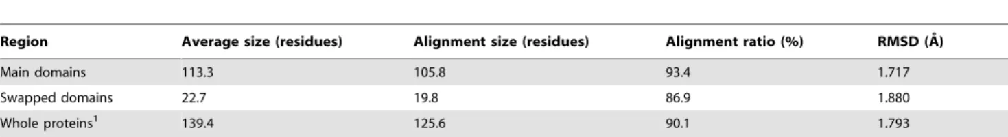
We previously developed a protein structural comparison technique based on angle-distance (A-D) image transformation [38], which has been shown to detect structural similarities between evolutionarily distantly related proteins and to identify structurally similar proteins with different connectivities of secondary structural elements (SSEs) [38]. The A-D image is first constructed based on protein secondary structural information, and is then separated into three different sub-images focusing on various types of SSEs. Structural similarities can subsequently be identified using modified cross-correlation approaches to recognize specific patterns in the corresponding sub-images of the query and target structures. Our current work finds that homologous proteins with and without DS relationships reflect significantly different patterns in the matched A-D images through SSE matching algorithms (see Fig. 1). This finding confirms the feasibility of applying A-D images to develop automated DS detection procedures, which appear to be highly promising for DS-related researches.
Overview of the Proposed Method
Each point in an A-D image records the angular difference between representative vectors and the distance between the centers of two SSEs in a protein [38]. In this work, as two structures are transformed into corresponding A-D images, the pairings of points from the two images are then analyzed and scored using a pair graph. Following an optimization procedure, the equivalence of SSEs between the two proteins is extracted based on the scores of the identified pairs. These processes, the pair graph analysis and extraction of SSE equivalence, are referred to as ‘‘matching’’. Notably, SSEs in the protein bodies and swapped domains of a DSCO pair can be matched
simultaneously because the matching process does not depend on structural superposition. This feature markedly differs from that of most conventional structural alignment methods, which can only align one region at a time. Exploiting this significant difference allows us to compare the results of the SSE matching with those of a typical protein structural alignment and thus locate the boundary between the protein body and the swapped domain,i.e., the approximate location of the hinge loop. Next, superimposing the two structures based on their protein bodies enables us to measure and normalize the conformational difference between these two structures with respect to the swapped domains into the DS score defined here. Two proteins with a DS score higher than a specific cutoff trained on known data are identified as a DSCOpair. Finally, the superpositions of
users simultaneously, separately or in a fused form. Meanwhile, the location and range of the hinge loop is refined according to the results of structure superpositions based on an improved version of the hinge loop determination algorithm by Eisenberg et al.[1], who created the term 3D domain swapping.
Given the lack of a DS database, we collected many DS cases either reported in the literature or annotated in the Protein Data Bank (PDB) [39] to train and test our DS-scoring system. Additionally, a significantly larger number of DS cases were retrieved and identified manually from the PDB and Protein Quaternary
Structure database [40] to perform more detailed assessments and experiments, including those on the quality and stability of binary classifications, the database independence of the discriminatory model, the performances for various DS types and sequence identities, and the quality of domain-swapped alignments and hinge loop determinations. The results revealed the uniqueness of the proposed method. In all experiments, most MCC (Matthews correlation coefficient), sensitivity and specificity values were considerably greater than 0.80, even when the sequence identities of the examined proteins were lower than 10%. On average, the alignment percentage and root-mean-square distance (RMSD) computed by the proposed method were 90% and 1.8A˚ for a set of 1,211 DS-related pairs of proteins, which is the largest DS dataset available. In addition, the range of hinge loops determined by the proposed method corresponded well to the results of manual inspections. To our knowledge, this work presents for the first time a detection and alignment method specifically developed for 3D domain swapping. The unique evaluation system and developmental processes of the proposed method and some unusual properties of DS for structure/sequence comparison methods that are observed for the first time are described in detail in this report, which concluded with some future perspectives on the post-genomic researches and applications of DS that may be enabled or facilitated by computational methods with high performances.
Results
Feasibility of Conventional Protein Structure Comparison Methods for Detecting the 3D Domain Swapping Phenomenon
Although it has been noticed that conventional protein structural comparison (PSC) methods may not be adequate for detecting DS relationships among proteins [29], extensive evaluations of the DS detection abilities of PSC methods have not been performed, probably due to the limited DS data and no standard evaluation mechanism for DS detection. This experiment involves Dataset L, which consists of literature-derived DS cases and their DS-related homologs, common structural homologs, and structurally non-homologous proteins (see MATERIALS AND METHODS for the preparation procedure). More specifically, the structural alignment ratio, that is, the percentage of structurally aligned/equivalent residues (see Experimental Parameters Sub-section), calculated by FAST [30] was used to gradually filter out common global structural homologs, which are not DS cases in general (see Figure S1 for more information about the composi-tions of the filtered dataset at various structural alignment ratio cutoffs). The classification qualities of various PSC methods based on the structural diversity (S-div) [41], a general structural similarity measure, were therefore monitored over the increasing ratio of indistinguishable cases,i.e., partial structural homologs and DS-related homologs, remaining in the test set by calculating the average MCC, sensitivity and specificity over the five-fold cross-validation. As an extensively adopted measure in machine learning for evaluating the quality of binary classifications, the MCC ranges from 21 (inverse prediction) to 0 (random prediction) to +1 (perfect prediction) and is generally considered to be a balanced measure even if the size of classes varies remarkably.
According to Fig. 2 and Table S2, all of the tested structural comparison methods could well discriminate homologs, both common ones and DS-related ones, from non-homologs (Fig. 2a). They could also well distinguish DS-related homologs from non-homologs (Fig. 2b). Their MCC values were generally greater than 0.80. However, their abilities to distinguish domain-swapped homologs from common homologs were low. The MCC values
were all lower than 0.54, and they declined dramatically as the cutoff of the structural alignment ratio became lower. When the structural alignment ratio cutoff was lower than 98%, the MCC values of most methods approached zero (Fig. 2c). Besides, their specificity values were mostly less than 0.20 (Table S2). Interestingly, those methods with good performance for separating homologs from non-homologs (MCC.0.86) were much weaker at distinguishing DS from common homologs (MCC,0.24) than those methods with relatively poor performance (Fig. 2c). The widely-used protein sequence comparison method BLAST [42] was also employed here for comparison, and its DS-detecting performance was unusually better than many structure-based alignment methods. See the DISCUSSION Section for explanations of these observations.
Performance of the Proposed A-D Image Based DS-detecting Method Combined with Conventional Structural Measures
We supposed that the weakness of most conventional PSC methods at detecting DS relationship lies in the fact that they can only identify the structural similarities of a part, i.e., the main domains or swapped domains, of DS-related proteins (see DISCUSSION for supporting information). Based on this suppo-sition, a delicate A-D image-based PSC procedure was designed to align the structures, identify possible hinge loops and determine the global structural similarities between a DSCOpair (see
MATERI-ALS AND METHODS). The output of this procedure includes a ‘‘virtual structure alignment’’ produced by allowing the two domains of a DSCOpair to be independently rotated and translated
and thus superimposed simultaneously. The alignment size and RMSD based on this virtual alignment are termed the ‘‘virtual alignment size’’ and ‘‘virtual RMSD (vRMSD)’’, respectively. Moreover, most well-defined protein structural similarity measures, e.g., the Q-score [43], S-score [44] and S-div [41], can be computed based on this alignment as well and are hence termed the ‘‘virtual (v)’’ structural similarity measures, such as the vQ-score, vS-score and vS-div. These flexible virtual structural similarity measures are more feasible for describing the structural similarity of DS-related proteins than their conventional rigid versions (see Experimental Parameters Subsection for more information).
This experiment attempts to evaluate this DS-detecting proce-dure and examine the capabilities of various virtual structural similarity measures as suitable discriminators for 3D domain-swapping proteins and common homologous proteins. As shown in Fig. 2d and Table S2, all tested virtual measures adequately discriminated homologous from non-homologous proteins. The obtained MCC values ranged stably between 0.82 and 0.89, with average sensitivities$0.87. When only DS-related homologs were used as the positive data while non-homologous proteins were the negative data, their classification performances were good as well (all MCCs = 0.90+0.03 and all average sensitivities $0.89; see Fig. 2e). However, these measures were not as effective in separating DS-related homologs from common homologs, as evidenced by the relatively low MCC and sensitivity for this part (MCCs,0.80 and average sensitivities#0.88), which declined as the alignment ratio cutoff became lower (Fig. 2f). Because the DS detection power of the S-div calculated by many PSC methods was verified, and most of the MCC values were lower than 0.24 (Fig. 2c), the high MCC values (all MCCs.0.72) achieved by vS-div shown in Fig. 2f confirmed the feasibility of the proposed DS detection procedure.
Definition and Evaluations of a Novel DS Score
designed to assist the determination of the likelihood that the proteins under examination are related by DS. Because conven-tional protein (virtual) structural similarity measures are relatively weak at distinguishing DS-related proteins from other structural homolog types, we hypothesized that a practical scoring system for DS should be defined in a more complex manner in which the different properties of the DS and non-DS homologs revealed by the A-D image-based PSC method could be fully exploited. We observed that the A-D image transformation and matching allowed us to match SSEs from corresponding domains of a DSCO pair (Fig. 1 and the MATERIALS AND METHODS).
However, in an optimized structural superposition using conven-tional alignment algorithms, SSE pairs from the swapped domains were still orientationally and spatially different/distant (examples can be found in Fig. 3 and Fig. 4), implying a large product of the angle and distance (angle|distance, or A-D product) between the matched SSEs. Therefore, we postulate that, in a profile of the A-D product (or ‘‘A?D profile’’ for simplicity) of a DSCOpair, a
high-valued region can be considered as a swapped domain, and the transition zone between a low-valued and a high-valued region represents the location of a hinge loop (see MATERIALS AND METHODS). The existence of a candidate hinge loop can be described by a binary function; in addition, other DS-specific structural properties can be analyzed and quantified. A DS score is defined here by integrating several such properties with the following formulas:
DS score~S0fpg ð1:1Þ
fp~
ml0{1
m0{1
ð1:2Þ
g~ 1, if a hinge loop is detected 0, otherwise
ð1:3Þ
l~ (ch
zcd)ma1, 0ƒlv1
1, l§1
ð1:4Þ
a~m2{msd ð1:5Þ
m0,m1,m2[R; m0w1; m1§1; m2§0 ð1:6Þ
wherem0, m1, andm2 are parameters that can be trained by a
dataset with both known DS and non-DS protein pairs.S0can be
any normalized structural similarity measure ranging from 0 (low similarity) to 1 (high similarity); fp represents an exponential
penalty function that reducesS0for common structural homologs
while maintaining the high value of S0 for domain-swapped
homologs;gdenotes the binary function determined by whether a suspected hinge loop was detected or not. Functionfpconsists of
three DS-specific factors, i.e., the angular difference factor ch,
displacement factorcdand minimal structural diversitymsdfor the
swapped domains, the last of which is defined as,
msd~
RMSD Ne,sd min (No,sd,Nc,sd)
1:5,RMSD~
ffiffiffiffiffiffiffiffiffiffiffiffiffiffi
P
Ne,sd
i~1
d2 i
Ne,sd
v u u u u
t ð2Þ
whereNe,sdrepresents the number of equivalent residues between
two swapped domains, whileNo,sdandNc,sdrefer to the size of the
swapped domain of the open oligomer and closed monomer, respectively. A greater similarity between the two domains implies a smallermsd. Two ‘‘identical domains’’ result in a zeromsd.
TheS0value selected in this work was the vQ-score. As long as a
candidate hinge loop is identified, two proteins can be aligned by the main domains in general (see MATERIALS AND METHODS). In this case, the conformational difference in a DSCO pair can be
viewed as a large swinging movement of the swapped domain, which can be further described as an angular motion resulting in the displacement of all residues in the swapped domain. The angular difference (ch) and average displacement of residues (cd) of the
swapped domains are thus, respectively, determined and normal-ized within a range of 0 to 1. Fig. 3 provides further details.
The DS score has a theoretical minimum value of zero and maximum value of one. According to these algorithms, two proteins with a low structural similarity possess a low DS score owing to their low vQ-score. Interestingly, a pair of common structural homologs with a high structural similarity also has a low DS score as well because itschandcdcannot be large, in addition
to the fact thatgcan be zero when no hinge loop is detected. By using this scoring scheme, only proteins with solid DS relationships yield a high DS score, which greatly facilitates the development of the DS detection method. Indeed, a repeat of the experiment stated in the previous subsection demonstrated that the DS score performed better than most conventional structural similarity measures at separating DS-related homologs from non-homologs (Fig. 2e) and clearly outperformed all conventional measures for distinguishing DS-related from common structural homologs
Figure 2. Performance of various alignment methods and similarity measures in identifying common homologs and/or DS-related homologs.The binary classification performance of several conventional alignment methods in distinguishing homologous from non-homologous structures is shown in (a), while their performances in distinguishing DS homologs from non-homologs and common homologs are shown in (b) and (c), respectively. The results of the binary classification tests for several similarity measures to distinguish homologs from non-homologs are summarized in (d). The results of distinguishing DS from non-homologs and common homologs by those measures are shown in (e) and (f), respectively. In these experiments, which involve Dataset L, a number of known DS-related homologous pairs (Lds), common homologous pairs (Lch)
and non-homologous pairs (Lnh) of protein structures were used as positive or negative data for different purposes. In (a) and (d) both Ldsand Lch
were used as positive data, and Lnhserved as the negative data, in (b) and (e) Ldswas the positive and Lnhwas the negative data, whereas in (c) and (f)
Ldsand Lchwere respectively viewed as the positive and negative data. The x-axes indicate that proteins pairs with globally superimposable
(Fig. 2f). The MCCs were generally .0.80, while the sensitivity and specificity values were all.0.89 (Table S2). As expected, the DS score is relatively weak at binarily classifying homologs and non-homologs (Fig. 2d), for which the MCC was 0.57 at a 100% alignment ratio cutoff and gradually increased to 0.71 as the cutoff decreased to 85%. This unique increase in the performance of the DS score also reveals its specific nature for detecting 3D domain swapping relationships. See DISCUSSION for explanations.
Evaluations of the Proposed Method Using Literature-derived and Manually Identified 3D Domain Swapping Cases
With a novel DS score defined based on the structural properties of DS, this study developed a complete A-D image-based DS detection method (refer to the MATERIALS AND METHODS). To more extensively evaluate this method and perform larger scale experiments in the following context than supported by the current literature-derived data, Dataset M was manually established utilizing the Protein Quaternary Structure database [40]. Similar to Dataset L, Dataset M consists of a number of DS-related homologs, common structural homologs and non-homologs (see MATERIALS AND METHODS). Because the proposed method is a DS detection method, the following experiments were all performed based on the condition that only DS homologs were treated as positive data, whereas common homologs and non-homologs were both regarded as negative data.
The dependency of the proposed method on the collected datasets is evaluated here. The parametersm0,m1, andm2required
in the formulas for the DS score and the discriminatory cutoff for the DS score were first determined by taking Dataset M as the training set, and the proposed method was evaluated by Dataset L. This procedure was then repeated by switching the roles of these two datasets. Receiver operating characteristic (ROC) analyses indicate that, regardless of whether trained or tested by literature-derived DS cases or manually identified DS candidates, the effectiveness of the proposed method remained high. The area under the ROC curve (AUC) in each experiment was greater than 0.95. Moreover, the MCC, sensitivity and specificity values all exceeded 0.80 (see Table S3). As Dataset M is involved in this experiment, in addition to verifying the performance of the proposed method with a larger range of data, the above results demonstrate that a classification made by this method generally corresponds to that by manual examination, which involves a considerable amount of manual labor and time.
The stability of the proposed discriminatory model related to the DS score was tested byk-fold cross-validations, wherekranged from 3 to 10. According to Figure S2, the classification quality of the discriminatory model remained high and stable in both datasets, even though Dataset L was much smaller than Dataset M. Combining these performance data obtained by both the inter-dataset (Table S3) and intra-inter-dataset (Figure S2) training and testing, the feasibility and robustness of the proposed DS detection
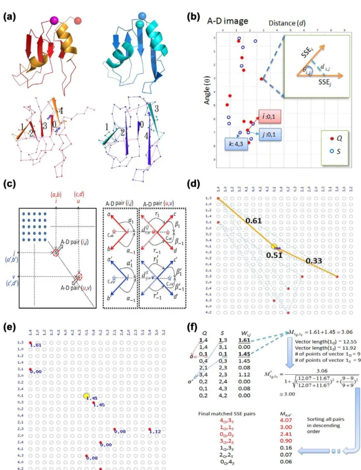
Figure 3. Measurement and normalization of the angular difference and displacement of swapped domains.(a) Structure superposition of a bona-fide DSCOpair, the monomeric (red; PDB entry 5rsaA) and dimeric (blue; PDB entry 1a2wA) forms of ribonuclease A from bovine. This
superposition was performed with a typical structural alignment algorithm that treats protein structures as rigid bodies. In this case, the main domains of two proteins were superimposed well, but the swapped domains (in dotted ellipses) were not superimposed or aligned because of the great difference in the orientation and position. The hinge loops (shown as thin strands) also could not be aligned because of the different conformations. (b) Computation of the normalized angular difference (ch). Using vector transformation techniques, the swapped domains can be represented as two vectors,vcandvo. The anglehbetweenvcandvocan be determined based on the law of cosines. Thechof two swapped domains is thus normalized as
h/180u. (c) Computation of the normalized average residue displacement (cd). In the process of hinge loop detection, the equivalent residues of two
swapped domains,e.g.,aanda9, were determined (see MATERIALS AND METHODS). Thecdwas calculated by dividing the average Euclidean distance of
all equivalent residue pairs (davg) bydmax,, which is the length of the diagonal of the virtual box defined by the boundary of two swapped domains.
methods was confirmed again. In this report, all experiments described hereafter were performed with a discriminatory model trained using all data from Datasets L and M.
Quality of Structural Alignments
Because the proposed method is the first DS-specific detection and alignment approach, the quality of its structural alignments may serve as a standard for the evaluation of related future works. There are 1,211 DSCOpairs in total in Datasets L and M, among
which 1,093 pairs could be successfully identified by the proposed
method. The results of the structural alignment, superposition and hinge loop determination for the successful cases are listed in detail in Table S4. In addition, the (structure-based) sequence alignments of these DSCOpairs calculated by the proposed and several other
PSC methods are listed in Table S5. It is notable that our method correctly distinguished main domains from swapped domains for almost all of these cases, such that the primary alignment of every DSCOpair was generally based on the main domains. The average
(structural) alignment ratio and RMSD of the main domains and swapped domains and the average virtual alignment ratio and
Figure 4. 3D domain-swapping homologs with special swapped domains.Given that the proposed method can align two DS-related proteins along the full length, it is more capable of detecting the global structural and sequence similarities of a DSCOpair than conventional PSC
methods. (a) A pair of ribonucleases (RNases) exhibiting DS phenomena at both termini. The RNase A fromBos taurus(PDB entry 1f0vA) and the RNase from human pancreas (PDB entry 1h8xA) are both domain-swapped dimers; however, their swapped domains are respectively located at the C- and N-terminus. Therefore, when they are superimposed in a conventional manner (the upper right region), both termini appear swapped and unaligned, even if the individual terminal domains are structurally similar (superpositions are shown in the lower right region). The virtual superposition made by the proposed method (left) revealed the actual structural similarity between these RNases, which share a sequence identity of 69% calculated based on the structure-based sequence alignment. In this figure, hinge loops determined by our method are highlighted in the alignment text. (b) Structural alignment of the same RNases performed by DALI [32]. The alignment size and sequence identity computed by DALI are clearly smaller than those by the proposed method. (c) Structural alignment of the snake venom protein Aa-X-bp-I (PDB entry 1yttA) and a subtilisin fragment of mannose binding protein A fromRattus norvegicus(SUB-MPB-A; PDB entry 1wt9A), a quasi-domain swapping case. Aa-X-bp-I is a domain-swapped dimer, while SUB-MPB-A possesses a closed conformation. Interestingly, their domain-swapped domains are located in the middle of the structures. The virtual superposition of the whole proteins is shown on the left side followed by superpositions of the main and swapped domains to the right. Comparing these proteins structures by DALI, the alignment ratio, RMSD and sequence identity were 78%, 2.4 A˚ and 10%, respectively. All of these values are worse than those calculated by the proposed method.
vRMSD of whole proteins were calculated and are listed in Table 1, where the alignment ratios were calculated according to Formula3.
Alignment ratio of X~ Ne,X min (Nc,X,No,X)
ð3Þ
whereXcan be the main domain, swapped domain or the whole protein;Nedenotes the number of equivalent residue pairs,i.e., the
alignment size;Ncand Norefer to the number of residues in the
closed-form and open-form homologs, respectively.
On average, 90% of the residues of a DSCO pair could be
aligned with a vRMSD of 1.8 A˚ . The average alignment ratio calculated by the proposed method for main domains were 7% smaller than that for swapped domains. Detailed analyses revealed that the alignment ratios calculated by the proposed method for the two domains of DS-related proteins differed more as the sequence identity decreased, although the differences were not very large (,9%; see Figure S3). The average running time for a pairwise comparison by the web-based Java implementation of the proposed method was 5.3 seconds with a 2.66GHz processor.
Detecting Various 3D Domain Swapping Types
There are three types of DS regarding the location of the swapped domain, i.e., N-terminal-, C-terminal- and middle-domain swapping [2]. This work evaluated the performance of the proposed method for these three DS types by separating all of the DSCOpairs from Datasets L and M into three groups. Table 2
lists the classification sensitivities of the proposed method for these groups. Because the sensitivities were all .0.88, the proposed method performed satisfactorily for all reported DS types.
Effects of Sequence Identity on the Performance of the Proposed Method
As is well known, protein structural homologs may have low amino acid sequence identities [45]. This phenomenon is also observed in 3D domain-swapping proteins [2,28]. This work evaluated how sequence identity affects the performance of the proposed method by classifying all protein pairs from Datasets L and M into ten groups with decreasing levels of sequence identity
and then examining the quality of the binary classification and structural alignment of the proposed method. According to Table 3, the MCC, sensitivity and specificity varied slightly and still remain high as the sequence identity reduced. Even when the identities were lower than 10%, the values of MCC, sensitivity and specificity remained at the level of 0.83, 0.81 and 0.98 on average for DS detection. The alignment ratio and RMSD also varied with the sequence identity; however, because the variations were moderate, and it is reasonable that evolutionarily more distantly related proteins develop more structural differences, these results might just reflect the nature of the aligned proteins and should not be considered as a decrease in performance. Although it appeared that the decreasing sequence identity had minor influences on the performance of the proposed method, we did observe that the alignment qualities of this method became a little unbalanced as the identity decreased. As shown in Figure S3, the average difference of the alignment ratios calculated by this method for the two domains in a DSCOpair with ,10% sequence identity was
9%. It is not clear yet whether this unbalance revealed a natural property of DS or whether it stood for a decrease of alignment quality; nevertheless, this extent of unbalance brought little effect on the DS detection power of the proposed method for proteins with low sequence identities, as demonstrated in Table 3. Notably, in accordance with Table 3,,40% of the DSCOpairs identified in
this study shared less than 20% sequence identities. The DS relationships of these low-identity DSCO pairs would normally
escape detection by conventional structure/sequence comparison methods (see Figure S3). More importantly, by referring to Table S4,,25% of these low-identity DSCOpairs involved hypothetical
proteins with unknown or putative functions. These facts imply that the proposed method can be applied to suggest possible functions for functionally-unknown or hypothetical proteins, which are increasing rapidly because of many high throughput structural genomics efforts.
Identification of Hinge Loops
The hinge loop is the region linking the main domain and the swapped domain, and it is barely aligned in the structural alignment of a DSCOpair [1]. Eisenberget al.proposed that, after
the approximate location of a hinge loop is assigned as the segment
Table 1.Average alignment size and RMSD of all available DSCOpairs calculated by the proposed method.
Region Average size (residues) Alignment size (residues) Alignment ratio (%) RMSD (A˚ )
Main domains 113.3 105.8 93.4 1.717
Swapped domains 22.7 19.8 86.9 1.880
Whole proteins1 139.4 125.6 90.1 1.793
1
The alignment size/ratio and RMSD between whole proteins were calculated as the average virtual alignment size/ratio and average vRMSD. See the text for the definition of the virtual measures.
doi:10.1371/journal.pone.0013361.t001
Table 2.Sensitivity for the detection of various DS types.
DS type (swapped domain) Total No. (pairs) No. of true positive predictions (pairs) Sensitivity
N-terminal 433 384 0.887
C-terminal 676 614 0.908
Middle 102 91 0.892
not superimposable between the open and closed homologs, its full length can be determined by extending both ends to include residues with large phi (Q), psi (y) differences until two consecutive residues have a torsion angular difference smaller than a cutoffh0
(see Formula10), which was empirically set as 20ufor bona fide and 30ufor quasi domain swapping in their report [1].
While attempting to develop a fully automated DS-detecting procedure, this work designed a novel method for identifying the location and range of hinge loops by improving Eisenberg’s method [1,2] with a procedure dependent on the information extracted from the A-D image and A-D image-based structural alignments (see MATERIALS AND METHODS). Table 4 compares the hinge loops identified by the improved method with those reported in [2]. The automated determinations correlated well with those semi-manual identifications. The average difference in length between the hinge loops determined by the improved and original methods was small (1.4 residues), and the centers of the determined hinge loops only differed by 0.8 residue on average. However, the original method tended to lengthen the hinge loop identifications. For circumstances in which the two methods did not correlate, only five were determined longer by the improved method than by the original one; meanwhile, the original method determined longer hinge loops than the proposed method in 19 cases. For a detailed comparison between the two methods, a larger-scaled experiment than the above one was performed by computing the locations of the hinge loops of all DSCOpairs available in Datasets L and M. According
to Table S4, the improved version of Eisenberg’s method determined the ranges of the hinge loops more strictly than the original method in general. The hinge loops determined by the improved method were shorter than those calculated by the original formula (Formula10) by 51% and 35% on average when h0was set as 20uand 30u, respectively. A close examination of the
determined hinge loops revealed that by using either 20uor 30uas the cutoff, Formula10 is likely to over-extend the boundary of hinge loops. Calculated hinge loops with obviously excessive or insufficient ranges judged by manual verifications are highlighted in this table. See the DISCUSSION Section for more information.
Implementation and Illustrative Examples of Structural Alignments
The proposed DS-detecting method has been implemented as a web-based tool [http://ADiDoS.cs.nthu.edu.tw/]. The basic
output on the web interface includes graphical and interactive Jmol [46] objects for the superpositions of the input protein structures, a table listing the detailed results of the structural alignments, and the DS score. If a DS relationship is identified, superpositions of the main and swapped domains and a virtual superposition of the whole proteins are generated. Additionally, the determined range of hinge loops and some novel structural measures defined in this study, such as the virtual alignment size, vRMSD and vS-div, are also provided. The alignment results of two DSCOpairs identified in this work performed using the web
interface are shown in Fig. 4. In the case of ribonucleases, our method precisely detected their overall structure and sequence similarities whereas DALI [47] only aligned them partially. As for the Aa-X-bp-I, a snake venom protein fromAgkistrodon acutus, and SUB-MPB-A, a subtilisin fragment of mannose binding protein A from the rat, their DS relationships were well identified by the proposed method even though their overall sequence identity is only 12%.
Discussion
Difficulties in DS-detection for Conventional Alignment Approaches
The fact that conventional protein structural comparison (PSC) methods are weak at specifically identifying domain-swapped homologs implies that detecting DS relationships is a very different problem from detecting common structural similarities between proteins. As shown in Fig. 2c, at the 100% alignment ratio cutoff, which actually means that no protein was filtered out from the testing set, the best MCC value achieved was only 0.54 by SARST [34]. Because a lower alignment ratio cutoff means a more thorough exclusion of proteins with global similarities, the dramatic decline of the MCCs of the tested methods suggests that they are much less able to distinguish between DS homologs and other homolog types remaining in the testing dataset, which might be ‘‘partial homologs’’ (proteins with only local structural similarities) or ‘‘low-similarity homologs’’ (proteins with similar overall topologies but large displacements of the corresponding residues/SSEs). Difficulties in detecting DS relationships for conventional structural alignment approaches originate in the nature of the algorithms. A common goal of PSC is to determine a possible largest set of equivalent residues accompanying the possible smallest RMSD of a superposition. This can be visualized
Table 3.Performance of DS-detection over various sequence identities.
Identity (%) No. of DS cases (pairs) No. of non-DS cases (pairs) MCC Sensitivity Specificity Alignment ratio1(%) RMSD1(A˚ )
0–10 242 2478 0.825 0.810 0.988 85.824 2.339
10–20 253 1284 0.864 0.893 0.976 88.237 2.178
20–30 216 456 0.902 0.944 0.963 90.983 1.955
30–40 126 190 0.901 0.929 0.968 92.788 1.613
40–50 67 64 0.783 0.806 0.969 96.164 1.532
50–60 14 56 0.863 0.857 0.982 97.366 1.294
60–70 38 42 0.836 0.816 1.000 98.989 1.127
70–80 52 26 0.972 0.981 1.000 98.038 1.199
80–90 69 40 1.000 1.000 1.000 94.391 0.957
90–100 134 195 0.987 0.993 0.995 99.400 0.825
1The alignment ratio and RMSD listed here are the average virtual alignment ratio and average vRMSD. Only DS cases with true positive predictions were included in the
as if one structure were translated and rotated with respect to the other to make as many residues aligned and as close as possible. Under this circumstance, the protein structure is treated as a rigid body that always moves as a whole. However, the two objectives, a large number of aligned residues and a small RMSD, cannot be achieved simultaneously in a DSCO pair due to a significant
orientational difference between the swapped domains. A situation in which a method highly prioritizes RMSD implies its feasibility in detecting local structural similarities, but it is subsequently less applicable to the detection of DS relationships. Among the PSC methods assessed in the experiments of Fig. 2c that treated protein structures as rigid bodies, TM-align performed best. An
Table 4.Comparison of manually examined hinge loops and hinge loops identified by the proposed method.
Closed form Open form
Hinge loops examined by [2]
Hinge loops identified by the proposed method
Length difference of hinge loops1(residues)
Shift of the centers of hinge loops2(residues)
Rangee* Lengthe Rangei Lengthi
1msbA 1ixxA 72-75 4 72-80 9 25 2.5
2ezmA 3ezmA 50-53 4 49-54 6 22 0
1hz5A 1jmlA 52-55 4 52-56 5 21 0.5
1wwwX 1wwbX 299-301 3 297-300 4 21 1.5
1orcA 5croA 55-55 1 55-56 2 21 0.5
1orcA 5croA 55-56 2 55-56 2 0 0
1sncA 1sndA 112-120 9 112-120 9 0 0
1mupA 1obpA 126-130 5 126-130 5 0 0
1fynA 1aojA 112-118 7 112-118 7 0 0
1sncA 1sndA 112-120 9 112-120 9 0 0
1mupA 1obpA 121-124 4 121-124 4 0 0
1fynA 1aojA 34-49 16 34-49 16 0 0
1brnL 1yvsA 37-41 5 37-41 5 0 0
5rsaA 1bsrA 15-22 8 15-22 8 0 0
1wwwX 1wwaX 297-299 3 296-298 3 0 1
1dksA 1cksA 60-65 6 60-65 6 0 0
1msbA 1ixxA 93-98 6 95-100 6 0 2
1griA 1fyrA 121-123 3 121-123 3 0 0
1nloC 1aojA 34-39 6 34-39 6 0 0
1mdtA 1ddtA 379-387 9 379-386 8 1 0.5
1qmpA 1dz3A 103-109 7 106-111 6 1 2.5
1wwwX 1wwcA 317-319 3 316-317 2 1 1.5
1k3sA 1k3eA 33-36 4 34-36 3 1 0.5
1eydA 1sndA 112-120 9 113-120 8 1 0.5
1pv3A 1k04A 943-948 6 943-947 5 1 0.5
1qd0A 1sjvA 95-100 6 94-98 5 1 1.5
1cunA 2spcA 72-75 4 72-73 2 2 1
1gmfA 1hulA 87-99 13 87-97 11 2 1
1qlxA 1i4mA 188-198 11 189-197 9 2 0
5rsaA 1f0vA 112-115 4 112-113 2 2 1
5rsaA 1js0A 112-115 4 112-113 2 2 1
1cewI 1g96A 55-59 5 57-59 3 2 1
5rsaA 1a2wA 15-22 8 18-22 5 3 1.5
1a5pA 1a2wA 15-22 8 18-22 5 3 1.5
1gmfA 1hulA 82-89 8 82-85 4 4 2
1hngA 1cdcA 44-50 7 44-46 3 4 2
4icbA 1ht9A 38-47 10 41-45 5 5 0.5
4gcrA 1blbA 79-87 9 86-87 2 7 3.5
Unsigned average 1.4 0.8
*According to [2], this range was determined for the protein indicated by the bold italic text. 1The length difference of the hinge loops was calculated as Length
e– Lengthi. 2This shift was calculated as the distance between the center of Range
eand the center of Rangei.
experiment based on all identified DS cases listed in Table S4 and Table S5 was performed to determine the simultaneous alignment quality of TM-align on both domains of DSCOpairs. Figure S3
clearly verifies that for most DSCOpairs TM-align could only align
one domain and thus could not detect their global structural similarities. On the other hand, despite the availability of several more flexible PSC methods that do not completely treat protein structures as rigid bodies and may detect the global structural similarity of 3D domain-swapping proteins by determining more aligned residues, their scoring systems prevent them from distinguishing between DS-related homologs and common structural homologs. Take SARST for example. It is a PSC method working based on a structural linear encoding method-ology [34]. By transforming protein local backbone conformations into a conformational alphabet [34], it converts geometric structural comparison problems into string comparison problems that can be solved by conventional sequence alignment algorithms. As a hybrid, SARST was almost as precise as conventional PSC methods like CE in structural similarity searches while at the same time it possessed some properties of sequence alignment methods such as the high running speed [34]. Just like a sequence alignment algorithm, as SARST aligns structural strings, some minor local differences will result in mismatches and/or gaps but will not terminate the alignment as long as the score reduction effects of those local differences can be compensated by the score increasing effects of nearby string similarities. Besides, SARST does not consider the RMSD of structure superposition during its alignment but focuses completely on local backbone conforma-tional similarities. As a result, when SARST deals with DSCO
pairs, in many cases the hinge loop will only cause some gaps but not prevent it from simultaneously aligning the main and swapped domains (see Figure S3 and Table S5 for experimental results demonstrating this property). These algorithmic features of SARST were the reasons that it could detect the overall structural similarity of many DS-related proteins (Fig. 2c). For a DSCOpair and a pair of
common homologs, when SARST reports similar alignment ratios for both cases, usually the RMSD of the former will be much larger than that of the latter, resulting in very different structural diversity (S-div) [41] values. In many DS cases, the RMSD values reported by SARST can be larger than 12 A˚ (,28% in Table S5), a very
extreme value that most conventional PSC methods barely report. For instance, the highest RMSD reported by TM-align in Table S5 was only 4.1 A˚ . Although this extreme difference in RMSD and S-div values has made SARST more capable of detecting DS relationships than most conventional PSC methods, it is not extreme enough to efficiently distinguish between DS-related and common homologs. As compared with SARST, the DS scoring system of the proposed method gives DS-related and common homologs much more different scores. A bona fide DSCOpair usually has a DS score
around 1 while a pair of common global homologs possesses a DS score close to 0 (note that the range of DS score is between 0 and 1 by definition).
Although it is normally considered that structure-based alignment methods are better than sequence-based ones at detecting protein structural similarities [45], very interestingly, in the case of DS-detection, the widely-used sequence alignment method BLAST outperformed most PSC methods, especially when the cutoff of structural alignment ratio (calculated by FAST [30]) was lower than 95%, forming an MCC curve with very different tendency from those of the PSC methods in Fig. 2c. This novel discovery also resulted from the nature of the alignment method. Sequence alignment does not consider any 3D structural information and is thus not affected by the conformational difference between closed and open homologs in the comparison
processes. Similar to the situation shown by SARST, in many cases hinge loops only cause gaps but do not terminate the alignment. Provided that there are sequence similarities detectable by BLAST both in the main and swapped domains, a global alignment can be made and scored. As shown in Table S5 and Figure S3, for those DS-related proteins with global sequence identities lower than 20%, in most cases BLAST aligned them with only one domain. As the sequence identity increased, more and more protein pairs were aligned with two domains. At sequence identities$20%, the alignment ratio calculated by BLAST for the two domains of domain-swapped proteins differed less than 30%; at sequence identities $50%, the difference reduced to ,10%. Although BLAST is capable of making global alignment for many DSCO pairs, unfortunately it makes no difference to BLAST
whether a high alignment score is achieved by a DSCOpair or by a
globally superimposable pair of common homologs. In the experiment of Fig. 2c, at a high structural alignment ratio cutoff, only those DSCOpairs with small swapped domains and common
homologous pairs with a small number of non-superimposable residues were eliminated. Thus, there were still many highly globally superimposable homologs, which were not distinguishable by BLAST from the DSCOpairs, remaining in the testing set. As a
result, the MCC of BLAST was only ,0.3. However, as the
alignment ratio cutoff decreased, the number of common global homologs in the testing set decreased much more rapidly than that of DSCOpairs (refer to Figure S1) and the remaining DSCOpairs
maintained high scores that became relatively higher and higher than the scores of the partial and low-similarity homologs remaining in the testing set. Subsequently, BLAST registered higher MCC values at lower structural alignment ratio cutoffs than at the high cutoffs, and it achieved the highest MCC of 0.46 at the 85% cutoff; nevertheless, it was still inadequate to serve as an accurate DS-detecting method.
Crucial Factors for the DS-detecting Ability of the Proposed Method
ratio cutoff decreased, the relative amount of DS-related homologs remaining in the testing set increased, while that of the common homologs decreased. Because the DS score is specifically designed for detecting DS, its MCC for distinguishing homologs (both DS and common homologs) from non-homologs is supposed to increase as the alignment ratio cutoff declines. Moreover, by integrating the three DS-specific factors, which describe the angular difference (ch), spatial displacement (cd) and structural
similarity (msd) of the swapped domains (see Formulas1.1to1.6),
the DS score has a theoretical minimum value of zero (no DS relationship) and maximum value of one (definite DS relationship). In addition to simplifying the development of an automated procedure, this normalized score also offers users an easy way to recognize the DS relationship between proteins.
Precision of Hinge Loop Determinations
Both the current two evolutionary mechanisms proposed for DS involve the hinge loop. Deletions may shorten the hinge loop and turn a closed monomer into an open oligomer [24,25]; besides, mutations in the hinge loop and/or the contact interface of domains may promote swapping conditions [26,27]. However, which proposed mechanism plays the major role or whether there is any undiscovered mechanism for DS remain uncertain. Examinations of the lengths and amino acid compositions of hinge loops may help reveal detailed evolutionary mechanisms of DS; additionally, the results of such examinations can provide important information for protein engineering studies utilizing 3D domain swapping. Precise examinations of the hinge loops depend on precise determinations of their positions and ranges. It is conceptually clear to define a hinge loop as the non-superimpos-able region linking the protein body with the swapped domain [1]. However, the numerous factors that can greatly complicate the implementation of this concept include the limitations of conventional PSC methods, effects of sequence identity and lack of robust ways to identify the boundary of a hinge loop. Consequently, manual labor is usually an indispensable factor in determining the location and range of hinge loops.
First, regardless of whether a conventional PSC algorithm is used, including the one utilized by the proposed method, superimposing the main and swapped domains simultaneously such that the non-superimposable portion of a DSCOpair can be
easily identified is very improbable. The proposed method bypasses this difficulty by using the A?D profile along with a morphological smoothing technique (see MATERIALS AND METHODS), which allows the preliminary identification of the hinge loop to be fully automated because the approximate location of a hinge loop can be simply recognized by a sudden decline or increase within the profile.
After the approximate location of a hinge loop is identified, the boundary must be determined. Eisenberget al.suggested extending the hinge loop at both ends until two consecutive residues haveQ, y differences lower than a cutoff (h0). According to their
algorithm, a higher h0 results in a more restricted extension,
allowing us to infer that they reasonably set a higher cutoff for quasi-DSCOpairs (h0= 30u), which are evolutionarily more distant
than bona fide cases (h0= 20u). Such an extension step is assumed
here to be an excellent design and is thus applied as the fundamental hinge loop determination procedure in our DS-detecting system. However, as the discrimination of bona fide and quasi-domain swapping is somewhat empirical as well, manual inspections may be unavoidable to handle ambiguous cases. Although Eisenberg’s algorithm was shown to be feasible based on a dataset of 33 DSCOpairs, according to the large-scale test results
shown in the Table S4, by setting h0 as either 20u or 30u, the
algorithm tended to over-extend the hinge loop of DSCOpairs,
especially for those with low sequence identities. This work also attempts to simplify the requirement for manual examinations and increase the precision of hinge loop identification by, first simply unifying the cutoff for bona fide and quasi-DS cases as 25u
multiplied bynhl, the number of hinge loops detected in the same
swapped domain (see Formula 11). Then, the extension is restricted by a distance constraint of aligned residues in the front of the extending region. Given that the estimated range of hinge loops agrees well with the semi-manually verified ones reported in [2] (Table 4), and the over extension is well prevented (as also shown in the Table S4), we conclude that the proposed procedure is a fully automated, precise, and generally applicable method for hinge loop detection.
Sensitivities to Middle-domain Swapping Cases
The experiment for detecting the three types of DS, N-terminal-, C-terminal-N-terminal-, and middle-domain swappingN-terminal-, discloses the robustness of the proposed method. Although the sensitivities for these three DS types seem evenly high (Table 2), the detection of middle domain swapping is actually more difficult than the other types. It is noteworthy that, as mentioned in the above subsection, a parameter nhl was introduced to help define the boundary
condition (h0) of a hinge loop. Because a middle-swapped domain
has two hinge loops, itsh0is twice as large as that of N-terminal- or
C-terminal-swapped domains, resulting in a situation in which a higher restriction is imposed upon middle-DS than upon other DS types. Without the parameternhl, the hinge loop(s) of a candidate
middle-swapped domain may be determined to be invalid because of its over-extension (see MATERIALS AND METHODS), and thus the candidacy of the swapped domain is incorrectly rejected. Actually, according to our preliminary tests, the sensitivity to middle-DS of the proposed method without nhl was obviously
lower. Two phenomena may explain the difficulty in detecting middle-DS.
First, a higher complexity of the ‘‘swapping movement’’ may hinder the calculation of the orientational difference (estimated by factor ch) between the swapped domains. The swapping of
domains of an N-terminal- or C-terminal-DSCO pair can be
visualized as a swinging movement, during which the single hinge loop might bend and slightly twist, but the overall conformation of the swapped domain is preserved. Differently, the swapping movement of a middle-DSCOpair could lead to some torsion of
the swapped domain, especially when the extents or directions of bending of the two hinge loops differed. In this case, the way we transform the swapped domains into representative vectors (refer to Fig. 3) may not be adequate. To resolve the problem, we plan to design a multiple vector transformation technique to more precisely estimate the orientational difference between swapped domains.
Second, structural dissimilarities between small equivalent swapped domains can blur the boundary of hinge loops. Many of the swapped domains of known middle-DSCOpairs are small
and/or have few regular SSEs (see Table S6), meaning that they are either prone to be affected by the complicated type of swinging movement, or they are structurally quite flexible. Therefore, the structure of these swapped domains, including the hinge loops, may tend to be variable. It was observed that the structures of hinge loops of some small pairs of middle-swapped domains and/ or the middle-swapped domains themselves were very varied that and thus boundaries of hinge loops were greatly over-extended when the parameternhlwas not applied. In some cases, they were
matter how visually apparent the middle-DS relationship between two proteins is, when a candidate middle-swapped domain is overwhelmed, or when the boundary of its hinge loop(s) cannot be determined, its candidacy is inevitably rejected based on our methodology. This is whynhlwas introduced into Formula11to
make the extension step more restricted in the determination process of hinge loops for middle-DS cases.
Conclusions
We have designed the first specific detection method for 3D domain swapping. This pairwise comparison method does not work as anab initiopredictor for DS but determines the existence and type of DS relationship between two given protein structures. The A-D image-based algorithm proposed and the DS score defined here achieved a satisfactory performance,i.e., an average MCC.0.80 in every experiment (Fig. 2, Figure S2 and Table S3). In addition, the robustness of this DS-detecting method have been evidenced by (1) the high true positive prediction rates for all three types of DS (Table 2), (2) the high sensitivity, specificity and structure alignment quality at low sequence identities (Table 3 and Figure S3), and, (3) the high precision of hinge loop determination (Table 4 and Table S4). With good performances, this method can greatly reduce the requirement for manual examinations for the identification of DS relation-ships among proteins, making it possible to develop automated procedures. As revealed by the fact that the structural similarities of DS homologs were prone to be underestimated by conven-tional PSC methods (Fig. 2 and Figure S3), the proposed method may also serve as a functional assignment system for novel hypothetical proteins which escape typical sequence and structural similarity searches/detections. Through several forms of structural alignments, the proposed method can present the actual structural similarities of main domains, swapped domains and the overall structures of DSCOpairs, helping users study the
structural and functional relationships among DS-related pro-teins. Changes in the hinge loops may profoundly impact the formation of 3D domain swapping [24,25]. Thoroughly eluci-dating the hinge loops may help scientists to clarify the evolutionary mechanisms of DS, and the proposed method may be applicable to this field because of its well-developed function for determining the location of hinge loops. At present, several DS-related research fields appear to advance slowly or pause at theoretical stages. There is still much uncertainty about the natural prevalence of DS, the dominance of possible mechanisms for DS and how/why Nature achieves evolutionary diversities and functional regulations of proteins by using 3D domain swapping. Studying DS can deepen our knowledge about the structural dynamics and folding of proteins. Understanding the mechanisms of DS may help find new treatments for several protein conformational diseases like Alzheimer’s disease and bovine spongiform encephalopathy [9,19,21]. Biotechnological applications of DS, such as the production of auto-assembling biomaterials and artificial biopolymers [20,22], also require enough background knowledge. A key to solving those uncer-tainties and facilitating those medical and bioengineering applications shall be a comprehensive DS database. By manually screening only a small fraction of PDB, over a thousand DS cases had been identified in this study. As the number of protein structures is increasing at an unprecedented rate in this post-genomic era, we believe that the proposed method can greatly contribute to retrieving a much larger amount of relevant DS-related data than before from protein structural databases and thus move related fields forward.
Materials and Methods
The experiments were performed using a Linux computer with a 2.66GHz Intel processor and 4 GB RAM. The source of protein structure files was a snapshot of the protein data bank (PDB) from August 2008. Specifically, the 90% sequence identity non-redundant subset of this PDB snapshot (abbreviated as nrPDB-90) was used. Programs were written in the Java, Asp.net and PHP languages. The structures shown in the figures were rendered using PyMol [48], Jmol [46], or Java OpenGL [49].
Preparation of Experimental Datasets
This work established two main datasets for the 3D domain swapping, one based on literature-derived information (Dataset L) and the other based on manual inspections (Dataset M).
Dataset L. The ‘‘bona fide’’ and ‘‘quasi-domain swapping’’ DSCOpairs summarized in [2], the domain-swapped dimers listed
in [37] and a number of PDB entries with 3D domain swapping annotations located by keyword searches were collected into a primary dataset consisting of 263 proteins. Additional relevant data was retrieved by using each protein in this primary dataset as a query to search nrPDB-90 for DS-related homologs, common structural homologs and non-homologous structures following the procedure below:
(1) For each proteinQ, a rapid protein structural similarity search service,iSARST, was applied to search against nrPDB-90 for its structural neighbors with an E-value cutoff of 10 [50]. (2) ProteinQand any protein Sretrieved byiSARST in the hit
list were defined as a neighboring structural pair (NS pair). (3) DaliLite v.3 [47] was utilized to perform structure
superpo-sition and to compute the Z-score of every NS pair. (4) According to the suggestion of [47], NS pairs with a Z-score
$2 were provisionally considered as homologous structural pairs, which were classified into ten groups with decreasing sequence identities: 100–90%, 90–80%, 80–70%, etc. (5) Each NS pair belonging to the ten groups was then carefully
examined by manual inspection. A DS relationship was identified when structural complementarities were observed. For instance, assume thatQis an open-form dimer; whenS was found to have a similar structure to the known closed monomer ofQ,QandSwould be considered a DSCOpair.
(6) After a DSCOpair was identified, homologs of the closed and
open forms were paired and examined to identify additional DSCOpairs.
(7) While all identified DSCOpairs were selected into a dataset
Lds, those homologous structural pairs without DS
relation-ships were randomly selected into another dataset Lch, where
the subscript ch refers to ‘‘common homologs’’.
(8) NS pairs with a Z-score,2 were generally considered as non-homologous (nh) structures, from which some were randomly selected into dataset Lnh.
(9) Finally, Dataset L consisted of datasets Lds (737 pairs), Lch
(499 pairs) and Lnh(720 pairs). Dataset S1 provides a full list.