Hypermetabolism and Clinical Outcomes
Marc G. Jeschke1,2,5., Felicia N. Williams1,2., Celeste C. Finnerty1,2,3., Noe A. Rodriguez1,2 , Gabriela A. Kulp1,2, Arny Ferrando4, William B. Norbury1,2, Oscar E. Suman1,2, Robert Kraft1,2, Ludwik K. Branski1,2, Ahmed M. Al-mousawi1,2, David N. Herndon1,2*
1Shriners Hospitals for Children, University of Texas Medical Branch, Galveston, Texas, United States of America,2Department of Surgery, University of Texas Medical Branch, Galveston, Texas, United States of America,3Institute for Translational Sciences and the Sealy Center for Molecular Medicine, University of Texas Medical Branch, Galveston, Texas, United States of America,4University of Arkansas for Medical Sciences, Little Rock, Arkansas, United States of America,5Sunnybrook Health Sciences Centre, University of Toronto, Toronto, Ontario, Canada
Abstract
Background:Hypercortisolemia has been suggested as a primary hormonal mediator of whole-body catabolism following severe burn injury. Ketoconazole, an anti-fungal agent, inhibits cortisol synthesis. We, therefore, studied the effect of ketoconazole on post-burn cortisol levels and the hyper-catabolic response in a prospective randomized trial (block randomization 2:1).
Methodology/Principal Findings:Fifty-five severely burned pediatric patients with.30% total body surface area (TBSA) burns were enrolled in this trial. Patients were randomized to receive standard care plus either placebo (controls, n = 38) or ketoconazole (n = 23). Demographics, clinical data, serum hormone levels, serum cytokine expression profiles, organ function, hypermetabolism measures, muscle protein synthesis, incidence of wound infection sepsis, and body composition were obtained throughout the acute hospital course. Statistical analysis was performed using Fisher’s exact test, Student’s t-test, and parametric and non-parametric two-way repeated measures analysis of variance where applicable. Patients were similar in demographics, age, and TBSA burned. Ketoconazole effectively blocked cortisol production, as indicated by normalization of the 8-fold elevation in urine cortisol levels [F(1, 376) = 85.34,p,.001] with the initiation of treatment. However, there were no significant differences in the inflammatory response, acute-phase proteins, body composition, muscle protein breakdown or synthesis, or organ function between groups.
Conclusions:Both groups were markedly hypermetabolic and catabolic throughout the acute hospital stay. Normalization of hypercortisolemia with ketoconazole therapy had no effect on whole-body catabolism or the post-burn inflammatory or hypermetabolic response, suggesting that hypercortisolemia does not play a central role in the post-burn hypermetabolic catabolic response.
Trial Registration:ClinicalTrials.gov NCT00675714; and NCT00673309
Citation:Jeschke MG, Williams FN, Finnerty CC, Rodriguez NA, Kulp GA, et al. (2012) The Effect of Ketoconazole on Post-Burn Inflammation, Hypermetabolism and Clinical Outcomes. PLoS ONE 7(5): e35465. doi:10.1371/journal.pone.0035465
Editor:Ioannis P. Androulakis, Rutgers University, United States of America
ReceivedFebruary 2, 2011;AcceptedMarch 19, 2012;PublishedMay 11, 2012
Copyright:ß2012 Jeschke et al. This is an open-access article distributed under the terms of the Creative Commons Attribution License, which permits unrestricted use, distribution, and reproduction in any medium, provided the original author and source are credited.
Funding:This study was supported by grants from Shriners Hospitals for Children (8660, 8490, 8640, 8760, and 9145), National Institutes of Health (2T32-GM008256-11, 1P50-GM060338-01, R01-GM56687, R01-HD049471, U54-GM062119, R01 GM087285-01), National Institute on Disability and Rehabilitation Research (H133A020102, H133A70019), and the American Surgical Association. The funders had no role in study design, data collection and analysis, decision to publish, or preparation of the manuscript.
Competing Interests:The authors have declared that no competing interests exist.
* E-mail: [email protected]
.These authors contributed equally to this work.
Introduction
The hypermetabolic response to a severe burn, defined as a burn encompassing greater than 40% of the total body surface area (TBSA), evokes a catabolic state that persists long after the initial insult [1]. This response is characterized by futile substrate cycling, increased oxygen consumption, glycogenolysis, proteoly-sis, and lipolysis [1–3]. The primary mediators of this deleterious response have been thought to be endogenous catecholamines and cortisol [1,4]. Urine cortisol levels are elevated 7–10 fold after severe burn and remain elevated beyond the acute phase [4,5].
levels of cortisol activate glucocorticoid receptors, which terminate the release of ACTH [9]. After severe burn injury, the negative feedback loop between cortisol and ACTH is deranged [10]. This physiologic, metabolic disruption leads to persistent elevations in cortisol in severely burned pediatric patients.
Ketoconazole is an imidazole anti-fungal agent that has been shown to diminish steroid synthesis by blocking P450-dependent enzyme systems [11,12]. It has been shown to be effective in reducing both stimulated and basal cortisol secretion in both normal and Cushing’s (hypercortisolemic) patients [13,14]. Keto-conazole also reduces the incidence of acute respiratory distress syndrome (ARDS) in critically ill patients and shortens length of ICU stay [15]. Preliminary studies in adult burn patients have shown that urinary cortisol excretion is decreased after one week of ketoconazole administration [8]. Although patients do not have completely normal urinary cortisol excretion, they have reduced muscle protein turnover and improved muscle protein balance [8]. The effect of ketoconazole administration on immune function, organ function, or hormonal balance has not been fully investigated in this patient population. Moreover, to our knowl-edge, the effect of inhibiting excess cortisol production has not been evaluated in the pediatric burn population. We hypothesized that the administration of ketoconazole to block excess cortisol production in severely burned pediatric patients during the acute hospitalization would attenuate inflammation, hypermetabolism, and protein wasting.
Results
Demographics
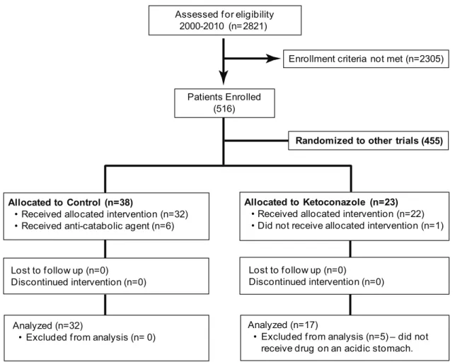
Two-thousand eight-hundred twenty-one patients were assessed for eligibility to be enrolled in our research studies. We enrolled 516 patients (Fig. 1, Consort Diagram), 455 of whom were enrolled in studies of other anti-catabolic agents. Of the 38 patients allocated to placebo, 6 were excluded because they received anti-catabolic agents, leaving 32 patients in the standard of care/placebo group. Of the 23 patients randomized to ketoconazole, 6 were excluded. One patient did not receive the drug, while 5 were given drug under non-optimal conditions, leaving 17 patients that received standard of care plus ketocona-zole treatment [per os (p.o.) on an acidic stomach] in the treatment group. There were no differences in age, gender, length of ICU stay, burn size, third-degree burn, length of ICU stay per percent burn, number of required operations, or time between operations between groups (Table 1). Incidence of inhalation injury was comparable in both groups (Table 1). Ketoconazole administra-tion did not decrease the incidence of pneumonia, sepsis, or multi-organ failure (MOF) (Table 1).
Cytokines, Hormones, and Proteins
Urinary cortisol was increased 8 fold after severe burn injury (Fig. 1). Ketoconazole treatment decreased urinary cortisol
excretion to normal levels [F(1, 376) = 85.34,p,.001]. This effect
was evident by post-burn day 8 [F(7, 376) = 8.21,p,.001] (Fig. 2).
Catecholamine levels were significantly elevated after severe burn. Ketoconazole treatment had no effect on urine catecholamine
levels [F (1, 329) = 1.08,p= .30] (norepinephrine shown in Fig. 3).
Confirming previous studies, we found that severe burn injury induced a vast inflammatory response. Ketoconazole did not alter any of the 17 serum cytokines measured (not shown). Serum acute-phase proteins were physiologically deranged throughout the acute hospitalization, but were not different based on treatment group. There were no differences in serum IGF-1, IGFBP-3, or rhGH between the groups. There were no differences in estrogen, free or
bound testosterone, or free or bound progesterone levels between the groups. There were no significant or sustained differences in
serum complement C3, a2-macroglobulin, haptoglobin, or
C-reactive protein. Ketoconazole had no effect on triglycerides or free fatty acids.
Adrenocorticotropic hormone (ACTH or Cosyntropin) chal-lenge tests showed no differences in responses in either patient group (Table 2). Both treatment groups were adrenally competent and responded to the challenge according to established guidelines [16].
Indirect Calorimetry
As previously reported, burn injury increases resting energy expenditure (REE), indicating a vast hypermetabolic response. In this study, control patients demonstrated a decrease in REE, as
predicted by the Harris-Benedict equation, from 14867%
predicted at admission to 13967% predicted at discharge (delta
29% REE) (Fig. 4a). Ketoconazole-treated patients exhibited an
increase in predicted REE from 143611% at admission (before
treatment) to 144611%, although this was not statistically
significant (delta +1% REE), indicating that ketoconazole
treat-ment had no effect on REE from admission to discharge when compared to controls.
Muscle Protein Synthesis
Stable isotope infusion studies were used to measure muscle protein synthesis and breakdown to determine net protein balance. Burned children had a negative net protein balance in skeletal muscle at the time of the first study, at one week post admission. Peripheral muscle catabolism further increased towards the second study, which was conducted around 3–4 weeks post admission. There were no differences between control and ketoconazole for net protein balance, protein synthesis, or the fractional synthetic rate of muscle protein synthesis (Fig. 4b).
Body Composition
Severe burn causes marked changes in body composition during acute hospitalization. Both control and ketoconazole patients experienced a 1% loss in lean body mass (LBM) during the study period. Control patients lost 2% of bone mineral density (BMD) and 4% of bone mineral content (BMC), while ketoconazole-treated patients lost 4% of their BMD and 8% of BMC (Fig. 3c). Control patients experienced 6% gains in whole-body fat, though this was not significantly different than the 9% gains in whole-body fat experienced by drug-treated patients (Fig. 3c).
Organ Function
In the control group, liver size markedly increased after injury. Ketoconazole did not attenuate this increase in liver size compared to the control treatment (data not shown).
Predicted cardiac output (CO), cardiac index (CI), predicted heart rate (HR), predicted stroke volume (SV), and cardiac work were altered in burn patients. Ejection fraction (EF) was preserved in severely burned pediatric patients. Ketoconazole had no effect on predicted CO, CI, predicted HR, or predicted SV. Ketoco-nazole treatment had no effect on cardiac function (data not shown).
Discussion
Major thermal injury is followed by a profound catabolic response that persists for years after injury [1]. This catabolic state after injury leads to increased risk for infection, severe muscle wasting, morbidity, and mortality. The response has been thought to be mediated by increased plasma catecholamines, glucagon, and cortisol [4]. If left unabated, patients can have severe cardiac stress, insulin resistance, and persistent derangements in structure and function of vital organs [1,4,17]. It has been postulated that excess cortisol, compounded by prolonged muscular inactivity, increases skeletal muscle protein breakdown in this patient population [18–21]. This study was designed to reveal whether ketoconazole treatment attenuates the hypercatabolic and inflam-matory response to severe burn trauma by decreasing cortisol synthesis. The principal findings in this study were that ketoconazole successfully decreased the excretion of urinary cortisol. However, it did not improve the hypermetabolic or catabolic condition of the patient population studied.
Ketoconazole blocks the 14-demethylation of lanosterol, thus preventing its conversion to cholesterol [12,22]. It has been shown to block steroid production and cortisol production [12]. Based on the results of this study, it may be incorrect to say that ketoconazole blocks steroid production, but rather, it interferes with steroid synthesis [23]. Forty-nine patients were included in this study, with 17 receiving ketoconazole enterally at a dose of 5 mg/kg every 12 h. These 17 patients had a significant decrease in urinary cortisol excretion with ketoconazole treatment when
compared to controls. Serum cortisol levels were not affected by ketoconazole treatment (data not shown). Of note, the normal circadian rhythm for serum cortisol is physiologically deranged and lost after severe burn injury, making serum cortisol an inaccurate measure of the total daily production of cortisol in our patients [24,25]. Instead, we measured urine cortisol, a more reliable measurement of the HPA axis in our patient population [26]. There were no significant differences in the patient demographics in the two arms of the study. Adrenal function was assessed in all patients involved in the study by ACTH challenge tests. None of the patients in this study had clinical signs or symptoms of adrenal insufficiency (no response to ACTH challenge tests). Hypercortisolemia has been shown to contribute to reduced T-helper lymphocyte proliferation and immunocom-petence [27]. Here, patients receiving ketoconazole did not have a
lower incidence of minor infections (p..05). In addition, though
ketoconazole is an anti-fungal agent, there were no significant differences in the numbers of patients with sepsis or MOF.
Ketoconazole treatment did not decrease REE and thus, hypermetabolism. Both patient populations had REE that was significantly higher than normal values throughout the study period. Excess cortisol has been linked to increases in REE [5,18]. In this study, muscle protein catabolism was elevated in control
burn patients (237657 nmol/100 ml leg/min). In the
ketocona-zole-treated burn patients, cortisol was reduced to normal levels,
but muscle protein catabolism remained elevated (236654 nmol/
100 ml leg/min). Muscle protein synthesis is also elevated in burn patients to compensate for the increased catabolism, but the net Assessed f or eligibility
2000-2010 (n= 2821)
Allocated to Control (n=38)
• Received allocated intervention (n=32) • Received anti-catabolic agent (n=6)
Allocated to Ketoconazole (n=23)
• Received allocated intervention (n=22) • Did not receive allocated intervention (n=1)
Lost to f ollow up (n=0)
Discontinued intervention (n=0)
Lost to f ollow up (n=0)
Discontinued intervention (n=0)
Analyzed (n=32)
• Excluded f rom analysis (n= 0)
Analyzed (n=17)
• Excluded f rom analysis (n=5) – did not receive drug on an acidic stomach.
Enrollment criteria not met (n= 2305)
Patients Enrolled (516)
Randomized to other trials (455)
Figure 1. Consort Diagram.
muscle catabolism was similar with or without ketoconazole
treatment (259641 vs.224623 nmol/100 ml leg/min, p= .57).
The data suggest that the increase in muscle catabolism seen with severe burn injury is not mediated by elevated cortisol levels. Table 1.Patient demographics and the effect of ketoconazole.
Control (n = 32) Ketoconazole (n = 17) p,.05 pvalue
Length of ketoconazole administration (days) NA 3468* S ,.001
Demographics
Age (yrs) 961 761 NS .35
Gender (F/M) 11/21 3/14 NS .22
XHispanic (%) 94 76 NS .16
Caucasian (%) 6 24 NS .16
Time to admission (days) 461 461 NS .65
LOS ICU survivors (days) 3564 4067 NS .48
TBSA burned (%) 5763 6366 NS .19
3rd degree (%) 4965 5368 NS .74
Flame burn (%) 92 73 NS .97
Electrical burn (%) 4 9 NS .97
Scald burn (%) 4 18 NS .97
LOS/% TBSA burned survivors (days/%) 0.660.06 0.660.09 NS .94
No. of surgeries in survivors 561 561 NS .68
Inhalation injury (%) 13 (41%) 9 (53%) NS .41
Infections, Sepsis
Number of minor infections 10 (31%) 5 (29%) NS 1.0
Sepsis (n) 5 (16%) 3 (17%) NS 1.0
Multi-organ failure (n) 7 (22%) 5 (29%) NS .72
TBSA = total body surface area. LOS = length of stay. Data are presented as means6SEM, counts, or percentages. *Significant difference for control versus ketoconazole for corresponding parameter,p,.05.
doi:10.1371/journal.pone.0035465.t001
Days Post Burn 0 to 7 8 to 16 17 to 2
2
23 to 28 29 to 34 35 to 4041 to 60
Urine Cortisol (
µ
g/day)
0 50 100 150 200 250 300
Control
Ketoconazole
*
*
*
*
Ketoconazole Therapy
Figure 2. Ketoconazole administration reduces urinary cortisol levels.Data are presented as mean6SEM. *Significant difference for control vs. ketoconazole at corresponding time point, p,.05. There were no significant differences between control and ketoconazole-treated patients before ketoconazole treatment. Ketoconazole therapy was initiated by the first week post burn. Urinary cortisol approached normal values in ketoconazole-treated patients and was significantly decreased during therapy.
doi:10.1371/journal.pone.0035465.g002
Days Post Burn
No n-burne
d 0 to
7 8 to
16 17 to
22 23 to
28 29 to
34 35 to
40 41 to
60
Norepinephrine (
µ
g/day)
0 50 100 150 200
Control Ketoconazole
Figure 3. Nor-epinephrine levels are not altered by ketocona-zole administration.Data are presented as mean6SEM. Norepi-nephrine was significantly elevated post-burn compared to normal values (p,0.05).
There were no significant differences in body composition between groups. Immobility confounded by hypermetabolism and the increased catabolic state led to losses in BMC and BMD. While excess cortisol has been linked to short-term bone loss [28], there were no gains in BMC or density despite ketoconazole treatment.
Severe burn injury induced a profound hyper-inflammatory response. Pro-inflammatory cytokines and acute-phase proteins
were elevated throughout the study period. Ketoconazole treat-ment did not attenuate the inflammatory response post burn. Ketoconazole has been used to block androgen steroid synthesis in prostate cancer and been shown to cause gynecomastia in male patients in other studies [29,30]. Ketoconazole treatment did not cause gynecomastia in these patients and did not inhibit androgen steroid synthesis, despite blocking steroid synthesis and cortisol. Ketoconazole had no effect on liver function, size, or weight Table 2.ACTH stimulation test results.
Time of measurements Control
Mean increase from
baseline Ketoconazole
Mean increase from
baseline pvalue
Baseline 2366 2 38616 2 .31
30 minutes 3666 1363 49624 2569 .23
60 minutes 5663 3263 79628 2664 .26
Data are presented as means6SEM. There were no significant differences between groups at baseline, 30 min, or 60 min and no differences in mean increases. doi:10.1371/journal.pone.0035465.t002
Non-burned
Body Composition Lean
Body Mass
Bone Mineral Density
Bone Mineral Content
Whole Body
Fat
Percent Change (%
)
-20 -10 0 10 20 30
A
C
B
Control KetoconazoleMuscle Protein Net Balance
(nmol/100 ml leg/min)
-140 -120 -100 -80 -60 -40 -20 0
Week 1 Week 3
Admission Discharge
Percent
Predict
e
d R
e
st
ing Energy
Expenditure (%
)
0 20 40 60 80 100 120 140 160
180 Control Ketoconazole
Control Ketoconazole
Figure 4. Ketoconazole administration does not alter hypermetabolism or catabolism.Data are presented as mean6SEM.A,Resting energy expenditure was significantly higher in control and ketoconazole-treated patients than in non-burned volunteers. There were no significant differences between control and ketoconazole-treated patients.B,Changes in net protein balance induced by burn injury, or more specifically changes in muscle protein synthesis and breakdown, were measured by stable isotope studies using d5-phenyalanine infusion. Black bars indicate week one post burn and white bars week three post burn. There were no significant differences between groups. Both groups were catabolic during the study period.C,There was severe whole-body catabolism post burn. There were no significant differences between groups.
compared to controls. In addition, it had no effect on cardiac function. Renal function measures did not differ between standard of care and standard of care with ketoconazole treatment.
Preliminary studies have shown an improvement in muscle protein synthesis with 7 days of ketoconazole treatment in adults [8]; however, this was not duplicated in this study, possibly due to the size of the burn studied, the pediatric population studied, or the dose of ketoconazole treatment used. Twenty-four-hour urinary cortisol in excess of 300 mcg/day is diagnostic of Cushing’s Syndrome [31]. Our patients have values that approach these levels and remain elevated long after the acute hospitaliza-tion [5]. Hypercortisolemia leads to profound muscle wasting and growth retardation [31]. Even after successful definitive pituitary surgery, patients with Cushing’s Syndrome have no significant improvement in fat mass or LBM [32]. Furthermore, urine cortisol levels return to normal levels without therapy at 3 months post burn, while muscle catabolism persists up to 9 months after injury [1,5]. Despite reversing hypercortisolemia acutely in severe burned pediatric patients, catabolism was not reversed or attenuated. These data indicate another cause for continued muscle proteol-ysis.
Conclusions
The data suggest that cortisol may not be the predominant mediator of the hypermetabolic, hypercatabolic response to severe burn injury. The effects of and elevations in catecholamines and cortisol persist well into the rehabilitative period–months to years after injury [1,4,17]. Significant gains in LBM, muscle protein synthesis, and multi-organ dysfunction have been achieved by blocking the effects of plasma catecholamines [17,33–35]. This study attempted to isolate the potential benefits of interrupting excess cortisol in severely burned children. We have shown that attenuating cortisol levels by decreasing newly synthesized cortisol during hospitalization after the initiation of the hypermetabolic response did not diminish inflammation and hypermetabolism or alter morbidity and mortality. We conclude that efforts to abate the hypermetabolic, hypercatabolic response to stress must not exclusively address hypercortisolemia, but must inhibit the effects of catecholamines or other factors such as glucagon, either jointly or solely.
Methods
The protocol for this trial and supporting CONSORT checklist are available as supporting information; see Checklist S1 and Protocol S1.
Ethics Statement
This study was reviewed and approved by the Institutional Review Board of the University of Texas Medical Branch, Galveston, Texas. Prior to the study, each subject, parent, or child’s legal guardian signed a written informed consent form. All thermally-injured children had burns over 30% of their TBSA, were admitted and consented to the study protocol between 2000 and 2008, and required at least one surgical intervention. After the patient or their parent or legal guardian consented to the study, the subjects were randomized to receive ketoconazole or placebo. Ketoconazole was given enterally at a dose of 5 mg/kg every 12 h on an acidic stomach and was administered throughout the initial acute hospitalization.
Participants
There were 38 patients randomized to control (standard of care) and 23 randomized to ketoconazole. Of the patients randomized
to standard of care alone, six were excluded because they received anti-catabolic agents. Of the patients randomized to receive ketoconazole, one did not receive the drug and five patients did not receive the drug on an acidic stomach. Data from 49 severely burned patients were analyzed in this study (32 Control and 17 Ketoconazole) (Fig. 1).
Within 24 h of admission, all patients underwent total burn wound excision. Wounds were covered with 4:1 expanded autograft or homograft [4]. After the first operative procedure, it took 5–10 days until the donor site healed and patients returned to the operating room. This approach was continued until all wound areas were covered with autologous skin material. Nutritional treatment was the same for all subjects; a caloric daily intake of
1500 kcal/m2body surface+1500 kcal/m2area was delivered as
previously published [36]. The nutritional route of choice for our
patient population was enteral, using Vivonex TENH.
Patient demographics (age, date of burn and admission, sex, burn size, and depth of burn) and concomitant injuries such as inhalation injury, sepsis, morbidity, and mortality were recorded. Inhalation injury was diagnosed by bronchoscopy along with a consistent history. Minor infection was defined as a positive culture
with less than 105 colony forming units per gram of tissue or
organisms. Wound infection was defined as.105colony forming
units per gram of tissue in a wound biopsy with the identification of the pathogen. Repeated counts of the same bacteria in the same location were counted as the same infection. Sepsis and infection were defined by the American Burn Association and Society of Critical Care Medicine guidelines [37–39]. MOF was defined as previously published [4]. Wound healing was evaluated by necessary time between operative interventions–defined here by the time needed for donor sites to heal so that further autografting of the burned patient is possible. Pulmonary function was evaluated from incidence of ventilation, length of ventilation, incidence of atelectasis, and ARDS as defined by the ARDS network [40]. Pneumonia was defined by guidelines recently published by the American Burn Association [37].
Cytokines, Hormones, and Proteins
Blood and urine were collected from each burn patient at admission, preoperatively, and 5 days postoperatively for 4 weeks and were used for analysis of serum hormone, protein, cytokine, and urine hormones. Blood was drawn in a serum-separator collection tube and centrifuged for 10 min at 1320 rpm; the serum
was removed and stored at270uC until assayed. Serum hormones
and acute-phase proteins were quantified using HPLC, nephe-lometry (BNII, Plasma Protein Analyzer Siemens Healthcare Diagnostics, West Sacramento, CA), and ELISA techniques [4]. The Bio-Plex Human Cytokine 17-Plex panel was used with the Bio-Plex Suspension Array System (Bio-Rad, Hercules, CA) to profile expression of 17 inflammatory mediators [41].
Cosyntropin challenge tests were performed using a high performance liquid chromatography (HPLC) method on a Beckmann Coulter instrument comprising a 508 autosampler, 125 pump system, 168 DAD (diode array detector), and 24 Karate software. The column was a Symmetry Shield C18 3.5 micron,
4?56150 mm from Waters Corporation. Mobile phase A consisted
of HPLC-grade methanol with 0.1% trifluoroacetic acid (TFA). Mobile phase B was HPLC-grade water (pH = 2) with TFA. An isocratic method with 18% over 10 min was used. The analytical range is 0.1 to 10000 ng/ml. Serum samples were extracted on a HLB 1 cc 30 mg Oasis solid-phase cartridge (Waters Corporation) by conditioning with 1 ml methanol followed by 1 ml water,
loading 200ml sample in 800ml acidified water, washing with 50%
Eluted samples were evaporated to dry under a gentle stream of air, reconstituted in 50% methanol water (pH = 2), and subjected to HPLC analysis. Patients were fasted at least 8 h prior to the test, and measurements were taken prior to 10 a.m. A baseline blood sample was drawn, and samples were subsequently drawn at 30 min and 60 min later to determine a patient’s response.
Hypermetabolism
Indirect calorimetry. All patients underwent REE mea-surements within one week following hospital admission and weekly thereafter during their acute hospitalization. All REE measurements were performed between midnight and 5 a.m. while the patients were asleep and receiving continuous feeding. REE was measured using a Sensor-Medics Vmax 29 metabolic cart (Yorba Linda, CA) as previously published [42]. REE was calculated from oxygen consumption and carbon dioxide produc-tion using equaproduc-tions described by Mlcak and colleagues [42]. Measured values were compared to predicted normal values, based upon the Harris-Benedict equation, and to body mass index [42].
Muscle protein synthesis. The degree of protein catabolism was quantified using stable isotope tracers. Protein kinetic studies were performed beginning between 5:00 and 7:00 a.m. on postoperative day five after the first excision and grafting procedure. All stable isotope studies consisted of a 5-h infusion
of 2H
5-Phenylalanine. Because phenylalanine is neither
synthe-sized nor degraded in the peripheral tissues (it is metabolized only in the liver), measurement across the leg reflects the net balance of protein synthesis and breakdown. Blood samples were taken simultaneously from an ipsilateral femoral artery and vein for this determination. Indocyanine green was used to determine leg blood flow. The blood concentration of unlabeled phenylalanine was determined by gas chromatography-mass spectrometry using the internal standard approach and tert-butyldimethylsilyl esters, as previously described [43]. Indocyanine green concentrations were
determined spectrophotometrically atl= 805 nm on a Spectronic
1001 (Bausch and Lomb, Rochester, NY). As phenylalanine is neither synthesized nor degraded in the periphery, the difference in concentration of this substrate in the femoral arterial and venous plasma pools reflects the net balance of leg skeletal muscle protein synthesis and breakdown. The net balance (NB) was calculated and standardized for leg volume by the following
equation: NB = (CA2CV)NBF, where CAand CVare free amino
acid concentrations in blood from the femoral artery and vein and BF is leg blood flow in cc/min/100 ml leg. Leg blood flow was determined from a modification of Fick’s equation. BF was normalized for each patient by leg volume. Subject weight, leg circumference at prescribed points relative to anatomic landmarks, and the distances between these points were used to mathemat-ically model leg volume [43]. Protein synthesis (PS) was calculated
from the formula PS = (EANCA2EVNCV)NBF/EM, where
EA, EV, and EM are the amino acid enrichments in artery, vein, and muscle, respectively. Protein breakdown (PB) was calculated
from the formula PB = PS2NB.
Body composition. Height and body weight were deter-mined clinically 5 days after admission and at discharge. Total
LBM, fat, BMD, and BMC were measured by dual energy x-ray absorptiometry (DEXA). A hologic model QDR-4500W DEXA (Hologic Inc, Waltham, MA) was used to measure body composition as previously published [44–48].
Organ Function
M-Mode echocardiograms were used to determine CO, CI, SV, resting HR, and EF. SV and CO were adjusted for body surface area and expressed as indexes. All cardiac ultrasound measure-ments were obtained using a Sonosite Titan echocardiogram, with a 3.5 MHz transducer. Three measurements were performed and averaged for data analysis. Recordings were performed with subjects in a supine position and breathing freely, as recommended by the American Society of Echocardiography [4,45]. Absolute values were then expressed as percent of normal based on published nomograms [49].
Liver ultrasound measurements were made with the HP Sonos 100 CF (Hewlett Packard Imaging Systems, Andover, MA). The liver was scanned using an Eskoline B-scanner, and liver size/ volume was calculated using a previously published formula [50]. Actual size was then compared to predicted size [44]. Serum proteins, e.g., creatinine, bilirubin, and total protein were determined using standard nephelometry to evaluate organ function [4].
Statistics
The distribution of the data was evaluated using QQ plots and the Kolmogorov-Smirnov normality test. To test for differences in normally-distributed data (cortisol, catecholamines), we conducted a two-way repeated measures ANOVA. To test for differences in non-normally distributed data (cytokines), we used two-way repeated measures ANOVA on Ranks. In either instance, we
determined group differences using a post-hoc Bonferroni-Dunn
correction to manage multiple comparisons. Two-sided equal-variance t-tests were used to compare continuous data. Fisher’s
exact test was used for frequency data.Pvalues less than.05 were
considered significant. Continuous data are presented as mean6
SD or SEM. Frequency data are presented as counts and percentages. SAS (version 9.2) was used for data analysis and hypothesis testing. SigmaPlot (version 11.0) was used for graphics.
Supporting Information
Checklist S1 CONSORT Checklist. (DOC)
Protocol S1 Trial Protocol. (DOC)
Author Contributions
Conceived and designed the experiments: MGJ DNH AF. Performed the experiments: MGJ FNW DNH OES CF NAR. Analyzed the data: MGJ FNW DNH OES CF. Contributed reagents/materials/analysis tools: MGJ FNW RK GAK DNH. Wrote the paper: MGJ FNW GAK OES WBN LKB AMA RK NAR. Obtained funding: CF MGJ DNH AF OES. Study supervision involved: MGJ FNW OES AF DNH.
References
1. Herndon DN, Tompkins RG (2004) Support of the metabolic response to burn injury. Lancet 363: 1895–1902.
2. Wilmore DW, Aulick LH (1978) Metabolic changes in burned patients. Surg Clin North Am 58: 1173–1187.
3. Wilmore DW, Long JM, Mason AD, Skreen RW, Pruitt BA (1974) Catecholamines: mediator of the hypermetabolic response to thermal injury. Ann Surg 180: 653–669.
4. Jeschke MG, Chinkes DL, Finnerty CC, Kulp G, Suman OE, et al. (2008) Pathophysiologic response to severe burn injury. Ann Surg 248: 387–401. 5. Norbury WB, Herndon DN, Branski LK, Chinkes DL, Jeschke MG (2008)
Urinary cortisol and catecholamine excretion after burn injury in children. J Clin Endocrinol Metab 93: 1270–1275.
7. Hasselgren PO (1999) Glucocorticoids and muscle catabolism. Curr Opin Clin Nutr Metab Care 2: 201–205.
8. Ferrando AA, Wolfe RR (2007) Restoration of hormonal action and muscle protein. Crit Care Med 35: S630–634.
9. Rhen T, Cidlowski JA (2005) Antiinflammatory action of glucocorticoids–new mechanisms for old drugs. N Engl J Med 353: 1711–1723.
10. Palmieri TL, Levine S, Schonfeld-Warden N, O’Mara MS, Greenhalgh DG (2006) Hypothalamic-pituitary-adrenal axis response to sustained stress after major burn injury in children. J Burn Care Res 27: 742–748.
11. Loose DS, Kan PB, Hirst MA, Marcus RA, Feldman D (1983) Ketoconazole blocks adrenal steroidogenesis by inhibiting cytochrome P450-dependent enzymes. J Clin Invest 71: 1495–1499.
12. Pont A, Williams PL, Loose DS, Feldman D, Reitz RE, et al. (1982) Ketoconazole blocks adrenal steroid synthesis. Ann Intern Med 97: 370–372. 13. Boscaro M, Sonino N, Rampazzo A, Mantero F (1987) Response of
pituitary-adrenal axis to corticotrophin releasing hormone in patients with Cushing’s disease before and after ketoconazole treatment. Clin Endocrinol (Oxf) 27: 461–467.
14. Engelhardt D, Dorr G, Jaspers C, Knorr D (1985) Ketoconazole blocks cortisol secretion in man by inhibition of adrenal 11 beta-hydroxylase. Klin Wochenschr 63: 607–612.
15. The ARDS Network (2000) Ketoconazole for early treatment of acute lung injury and acute respiratory distress syndrome: a randomized controlled trial. JAMA 283: 1995–2002.
16. Annane D, Maxime V, Ibrahim F, Alvarez JC, Abe E, et al. (2006) Diagnosis of adrenal insufficiency in severe sepsis and septic shock. Am J Respir Crit Care Med 174: 1319–1326.
17. Gauglitz GG, Herndon DN, Kulp GA, Meyer WJ, Jeschke MG (2009) Abnormal insulin sensitivity persists up to three years in pediatric patients post-burn. J Clin Endocrinol Metab 94: 1656–1664.
18. Brillon DJ, Zheng B, Campbell RG, Matthews DE (1995) Effect of cortisol on energy expenditure and amino acid metabolism in humans. Am J Physiol 268: E501–513.
19. Darmaun D, Matthews DE, Bier DM (1988) Physiological hypercortisolemia increases proteolysis, glutamine, and alanine production. Am J Physiol 255: E366–373.
20. Ferrando AA, Stuart CA, Sheffield-Moore M, Wolfe RR (1999) Inactivity amplifies the catabolic response of skeletal muscle to cortisol. J Clin Endocrinol Metab 84: 3515–3521.
21. Gore DC, Jahoor F, Wolfe RR, Herndon DN (1993) Acute response of human muscle protein to catabolic hormones. Ann Surg 218: 679–684.
22. Van den Bossche H, Willemsens G, Cools W, Cornelissen F, Lauwers WF, et al. (1980) In vitro and in vivo effects of the antimycotic drug ketoconazole on sterol synthesis. Antimicrob Agents Chemother 17: 922–928.
23. Deuschle M, Lecei O, Stalla GK, Landgraf R, Hamann B, et al. (2003) Steroid synthesis inhibition with ketoconazole and its effect upon the regulation of the hypothalamus-pituitary-adrenal system in healthy humans. Neuropsychophar-macology 28: 379–383.
24. Hobson KG, Havel PJ, McMurtry AL, Lawless MB, Palmieri TL, et al. (2004) Circulating leptin and cortisol after burn injury: loss of diurnal pattern. J Burn Care Rehabil 25: 491–499.
25. Molteni A, Warpeha RL, Brizio-Molteni L, Albertson DF, Kaurs R (1979) Circadian rhythms of serum aldosterone, cortisol and plasma renin activity in burn injuries. Ann Clin Lab Sci 9: 518–523.
26. Cohen J, Venkatesh B (2009) Assessment of tissue cortisol activity. Crit Care Resusc 11: 287–289.
27. O’Sullivan ST, Lederer JA, Horgan AF, Chin DH, Mannick JA, et al. (1995) Major injury leads to predominance of the T helper-2 lymphocyte phenotype and diminished interleukin-12 production associated with decreased resistance to infection. Ann Surg 222: 482–492.
28. Klein GL, Bi LX, Sherrard DJ, Beavan SR, Ireland D, et al. (2004) Evidence supporting a role of glucocorticoids in short-term bone loss in burned children. Osteoporos Int 15: 468–474.
29. De Coster R, Wouters W, Bruynseels J (1996) P450-dependent enzymes as targets for prostate cancer therapy. J Steroid Biochem Mol Biol 56: 133–143. 30. DeFelice R, Johnson DG, Galgiani JN (1981) Gynecomastia with ketoconazole.
Antimicrob Agents Chemother 19: 1073–1074.
31. Tsigos C, Chrousos GP (1996) Differential diagnosis and management of Cushing’s syndrome. Annu Rev Med 47: 443–461.
32. Pirlich M, Biering H, Gerl H, Ventz M, Schmidt B, et al. (2002) Loss of body cell mass in Cushing’s syndrome: effect of treatment. J Clin Endocrinol Metab 87: 1078–1084.
33. Barrow RE, Wolfe RR, Dasu MR, Barrow LN, Herndon DN (2006) The use of beta-adrenergic blockade in preventing trauma-induced hepatomegaly. Ann Surg 243: 115–120.
34. Herndon DN, Hart DW, Wolf SE, Chinkes DL, Wolfe RR (2001) Reversal of catabolism by beta-blockade after severe burns. N Engl J Med 345: 1223–1229. 35. Minifee PK, Barrow RE, Abston S, Desai M, Herndon DN (1989) Improved myocardial oxygen utilization following propranolol infusion in adolescents with postburn hypermetabolism. J Pediatr Surg 24: 806–811.
36. Hart DW, Wolf SE, Zhang XJ, Chinkes DL, Buffalo MC, et al. (2001) Efficacy of a high-carbohydrate diet in catabolic illness. Crit Care Med 29: 1318–1324. 37. Greenhalgh DG, Saffle JR, Holmes JHt, Gamelli RL, Palmieri TL, et al. (2007) American Burn Association consensus conference to define sepsis and infection in burns. J Burn Care Res 28: 776–790.
38. Hart DW, Wolf SE, Chinkes DL, Gore DC, Mlcak RP, et al. (2000) Determinants of skeletal muscle catabolism after severe burn. Ann Surg 232: 455–465.
39. Jeschke MG, Chinkes DL, Finnerty CC, Przkora R, Pereira CT, et al. (2007) Blood transfusions are associated with increased risk for development of sepsis in severely burned pediatric patients. Crit Care Med 35: 579–583.
40. The Acute Respiratory Distress Syndrome Network (2000) Ventilation with lower tidal volumes as compared with traditional tidal volumes for acute lung injury and the acute respiratory distress syndrome. N Engl J Med 342: 1301–1308.
41. Finnerty CC, Herndon DN, Przkora R, Pereira CT, Oliveira HM, et al. (2006) Cytokine expression profile over time in severely burned pediatric patients. Shock 26: 13–19.
42. Mlcak RP, Jeschke MG, Barrow RE, Herndon DN (2006) The influence of age and gender on resting energy expenditure in severely burned children. Ann Surg 244: 121–130.
43. Wolfe RR, Chinkes DL (2005) Principles and Practice of Kinetic Analysis. New Jersey: Wiley-Liss.
44. Barrow RE, Mlcak R, Barrow LN, Hawkins HK (2004) Increased liver weights in severely burned children: comparison of ultrasound and autopsy measure-ments. Burns 30: 565–568.
45. Jeschke MG, Finnerty CC, Suman OE, Kulp G, Mlcak RP, et al. (2007) The effect of oxandrolone on the endocrinologic, inflammatory, and hypermetabolic responses during the acute phase postburn. Ann Surg 246: 351–362. 46. Jeschke MG, Mlcak RP, Finnerty CC, Norbury WB, Gauglitz GG, et al. (2007)
Burn size determines the inflammatory and hypermetabolic response. Crit Care 11: R90.
47. Przkora R, Barrow RE, Jeschke MG, Suman OE, Celis M, et al. (2006) Body composition changes with time in pediatric burn patients. J Trauma 60: 968–971.
48. Przkora R, Jeschke MG, Barrow RE, Suman OE, Meyer WJ, et al. (2005) Metabolic and hormonal changes of severely burned children receiving long-term oxandrolone treatment. Ann Surg 242: 384–391.
49. Hazinski MF (1992) Cardiovascular disorders. In: Hazinski MF, ed. Nursing Care of the Critically Ill Child. Second ed. St. Louis: Mosby-Year Book. pp 117–394.