Ovarian Cancer Stem Cells Are Enriched in Side
Population and Aldehyde Dehydrogenase Bright
Overlapping Population
Kazuyo Yasuda1, Toshihiko Torigoe1*, Rena Morita1, Takahumi Kuroda1, Akari Takahashi1, Junichi Matsuzaki1, Vitaly Kochin1, Hiroko Asanuma2, Tadashi Hasegawa2, Tsuyoshi Saito3, Yoshihiko Hirohashi1*, Noriyuki Sato1
1Department of Pathology, Sapporo Medical University School of Medicine, Chuo-Ku, Sapporo, Japan,2Department of Surgical Pathology, Sapporo Medical University School of Medicine, Chuo-Ku, Sapporo, Japan,3Department of Obstetrics and Gynecology, Sapporo Medical University School of Medicine, Chuo-Ku, Sapporo, Japan
Abstract
Cancer stem-like cells (CSCs)/cancer-initiaiting cells (CICs) are defined as a small population of cancer cells that have self-renewal capacity, differentiation potential and high tumor-initiating ability. CSCs/CICs of ovarian cancer have been isolated by side population (SP) analysis, ALDEFLUOR assay and using cell surface markers. However, these approaches are not definitive markers for CSCs/CICs, and it is necessary to refine recent methods for identifying more highly purified CSCs/CICs. In this study, we analyzed SP cells and aldehyde dehydrogenese bright (ALDHBr) cells from ovarian cancer cells. Both SP cells
and ALDHBrcells exhibited higher tumor-initiating ability and higher expression level of a stem cell marker,sex determining region Y-box 2 (SOX2), than those of main population (MP) cells and ALDHLowcells, respectively. We analyzed an SP and
ALDHBroverlapping population (SP/ALDHBr), and the SP/ALDHBrpopulation exhibited higher tumor-initiating ability than
that of SP cells or ALDHBrcells, enabling initiation of tumor with as few as 102cells. Furthermore, SP/ADLHBrpopulation
showed higher sphere-forming ability, cisplatin resistance, adipocyte differentiation ability and expression ofSOX2than those of SP/ALDHLow, MP/ALDHBr and MP/ALDHLowcells. Gene knockdown of SOX2 suppressed the tumor-initiation of ovarian cancer cells. An SP/ALDHBrpopulation was detected in several gynecological cancer cells with ratios of 0.1% for HEC—1 endometrioid adenocarcinoma cells to 1% for MCAS ovary mucinous adenocarcinoma cells. Taken together, use of the SP and ALDHBroverlapping population is a promising approach to isolate highly purified CSCs/CICs and SOX2 might be a novel functional marker for ovarian CSCs/CICs.
Citation:Yasuda K, Torigoe T, Morita R, Kuroda T, Takahashi A, et al. (2013) Ovarian Cancer Stem Cells Are Enriched in Side Population and Aldehyde Dehydrogenase Bright Overlapping Population. PLoS ONE 8(8): e68187. doi:10.1371/journal.pone.0068187
Editor:Shannon M. Hawkins, Baylor College of Medicine, United States of America
ReceivedDecember 17, 2012;AcceptedMay 28, 2013;PublishedAugust 13, 2013
Copyright:ß2013 Yasuda et al. This is an open-access article distributed under the terms of the Creative Commons Attribution License, which permits
unrestricted use, distribution, and reproduction in any medium, provided the original author and source are credited.
Funding:This work was supported by Grants-in-Aid for Scientific Research from the Ministry of Education, Culture, Sports, Science and Technology of Japan (grant Nos. 16209013, 17016061 and 15659097) for Practical Application Research from the Japan Science and Technology Agency, and for Cancer Research (15-17 and 19-14) from the Ministry of Health, Labor and Welfare of Japan, Ono Cancer Research Fund (to NS) and Takeda Science Foundation (to YH). This work was supported in part by the National Cancer Center Research and Development Fund (23-A-44). The funders had no role in study design, data collection and analysis, decision to publish, or preparation of the manuscript.
Competing Interests:The authors have declared that no competing interests exist. * E-mail: [email protected] (TT); [email protected] (YH)
Introduction
Cancer stem-like cells (CSCs)/cancer-initiating cells (CICs) are defined as small population of cancer cells that have the properties of high tumor initiating ability, self-renewal ability and differentiation ability [1–3]. Furthermore, CSCs/CICs are shown to be resistant to standard cancer therapies including chemotherapy and radiotherapy; therefore, CSCs/CICs are responsible for cancer relapse after treatment [4,5]. Several approaches have been described to identify CSCs/CICs, including isolation by CSC/CIC-specific cell surface marker expression (e.g. CD44, CD133, CD166), detection of side population (SP) cell phenotype by Hoechst 33342 exclusion and detection of aldehyde dehydrogenase 1 (ALDH1) activity in the ALDEFLUOR assay [6]. However, the expression of cell surface markers, SP cells and the expression of ALDH1 are not related to tumor-initiating ability in some reports [7–9].
These observations thus suggest that these stem cell markers (cell surface markers, SP cells and ALDH1) are not functional and not necessary for maintenance of CSCs/CICs. These markers may not define high tumorigenic CSCs/CICs, and these markers are thus merely surrogate markers for CSCs/ CICs. Therefore, functional non-surrogate marker which is essential for maintenance of CSCs/CICs is expected.
To improve the methods for isolation of highly purified ovarian CSCs/CICs, we analyzed the combination of known ovarian CSC/CIC markers. We analyzed ovarian cancer cell lines by SP analysis and ALDEFLUOR assay and found that SP cells and ALDHBr cells were higher tumorigenic than those of main population (MP) cells and ALDHLowcells, respectively. We found that the overlapping population of SP cells and ALDHBrcells (SP/ ALDHBr) were more highly tumorigenic. And we found thatSOX2
was expressed in an SP/ALDHBrpopulation at higher level, and gene knockdown of SOX2 abrogated the tumor-initiation of ovarian cancer cells. Therefore, SOX2 might be a novel functional marker for ovarian CSCs/CICs and SP/ALDHBr population is more suitable population for analysis of ovarian CSCs/CICs than SP cells or ALDHBrcells.
Materials and Methods
Ethics Statement
Mice were maintained and experimented on in accordance with the guidelines of and after approval by the Committee of Sapporo Medical University School of Medicine, Animal Experimentation Center under permit number 08-006. Any animal found unhealthy or sick were promptly euthanized. Immunohistochem-ical staining study was approved by Institutional Review Boards (IRB) of Sapporo Medical University Hospital. We obtained written informed consent from all patients according to the guidelines of the Declaration of Helsinki.
Cell lines and culture
Human ovarian cell lines (MCAS, HTBoA, OVCAR3, OVSAHO) and human endometrial carcinoma (HEC-1) cells were obtained from ATCC (Manassas, VA, USA). MCAS and HEC-1 cells were maintained in Minimun Essential medium (MEM) (Life Technologies, Grand Island, NY, USA). HTBoA and OVCAR3 cells were maintained in Dulbecco’s modified Eagle’s medium (DMEM) (Sigma-Aldrich, St Louis, MO, USA). OV-SAHO cells were maintained in RPMI1640 medium (Sigma-Aldrich). Each cell line was supplemented with 10% FBS and cultured in a humidified 5% CO2incubator at 37uC.
Side population (SP) assay
Side population (SP) analysis was performed as described previously with some modifications [15,16]. Hoechst 33342 (Lonza, Walkersville, MD, USA) dye was used at the concentra-tion of 2.5 or 5.0mg/ml in the presence or absence of verapamil (50 mM; Sigma-Aldrich) as an inhibitor of the ABC transporter. The cells were incubated at 37uC for 60 min or 90 min with continuous shaking. One million of stained cells were analyzed by FACS Aria II (BD Biosciences, San Jose, CA, USA). The Hoechst 33342 dye was excited at 357 nm and its fluorescence was analyzed using dual wave-lengths (blue, 402–446 nm; red, 650– 670 nm).
ALDEFLUOR assay
Aldehyde dehydrogenase (ALDH) activity was detected using an ALDEFLUOR assay kit (StemCell Technologies) according to the manufacturer’s protocol [17]. Cells were stained by bodipy-aminoacetaldehyde (BAAA) at 1.5 mM and incubated for 30 min at 37uC. An inhibitor of ALDH1, diethylamino- benzaldehyde (DEAB), at a10-fold molar excess was used as a negative control. One million of stained cells were analyzed by FACS Aria II. The brightly fluorescent ALDH1-expressing cells (ALDH1Br) were detected in the green fluorescence channel (520–540 nm).
SP and ALDEFLUOR dual staining
The cells were stained by Hoechst 33342 dye and then stained by BAAA. One million of SP and ALDEFLUOR-dual-stained cells were analyzed by FACS Aria II. The cells were divided into three groups according to ALDH intensity (ALDHBr (ALDH bright), ALDHMid(ALDH middle), ALDHLow(ALDH low)), then analyzed by SP assay.
Immunohistochemical staining
Immunohistochemical staining using formalin-fixed paraffin-embedded sections of surgically resected ovarian carcinoma tissue was performed as described previously [18]. Anti-ALDH1 mouse antibody was used at 250-times dilution. Anti-ABCG2 rabbit polyclonal antibody (Sigma-Aldrich) was used at 5mg/ml. Peroxidase-labeled goat anti-rabbit polyclonal antibody (Nichirei, Tokyo, Japan) was used as manufacturer’s protocol and visualized by DAB. Alkaline phosphatase-labeled goat anti-mouse polyclonal antibody (Nichirei) was used as manufacturer’s protocol and visualized by New Fuchsin (Nichirei). Membrane brown staining was judged as positive staining for ABCG2, and cytoplasm red staining was judged as positive staining for ALDH1.
Xenograft transplantation
Sorted cells were collected and re-suspended at concentrations of 102–104cells per 50ml of PBS and then mixed with 50ml of matrigel (BD Biosciences). The cell-matrigel mixture was injected in the subcutaneous space of 6-week-old non-obese diabetic/ severe combined immune-deficiency (NOD/SCID) mice (NOD.CB17-Prdkcscid/J, Charles River Laboratory, Yokohama, Japan) under anaesthesia. Tumor growth was monitored weekly, and tumor volume was calculated by XY2/2 (X = long axis, Y = short axis).
Sphere formation assay
Spherical colony formation assay was performed using CSC Complete Recombinan Medium (Cell Systems Corporation, Kirkland, WA, USA). SP/ALDHBr, SP/ALDHLow, MP/ALDHBr and MP/ALDHLowcells were plated at 103cells per well in 6-well ultra-low attachment plates (Corning Inc., Corning, NY, 14831) and cultured for 10 days. The morphology of the cells was assessed and pictures were taken under a light microscope every day. Round cell clusters larger than 100mm were judged as spheres.
Cell viability assay
For cell viability assay, SP/ALDHBr, SP/ALDHLow, MP/ ALDHBrand MP/ALDHLowcells were isolated. Then, the cells were plated at 1000 cells per 96-well plate for 1 day and then were treated with cisplatin for 3 days under several concentrations. Subsequently, the cell viability was investigated using the Premix WST-1 Cell Proliferation Assay System (Takara Bio Inc., Otsu, Japan) according to the manufacturer’s protocol.
Adipocyte differentiation assay
staining. Lipid staining was observed using microscope, and lipid stained cells were counted.
SOX2mRNA knockdown by siRNA
A SOX2 gene knockdown experiment was performed using small interfering RNA (siRNA). SOX2 siRNA (NM003106) and negative control siRNA were purchased from Life Technologies. MCAS cells were seeded into a 24-well plate, and transfections were carried out using Lipofectamine RNAi max (Life Technol-ogies) in Opti-MEM according to the manufacturer’s instructions. Fourty-eight hours later, the cells were analyzed for expression of
SOX2,ALDH1A1andABCG2by RT-PCR.
Reverse transcription polymerase chain reaction (RT-PCR) analysis
Gene knockdown of SOX2 was confirmed by RT-PCR. Isolation of RNA and RT-PCR analysis were performed as described previously [20]. The thermal cycling conditions were 94uC for 2 min, followed by 35 cycles of 15 sec at 94uC, 30 sec at 60uC, and 30 sec at 72uC. Primer pairs used for RT-PCR analysis were 59-TGTTAGCTGATGCCGACTTG-39 and 59 -TTCT-TAGCCCGCTCAACACT-39 for ALDH1A1 with an expected PCR product size of 154 base pairs (bps), 59 -CACCT-TATTGGCCTCAGGAA-39 and 59- CCTGCTTGGAAGGC-TCTATG-39 for ABCG2with an expected PCR product size of 206 bps, 59-CATGATGGAGACGGAGCTGA-39 and 59 -AC-CCCGCTCGCCATGCTATT-39 for SOX2 with an expected PCR product size of 410 bps, 59 -GCAGTCAACAGTCGAA-GAAGG-39, and 59-ACCACAGTCCATGCCATCAC-39and 59 -TCCACCACCCTGTTGCTGTA-39 for glyceraldehyde-3-phosphate dehydrogenase (GAPDH)with an expected product size of 452 bps.
GAPDHwas used as an internal control.
Quantitative real-time PCR analysis (qPCR)
Quantitative real-time PCR was performed using the ABI PRISM 7000 Sequence Detection System (Applied Biosystems, Foster City, CA) according to the manufacturer’s protocol.
SOX2 (Hs01053049_s1), ABCG2 (Hs00184979_m1), CD44
(Hs01075861_m1), PROM1 (Hs01009250_m1) and ABCB1
(Hs00184500_m1) primers and probes were designed by the manufacturer (TaqMan Gene expression assays; Applied Bio-systems). Thermal cycling was performed using 40 cycles of 95uC for 15 seconds followed by 60uC for 1 min. Each experiment was done in triplicate, and normalized to the
GAPDHgene as an internal control.
Results
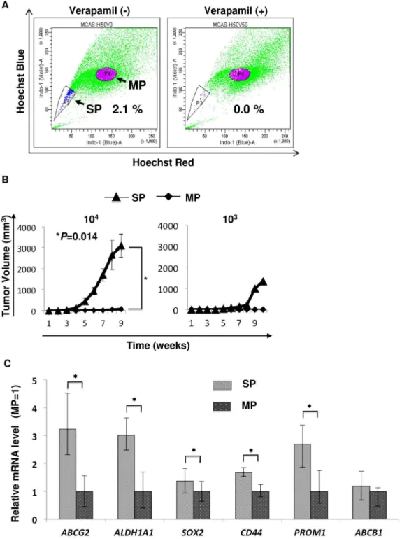
CSCs/CICs were enriched in SP cells
Ovarian CSCs/CICs have been isolated as SP cells from human and mice ovarian cancer line cells [11,21]. We analyzed several gynecological caner cell lines including human ovarian cell lines (MCAS, HTBoA, OVCAR3, OVSAHO) and human endometrial carcinoma (HEC-1) cell line (Figure 1A and Figure S1). SP cells could be detected in all line cells and the SP cell ratio were ranged from 1.2% to 2.6%. Since there is no report describing human mucinous adenocarcinoma line cell MCAS, we thus further analyzed SP cells derived from MCAS. CSCs/CICs have high tumor-initiating ability [22], we thus injected serially diluted numbers of SP cells and MP cells into the backs of three NOD/ SCID mice subcutaneously to examine the tumor-initiating ability. In all three mice, tumors were initiated with 104 SP cells, and tumors were initiated with 104MP cells in 2 of the 3 mice. In one mouse, a tumor was initiated with 103SP cells, while 103MP cells
did not initiate any tumor (Table 1). The size of tumors derived from SP cells was significantly larger than that of tumors derived from MP cells (Figure 1B). The expression levels of stem cell markers were investigated by qPCR, and SP cells derived from MCAS cells expressed higher levels of the stem cell markers
SOX2, CD44 and PROM1 and the ABC transporter gene
ABCG2, whereas ABCB1 was not (Figure 1C). These results indicate that CSCs/CICs were enriched in SP cells derived from MCAS cells. The results were reproduced in at least three independent experiments.
CSCs/CICs were enriched in ALDHBrcells
CSCs/CICs could be isolated as ALDHBr cells by the ALDEFLUOR assay [23]. We therefore examined whether CSCs/CICs can be successfully isolated by the ALDEFLUOR assay. MCAS, HTBoA, OVCAR3, OVSAHO and HEC-1 cells were analyzed by the ALDEFLUOR assay and we found that the ratio of ALDHBrcells was 8.1% to 11.3% (Figure 2A and Figure S1). ALDHBrcells and ALDHLowcells derived from MCAS were sorted and injected into the backs of five NOD/SCID mice to examine the tumor-initiating ability. In all five mice, tumors were initiated with 104ALDHBrcells, while tumors were initiated with 104ALDHLowcells in only 2 of the 5 mice (Table 1). The size of tumors derived from ALDHBr cells was significantly larger than that of tumors derived from ALDHLow cells (Figure 2B). The expression levels of stem cell markers were investigated by qPCR. ALDHBrcells derived from MCAS cells expressed higher levels of the stem cell markersSOX2,CD44andPROM1, ALDH1A1, and the ABC transporter geneABCG2andABCB1(Figure 1C). These results indicate that CSCs/CICs were also enriched in ALDHbr cell derived from MCAS cells. The results were reproduced in at least three independent experiments.
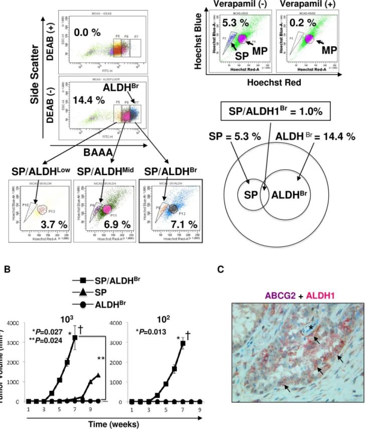
SP and ALDEFLUOR dual analysis
SP cells and ALDHBrcells show greater efflux of Hoechst 33342 dye and higher expression level of aldehyde dehydrogenese, which are different phenotypes, and hematopoietic stem cells were isolated as an SP and ALDHBrpopulation in a previous study [24]. We therefore investigated the overlapping population of SP cells and ALDHBrcells. After staining the MCAS cells with Hoechst 33342 dye, the cells were washed and then stained with ALDEFLUOR reagent and analyzed. In this experiment, the ratio of SP cells was 5.3% and the ratio of ALDHBr cells was 14.4%. 7.1% of ALDHBrcells showed SP population, and 6.9% of ALDHMid cells showed SP population, and only 3.7% of ALDHLowcells showed SP population (Figure 3A). ALDHBrcells exhibited partial overlapping, and only 1.0% of total cells (7.1% of 14.4% population) expressed both SP cell phenotype and ALDHBr phenotype (Figure 3A).
ovarian cancer cells were detectable in ovarian carcinoma tissue. Interestingly, some dual-positive cells exist next to vessels, might be indicating ovarian CSCs/CICs exist in vascular niche. SP/ ALDHBr cells were detected also from other ovarian serous adenocarcinoma line cells (OVSAHO, OVCAR3 and HTBoA) and an endometrial cell line (HEC-1), and the ratios of SP/ ALDHBrcells were 0.1% to 0.8% (Figure S1).
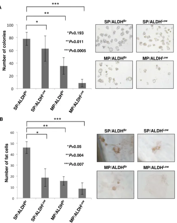
SP/ALDHBrcells have stem cell phenotypes
The SP/ALDHBr population was compared with other populations by the sphere forming assay. SP/ALDHBr, SP/ ALDHLow, MP/ALDHBrand MP/ALDHLowcells were isolated from MCAS cells and cultured in an ultra-low attachment condition for the sphere forming assay. SP/ALDHBr cells exhibited higher sphere formation efficiency than that of MP/
ALDHBrcells and MP/ALDHLowcells (Figure 4A). The difference between SP/ALDHBr cells and SP/ALDHLow cells was not significant; however, SP/ALDHBrcells tend to have higher sphere formation efficiency than that of SP/ALDHLowcells.
CSCs/CICs have been described to have pluripotency [25], we thus analyzed the adipocyte differentiation ability of SP/ALDHBr cells (Figure 4B). Isolated SP/ALDHBr, SP/ALDHLow, MPALDHBrand MP/ALDHLowpopulation were cultured in an adypocyte differentiation condition. SP/ALDHBr cells derived from MCAS cells revealed highest adipocyte differentiation ability compared with SP/ALDHLow, MP/ALDHBrand MP/ALDHLow cells derived from MCAS cells.
CSCs/CICs are resistant to chemotherapy [4], we thus analyzed drug resistance of SP/ALDHBr cells. Since cisplatin is a key drug for ovarian carcinoma chemotherapy, we used cisplatin. Isolated SP/ALDHBr, SP/ALDHLow, MPALDHBrand MP/ALDHLow population were cultured in an existence of cisplatin. SP/ALDHBrcells significantly higher cisplatin resistance compared with SP/ALDHLowcells, MP/ALDHBrcells and MP/ ALDHLow cells. And, MP/ALDHLow cells exhibited highest sensitivity to cisplatin (Figure 5A).
To analyze the molecular characteristics of SP/ALDHBr population, we performed qPCR (Figure 5B). ABCG2 mRNA was preferentially expressed in SP/ALDHBr cells and SP/ ALDHLow cells. ALDH1A1 was expressed in SP/ALDHBr cells and MP/ALDHBr cells. These expression profiles are consistent with the fact that the SP cell phenotype depends on the expression of ABCG2 and the ALDHBr cell phenotype depends on the expression ofALDH1A1. SOX2mRNA was expressed at highest level in SP/ALDHBr cells but not in SP/ALDHLow cells, MP/ ALDHBrcells or MP/ALDHLowcells (Figure 5B), indicating that the SP/ALDHBr population preferentially include a stem cell population. Since SOX2 has a role in the maintenance of lung CSCs/CICs [26], we investigated the relation of SOX2 and expressions ofABCG2andALDH1A1. The expressions ofABCG2
andALDH1A1were reduced inSOX2mRNA knocked down cells (Figure 5C). Furthermore, SOX2 knocked down cells showed lower tumor-initiation than that of control siRNA transfected MCAS cells (Figure 5D). Therefore, these results suggest that SP/ ALDHBr population express high level of stem cell gene SOX2, which may have role in the maintenance of CSCs/CICs and expressions ofABCG2andALDH1A1.
The expression levels of CD44, a representative marker for ovarian CSCs/CICs [27,28], was similar in all populations (Figure 5B). We therefore investigated the relation of SP cells, ALDHBr cells and CD44-positive (CD44+
) cells. The ratio of
CD44+
cells was 8.0%. The ratio of SP/CD44+
overlapping population was 0.9%, and the ratio of ALDHBr/CD44+
overlap-ping population was 3.3%. Furthermore, the ratio of SP/ ALDHBr/CD44+
overlapping population was 0.4% (Figure 6).
Discussion
The concept of CSCs/CICs was proposed long time ago [29]. Leukemia stem cells have been isolated from acute leukemic cells [30,31], and the first CSC/CIC population was isolated from breast carcinoma with the combination of CD44 and CD24 expression [32]. In the following works, CSCs/CICs were successfully isolated in several malignancies. However, since the molecular mechanisms of CSCs/CICs are still elusive, accurate markers for CSCs/CICs are still unknown. Therefore, improve-ments in methods for isolation of CSCs/CICs are still needed.
Combination methods with double or triple markers and with markers and ALDEFLUOR assay have been reported. The populations of ALDHBr and CD44+
/CD242 cells exhibited partial overlapping, and the ALDHBr/CD44+
/CD242population showed higher tumor-initiating ability than that of the ALDHBr population or CD44+/CD242 population [17]. The ALDHBr
/ CD44+
population and ALDHBr/CD133+
population derived from human primary colon carcinoma exhibited higher tumor-initiating ability than that of ALDHBr, CD44+
and CD133+ populations [33]. These findings indicate that the expressions of CSC/CIC markers are partially overlapped and that the overlapped population is highly enriched with CSCs/CICs. Indeed, our results also showed similar overlapping of ALDHBr cells and SP cells, and the overlapping population exhibited higher tumor-initiating ability. In an ovarian cancer study, ALDHBrcells were more enriched in CD44+
cells than in use of the CD133+ cells [23], but the ALDHBr and CD44+overlapping was partial. We identified SP/ALDHBr/CD44+ overlapping population from MCAS cells (Figure 6). Therefore, use of the overlapping population of SP/ALDHBr/CD44+
cells may be a better approach to identify CSC/CIC populations.
Glioma stem cells have been described to differentiate into endothelial cells [34]. Malignant methotelioma stem cells have been described to differentiate into endothelial cells, neural cells and adipocytes [25]. In this study, we confirmed that SP/ALDHBr cells have higher adipocyte differentiation ability. Therefore, ovarian cancer stem cells might have potential to differentiate into different lineage cells, suggesting that ovarian cancer stem cells are immature state. SOX2, a key factor for cell reprogramming [35], was expressed in SP/ALDHBr cells at highest level. And knockdown of SOX2 reduced the expressions of ABCG2 and
ALDH1A1. Thus SOX2 might have a role to sustain immature state of ovarian cancer stem cells.
CSCs/CICs are described to be resistant to chemotherapy [4]. Indeed SP/ALDHBrcells showed higher cisplatin resistance than did SP/ALDHLow cells, MP/ALDHBr cells and MP/ALDHLow cells. Several mechanisms of drug resistance have been described, such as CSCs/CICs are dormant state, CSCs/CICs express higher levels of transporters, CSCs/CICs express higher levels of inhibitor of apoptosis proteins (IAPs) and so on. In this study, only SP/ALDHBr population expressed both ABCB1 and ABCG2 transporters among SP/ALDHBr, SP/ALDHLow, MP/ALDHBr and MP/ALDHLowcells (Figure 6B). Thus, higher expressions of transporters might be one mechanisms of drug resistance of SP/ ALDHBrcells.
Dual SP and ALDHBrcells in hematologic stem cells have been reported [24]. The overlapping population of ALDHBrcells and CD133+
cells has prominent tumorigenicity [36]. However, SP/ Table 1.Summary of tumor initiation incidence.
MCAS Cell Tumor initiation (injected cell number)*
102 103 104
SP/ALDHBrcells 5/5 5/5 n.d.
SP cells 0/3 1/3 3/3
ALDHBrcells 0/5 0/5 5/5
MP cells 0/3 0/3 2/3
ALDHLowcells 0/5 0/5 2/5
MP/ALDHLowcells 0/5 0/5 0/5
*initiating abilities were evaluated at day 70 post cell injection. Tumor-initiation/injection.
n.d.: not determined.
ALDHBrcells have not been reported in solid tumors yet. SP cell phenotype represents the efflux of Hoechst 33342 dye due to the expression of ABC transporter, ABCG2, which may be involved in drug resistance [37]. ALDHBrcells represent the higher expression of ALDH1, which may be involved in detoxification [38]. SP cells
and ALDHBrcells thus have different molecular properties, and the overlapping of SP cells and ALDHBrcells were partial. And we found SP and ALDHBr overlapping population was the highest CSCs/CICs enriched population which exhibited higher sphere formation, higher tumor-initiation, higher adipocyte
differentia-Figure 2. MCAS CSCs/CICs are enriched in ALDHBr cells. A. Detection of ALDHBr cells from MCAS cells. MCAS ovarian mucinous
adenocarcinoma cells were stained with BAAA and analyzed. The percentage represents the ratio of ALDHBrcells. Inhibitor indicate ALDH1 inhibitor
(diethylamino- benzaldehyde (DEAB)). B. Tumor initiation of ALDHBrcells derived from MCAS cells. 104ALDHBrand ALDHLowcells derived from MCAS
cells were inoculated subcutaneously into the backs of NOD/SCID mice, and tumor growth was measured weekly. Data represent means6SD. Differences between ALDHBrand ALDHLowcells were examined for statistical significance using Student’s t-test. *P values. C. qPCR of CSC/CIC markers in MCAS SP and MP cells. Data represent means6SD. Asterisks indicate significant difference. *P,0.05. t-test.
Figure 3. SP and ALDEFLUOR dual assay.A. Summary of SP and ALDEFLUOR dual assay. MCAS cells were stained by Hoechst 33342 dye and then stained by BAAA and analyzed. The cells were divided into three groups according to ALDH intensity (ALDHBr(ALDH bright), ALDHMid(ALDH middle), ALDHLow(ALDH low)), then analyzed by SP assay. The ratio of ALDHBrcells was 14.4%, and the ratio of SP cells was 5.3%. The ratios of SP cells
in ALDHBrcells, ALDHMidcells and ALDHLowcells were 7.1%, 6.9% and 3.7%, respectively. The ratio of SP/ALDHBrcells in total cells was 1.0%. B. Tumor initiation of SP/ALDHBr, SP and ALDHBrcells. 102and 103SP/ALDHBr, SP and ALDHBrcells derived from MCAS cells were inoculated subcutaneously
into the backs of NOD/SCID mice, and tumor growth was measured weekly. Data represent means6SD. Differences between SP/ALDHBrand SP cells
or ALDHBr cells were examined for statistical significance using Student’s t-test. *P values. Daggers indicate mice death due to tumor. C.
Immunohistochemical staining of ABCG2 and ALDH1. Ovarian carcinoma tissue was stained by anti-ABCG2 antibody and anti-ALDH1 antibody. Brown membrane staining indicates ABCG2 and cytoplasm pink staining indicates ALDH1. Asterisk indicates vessel, and arrows indicate ABCG2 and ALDH1 double-positive ovarian carcinoma cells. Magnification,6400.
tion ability, higher drug resistance and higher expression level of SOX2, a representative marker of CSCs/CICs, which is related to the tumor-initiating ability of CSCs/CICs [26]. Therefore, SP/ ALDHBrpopulation is the better source of CSCs/CICs than SP cells or ALDHBr cells that have been previously described. We found that SOX2 is expressed at high level in SP/ALDHBr cells, and knockdown of SOX2 suppressed the expressions ofALDH1A1
andABCG2, and suppressed tumor-initiation. Thus, SOX2 might has role in the maintenance of both SP cell and ALDHBrcell population, and also has role in the maintenance of ovary CSCs/CICs.
In summary, SP/ALDHBrcells comprise a more highly CSC/ CIC- enriched popuration than do SP cells or ALDHBrcells, and further analysis of SP/ALDHBrcells should lead to elucidation of the molecular mechanisms of CSCs/CICs.
Figure 4. Characterization of SP/ADLHBrcells.A. Sphere formation assay. The numbers of colonies from four fractions (SP/ALDHBr, SP/ALDHLow, MP/ALDHBrand MP/ALDHLow) were evaluated at day 7. Data represent means
6SD. The differences were examined for statistical significance using Student’s t-test. *P values. Representative images of spheres are shown (6100). B. Adipocyte differentiation assay. The cells were cultured under
existence of trans-retinoic acid followed by adipocyte differentiation medium. Oli Red O-positive adipocytes were counted. Data represent means6
SD. The differences were examined for statistical significance using Student’s t-test. *P values. Representative images of Oil Red O-staining are shown (6200). Red-staining indicate adipocyte differentiation.
Figure 5. Charasterization of SP/ALDHBr cells.A. Cell viability assay. The cells were cultured under existence of serially diluted cisplatin. The viable cells were analyzed by WST-1 kit. Y-axis indicates the viability of cells. Data represent means6SD. The differences were examined for statistical significance using Student’s t-test. *P values. B. qPCR analysis. The expression of stem cell markers was examined using SP/ALDHBr, SP/ALDHLow, MP/
ALDHBrand MP/ALDHLowcells. Data represent means
6SD. Asterisks indicate significant difference. *P,0.05. t-test. C.SOX2knockdown suppress the expressions ofALDH1A1andABCG2.SOX2mRNA was knocked down by siRNA. Two days after transfection of SOX2 siRNA, the expressions of
ALDH1A1andABCG2were investigated by RT-PCR.GAPDHwas used as an internal control. Control siRNA (si-Cont) transfected cells were used as negative control. D. SOX2 knock down suppress the tumor-initiation.SOX2mRNA was knocked down by siRNA. Ten thousand si-SOX2 and control siRNA (si-Cont) transfected cells were inoculated subcutaneously into the backs of NOD/SCID mice, and tumor growth was measured weekly. Data represent means6SD. Differences were examined for statistical significance using Student’s t-test. *P values.
Supporting Information
Figure S1 SP and ALDEFLUOR dual assay. OVCAR3, OVSAHO, HTBoA and HEC-1 cells were analyzed by SP and ALDEFLUOR dual assay. Percentages indicate the ratios of ALDHBr, SP and SP/ALDHBrcells.
(TIF)
Acknowledgments
The authors thank Ms. Eri Saka for technical assistance.
Author Contributions
Conceived and designed the experiments: KY TT YH NS. Performed the experiments: KY RM TK AT VK HA. Analyzed the data: KY TT YH NS. Contributed reagents/materials/analysis tools: KY TK JM TS TH. Wrote the paper: KY TT YH NS.
References
1. Rosen JM, Jordan CT (2009) The increasing complexity of the cancer stem cell paradigm. Science 324: 1670–1673.
2. Ghaffari S (2011) Cancer, stem cells and cancer stem cells: old ideas, new developments. F1000 Med Rep 3: 23.
3. Hirohashi Y, Torigoe T, Inoda S, Morita R, Kochin V, et al. (2012) Cytotoxic T lymphocytes: Sniping cancer stem cells. Oncoimmunology 1: 123–125. 4. Dean M, Fojo T, Bates S (2005) Tumour stem cells and drug resistance. Nat Rev
Cancer 5: 275–284.
Figure 6. SP, ALDEFLUOR and CD44 triple staining.MCAS cells were stained by Hoechst 33342 dye, BAAA and anti-CD44 antibody, and analyzed. The ratios of SP, ALDHBr, CD44+
, SP/ALDHBr, SP/CD44+
, ALDHBr/CD44+
and SP/ALDHBr/CD44+
cells were 5.3%, 14.4%, 8.0%, 1.0%, 0.9%, 3.3% and 0.4%, respectively.
5. Rich JN (2007) Cancer stem cells in radiation resistance. Cancer Res 67: 8980– 8984.
6. Tirino V, Desiderio V, Paino F, De Rosa A, Papaccio F, et al. (2012) Cancer stem cells in solid tumors: an overview and new approaches for their isolation and characterization. FASEB J.
7. Lehmann C, Jobs G, Thomas M, Burtscher H, Kubbies M (2012) Established breast cancer stem cell markers do not correlate with in vivo tumorigenicity of tumor-initiating cells. Int J Oncol 41: 1932–1942.
8. Shmelkov SV, Butler JM, Hooper AT, Hormigo A, Kushner J, et al. (2008) CD133 expression is not restricted to stem cells, and both CD133+ and CD1332metastatic colon cancer cells initiate tumors. J Clin Invest 118: 2111– 2120.
9. Burkert J, Otto WR, Wright NA (2008) Side populations of gastrointestinal cancers are not enriched in stem cells. J Pathol 214: 564–573.
10. Pecorelli S, Favalli G, Zigliani L, Odicino F (2003) Cancer in women. Int J Gynaecol Obstet 82: 369–379.
11. Szotek PP, Pieretti-Vanmarcke R, Masiakos PT, Dinulescu DM, Connolly D, et al. (2006) Ovarian cancer side population defines cells with stem cell-like characteristics and Mullerian Inhibiting Substance responsiveness. Proc Natl Acad Sci U S A 103: 11154–11159.
12. Deng S, Yang X, Lassus H, Liang S, Kaur S, et al. (2010) Distinct expression levels and patterns of stem cell marker, aldehyde dehydrogenase isoform 1 (ALDH1), in human epithelial cancers. PLoS One 5: e10277.
13. Zhang S, Balch C, Chan MW, Lai HC, Matei D, et al. (2008) Identification and characterization of ovarian cancer-initiating cells from primary human tumors. Cancer Res 68: 4311–4320.
14. Friel AM, Sergent PA, Patnaude C, Szotek PP, Oliva E, et al. (2008) Functional analyses of the cancer stem cell-like properties of human endometrial tumor initiating cells. Cell Cycle 7: 242–249.
15. Goodell MA, Brose K, Paradis G, Conner AS, Mulligan RC (1996) Isolation and functional properties of murine hematopoietic stem cells that are replicating in vivo. J Exp Med 183: 1797–1806.
16. Inoda S, Hirohashi Y, Torigoe T, Morita R, Takahashi A, et al. (2011) Cytotoxic T lymphocytes efficiently recognize human colon cancer stem-like cells. Am J Pathol 178: 1805–1813.
17. Ginestier C, Hur MH, Charafe-Jauffret E, Monville F, Dutcher J, et al. (2007) ALDH1 is a marker of normal and malignant human mammary stem cells and a predictor of poor clinical outcome. Cell Stem Cell 1: 555–567.
18. Michifuri Y, Hirohashi Y, Torigoe T, Miyazaki A, Kobayashi J, et al. (2012) High expression of ALDH1 and SOX2 diffuse staining pattern of oral squamous cell carcinomas correlates to lymph node metastasis. Pathol Int 62: 684–689. 19. Billon N, Iannarelli P, Monteiro MC, Glavieux-Pardanaud C, Richardson WD,
et al. (2007) The generation of adipocytes by the neural crest. Development 134: 2283–2292.
20. Nakatsugawa M, Hirohashi Y, Torigoe T, Asanuma H, Takahashi A, et al. (2009) Novel spliced form of a lens protein as a novel lung cancer antigen, Lengsin splicing variant 4. Cancer Sci 100: 1485–1493.
21. Vathipadiekal V, Saxena D, Mok SC, Hauschka PV, Ozbun L, et al. (2012) Identification of a potential ovarian cancer stem cell gene expression profile from advanced stage papillary serous ovarian cancer. PLoS One 7: e29079.
22. Hirohashi Y, Torigoe T, Inoda S, Takahashi A, Morita R, et al. (2010) Immune response against tumor antigens expressed on human cancer stem-like cells/ tumor-initiating cells. Immunotherapy 2: 201–211.
23. Wang YC, Yo YT, Lee HY, Liao YP, Chao TK, et al. (2012) ALDH1-bright epithelial ovarian cancer cells are associated with CD44 expression, drug resistance, and poor clinical outcome. Am J Pathol 180: 1159–1169. 24. Pierre-Louis O, Clay D, Brunet de la Grange P, Blazsek I, Desterke C, et al.
(2009) Dual SP/ALDH functionalities refine the human hematopoietic Lin-CD34+CD382stem/progenitor cell compartment. Stem Cells 27: 2552–2562. 25. Varghese S, Whipple R, Martin SS, Alexander HR (2012) Multipotent cancer stem cells derived from human malignant peritoneal mesothelioma promote tumorigenesis. PLoS One 7: e52825.
26. Nakatsugawa M, Takahashi A, Hirohashi Y, Torigoe T, Inoda S, et al. (2011) SOX2 is overexpressed in stem-like cells of human lung adenocarcinoma and augments the tumorigenicity. Lab Invest 91: 1796–1804.
27. Shi MF, Jiao J, Lu WG, Ye F, Ma D, et al. (2010) Identification of cancer stem cell-like cells from human epithelial ovarian carcinoma cell line. Cell Mol Life Sci 67: 3915–3925.
28. Wang L, Mezencev R, Bowen NJ, Matyunina LV, McDonald JF (2012) Isolation and characterization of stem-like cells from a human ovarian cancer cell line. Mol Cell Biochem 363: 257–268.
29. Clevers H (2011) The cancer stem cell: premises, promises and challenges. Nat Med 17: 313–319.
30. Lapidot T, Sirard C, Vormoor J, Murdoch B, Hoang T, et al. (1994) A cell initiating human acute myeloid leukaemia after transplantation into SCID mice. Nature 367: 645–648.
31. Bonnet D, Dick JE (1997) Human acute myeloid leukemia is organized as a hierarchy that originates from a primitive hematopoietic cell. Nat Med 3: 730– 737.
32. Al-Hajj M, Wicha MS, Benito-Hernandez A, Morrison SJ, Clarke MF (2003) Prospective identification of tumorigenic breast cancer cells. Proc Natl Acad Sci U S A 100: 3983–3988.
33. Huang EH, Hynes MJ, Zhang T, Ginestier C, Dontu G, et al. (2009) Aldehyde dehydrogenase 1 is a marker for normal and malignant human colonic stem cells (SC) and tracks SC overpopulation during colon tumorigenesis. Cancer Res 69: 3382–3389.
34. Wang R, Chadalavada K, Wilshire J, Kowalik U, Hovinga KE, et al. (2010) Glioblastoma stem-like cells give rise to tumour endothelium. Nature 468: 829– 833.
35. Takahashi K, Yamanaka S (2006) Induction of pluripotent stem cells from mouse embryonic and adult fibroblast cultures by defined factors. Cell 126: 663– 676.
36. Silva IA, Bai S, McLean K, Yang K, Griffith K, et al. (2011) Aldehyde dehydrogenase in combination with CD133 defines angiogenic ovarian cancer stem cells that portend poor patient survival. Cancer Res 71: 3991–4001. 37. Goodell MA, Rosenzweig M, Kim H, Marks DF, DeMaria M, et al. (1997) Dye
efflux studies suggest that hematopoietic stem cells expressing low or undetectable levels of CD34 antigen exist in multiple species. Nat Med 3: 1337–1345.